neuro psychology
1/72
There's no tags or description
Looks like no tags are added yet.
Name | Mastery | Learn | Test | Matching | Spaced |
|---|
No study sessions yet.
73 Terms
occipital lobe
the area at the back of the brain which controls vision
often also known as the visual cortex as its main job is controlling all things related to processing visual information
weight of the brain
1.4kg
parietal lobe
the area at the top of the brain that plays an important role in perceptions, language and sensations of touch. ( gives the ability to identify faces)
At the front of the parietal lobe just behind the central sulcus is the somatosensory cortex which is responsible for sense of touch.
cerebellum
a part of the brain near the brainstem that controls motor movements
regulates movement, posture, coordination and balance
takes information from different senses. Our spinal cord and other parts of the brain combines them to coordinate behaviour
(For example, if we are running and see an object in our way, the cerebellum combines this information and sends a message back to the body, telling it to move to avoid the object. The message is sent via the spinal cord telling us to change direction while helping us keep our balance so we do not fall as we dodge the object in our way.)
temporal lobe
the area on the side of the brain that controls hearing, learning and feelings.
contains auditory cortex - controls hearing
frontal lobe
the area at the front of the brain responsible for decision making, impulse control, memory, behaviour and movement
at the back of the frontal lobe is the motor cortex which is just in front of the central sulcus.
The motor cortex plays a large role in voluntary movements.
cerebrum
the largest part of the brain where higher processing happens. Includes the cortex.
divided into 4 sections called lobes
a gyrus is a ridge on the surface of the brain (out) each ridge is surrounded by (openings/slices/indents) known as sulci.
Brain definition
an organ in your head made up of nerves that process information and control behaviour
cortex
the outer layer of the brain
the large surface area of the human brain allows it to have more nerve cells and allows it to control more functions. Animals have fewer behavioural functions than humans and therefore have ‘smoother’ (less surface area) brains.
hemisphere
half of the brain
brain stem
the part of the brain that connects the spinal cord to the upper brain.
responsible for breathing, heart rate + temperature
spinal cord
a pathway of nerves inside the spine which connects the brain with the rest of the body + / peripheral nervous system
reflexes vs reaction
reflex - actions that are automatic and do not require conscious thought. sensory nerves bypass the brain and go up the spinal cord. Faster.
reaction - voluntary. Takes place through sensory nerves which relay messages from the brain to the motor nerve
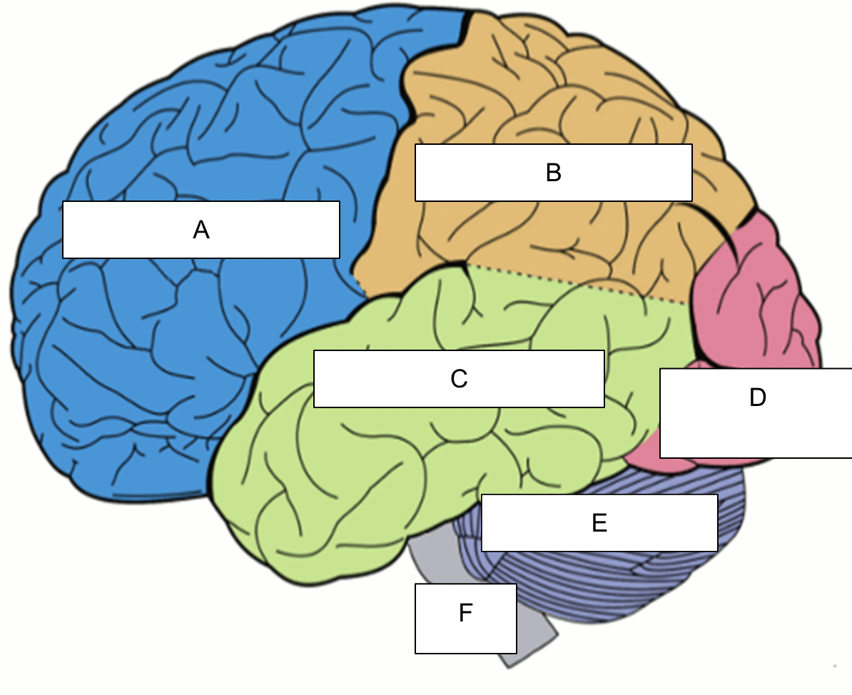
label
A - Frontal lobe
B - Parietal lobe
C - temporal lobe
D - occipital lobe
E - cerebellum
F - brain stem
central nervous system
the brain and the spinal cord which relays messages from the brain to the rest of the body to instruct it what to do
main roles -
acts as the command center of the body
interprets incoming sensory information
sends out instructions on how the body should react
how does the CNS work example - ruler falling
the eyes see the ruler is dropped
the visual system in the brain sends a message saying that the ruler is falling
the motor cortex in the brain (CNS) send a message telling the body to react to catch the falling ruler
the spinal cord sends a message to nevres (PNS) in the muscles telling them to react
The hand muscles contract allowing the fingers to catch the ruler
Peripheral nervous system
the system of nerves that connects to the central nervous system (mainly the spinal cord) to the skin, muscles and organs in the body
made up of motor and sensory neurons
it monitors the conditions inside and outside the body
motor and sensory neurons
motor - tells the peripheral nervous system what to move
sensory - tells the central nervous system about external stimuli
synaptic transmission
the process by which neurotransmitters are released by a neuron, move through the synaptic gap and get taken up by another neuron.
terminal button
the end of a neuron
vesicles
small sacks containing neurotransmitter molecues
neurotransmitters
chemicals found within the nervous system that pass messages from one neuron to another across a synapse
neurotransmitters are released from neurons when a nerve impulse reaches the end of a nerve fibre. The neurotransmitter is then picked up by another neuron to receive the message.
There are different types of neurotransmitters that have different jobs
types of neurotransmitters and examples of what they do
Dopamine - plays a role in attention and learning. Not enough dopamine can make it difficult to concentrate on tasks
Serotonin - Plays a role in mood. Not enough serotonin can make people depressed.
GABA - plays a role in calming us down. When we are stressed we produce GABA to relax us.
neuron
a nerve cell that transmits information
synapse
a gap between 2 neurons that allow messages in the form of neurotransmitters to pass from one cell to another
axon
the long structure that connects the cell body of a neuron to the terminal button at the end of the cell.
receptors
special sites on neurons that are designed to absorb neurotransmitter molecules
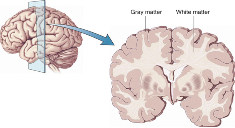
gray matter
nerve tissue in the CNS composed of neuron cell bodies, neuroglia and unmyelinated axons
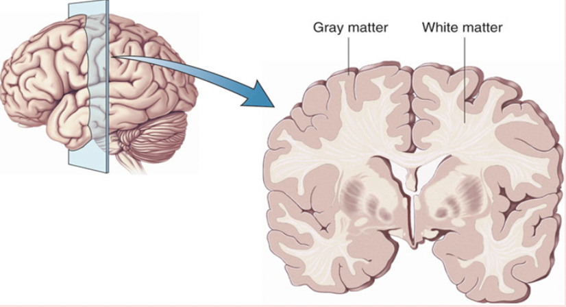
white matter
white matter is nerve tissue in the CNS composed chiefly of bundles of myelinated axons. This is where the neurons pass their messages. If this area is damaged messages cannot be passed.
Aims of damasio et al
to build a model of Gage’s skull so they could map out how the rod passed through his head
they aimed to create a 3D model on the computer
They wanted to identify which areas of the brain would have been damaged
Damasio et al. procedure
Damasio et al exhumed Gage’s skull and the rod
the team took pictures and measurements of the skull and built a 3D computer replica.
They narrowed down the entry and exit points from 36 to one and mapped which areas would have been damadged
damasio et al results
Brain damage suffered in the accident was likely to only have affected the frontal lobe
no other brain areas were harmed
the iron rod went through his left eye socket and up through the head
Damage to white matter and neurons meant that Gage was unable to pass neural messages in this part of his brain making it useless
damage in both hemispheres seemed to be worse in the middle of the underside while the top edges of the frontal lobe were less affected
12 patients with frontal lobe damage were compared to Phineas Gage and they all had similar issues with making decisions and processing emotions
Damasio et al. conclusions
the research conducted by damasio et al showed that only the frontal lobe (especially the ventromedial region) was damaged during the accident
Phineas Gage’s behaviour/personality changed after the accident
he found it difficult to control his impulses (he started gambling) and he found it difficult to make sensible decisions.
ventromedial area of the frontal lobe is important for making sensible decisions and controlling our impulses around people and also important for emotions
strengths of damasio et al.
able to use modern day technology to investigate data from 1848 so results so results can have scientific status (evidence could actually be seen rather than inferred)
can make predictions and use the study for similar frontal lobe damage cases in the future (predict behaviour of future patients - can help treatment)
weaknesses of damasio et al
had to use replica of skull - may not be reliable
Information may not be accurate as it was from 150y before the experiment
cannot be applied to future cases where frontal lobe is damaged (Phineas Gage case was unique and the exact same thing would have to happen to someone else)
lateralisation of function in the brain
each hemisphere of the brain has a different job.
some behaviour are controlled more from the right than the left and vice versa
asymmetrical function
the two hemispheres are not exactly the same
the difference between the two sides of the brain makes it look asymmetrical as although they look very similar they are not a mirror image
each side of the brain appears to control the functions of the opposite side of the body
( e.g hand control : the right hemisphere is in control of the left hand while the left hemisphere controls the right hand.)
corpus callosum definition
a thick layer of nerve fibers that connects the two hemispheres which allows the two hemispheres to send messages to eachother so the whole brain can work as a complete organ
functions of the left hemisphere
analytical, logical, precise, repetitive, organised, details, scientific, detached, literal, sequential
processing language
Broca’s area
writing + understanding language
functions of the right hemisphere
creative, imaginative, general, intuitive, conceptual, big picture, heuristic, empathetic, figurative, irregular
spatial awareness
recognising faces
music processing
processing visual information
Broca’s aphasia
when the Broca’s area is damaged and the patient has trouble producing speech.
symptoms - stuttering, finding it hard to identify an object verbally
Male brains
good spatial skills (right brain) (e.g imagining what a shape would look like if it were from a different angle)
using each hemisphere separately
Female brains
language skills (left brain) (e.g speech production)
thicker corpus callosum - both sides of the brain used more
sex differences theory strengths
Harasty 1997 said parts of the brain that process and produce language are slightly bigger in females. (evidence + explains why women are better at language tasks)
Scientific methods used to investigate the differences between male/female brains (e.g brain scans) (help to prevent the interference of extraneous variables)
sex differences theory weaknesses
Rilea found that males do not always do better than females at spatial tasks
Sommer said there was no strong evidence that females used both hemispheres for language tasks
(evidence that supports that there are no sex differences in lateralisation of brain function)
neurological damage definition
any damage to parts of the nervous system. It is caused by trauma or disease
Agnosia definition
problem with the way the brain processes sensory information. The brain is unable to make sense of the information.
what is visual agnosia and why does it occur
a disorder where a person can see perfectly well but cannot understand what they are seeing. Happens due to damage to the occipital or parietal lobe.
example : can see a kettle but cannot explain that it is a kettle
symptoms of visual agnosia
information sent from the eyes to the brain cannot be understood
patients might not be able to recognise the colour of an object or the object itself and name it
Patients might not be able to recognise places they are familiar with
what is prosopagnosia and why does it occur
Face blindness
unable to recognise faces even though they can be seen
even if they know the person really well
happens due to damage to the fusiform face area located at the back of the temporal lobe next to the occipital lobe
prosopagnosia symptoms
find it difficult to identify people from their faces
some people find that they see all faces the same and cannot tell faces apart
others have more trouble with matching up pictures of faces. they do not know
prefrontal lobe damage
(Role of the frontal lobe - helps control impulses, helps keep our emotions balanced)
when the frontal lobe becomes damaged people can become impulsive and aggressive
example - Rein et al 1997 studied the brains of murders and compared these to a similar group of people who had not committed murder. He found differences in the prefrontal lobe cortex. Murders had less activity in the prefrontal lobe making them more aggressive and impulsive
Sperry (1968) Aims
to investigate the cognitive functions that are linked to each hemisphere in the brain
to investigate the behavioural. neurological and psychological effects of the split-brain surgery on the patients
what is corpus callosotomy
when the corpus callosum is cut so the left and right hemispheres can no longer communicate this reduces seizures in epileptic patients
Sperry 1968 procedure participant information
11 participants who had corpus callosotomy
9 had recent surgery and 2 had surgery a while ago with excellent recovery
they had surgery because they had epilepsy
Sperry 1968 procedure tasks general
tasks to test the right and left halves of the visual field separately or together. And the right and left hands and legs with vision excluded
Sperry 1968 Task 1
Patients looked at a spot and projected words or pictures would show on either side of the sport for 1/10th of a second
information only went into the right or left hemisphere
patients either needed to say, draw or select the object behind the screen to identify what they have seen
Sperry Task 1 results
words projected to the right side could only be spoken and written but not with the right hand. Patients could find an item with their left hand when presented on the right
(this showed that the left visual field and hand was processed by the right hemisphere and vice versa)
Sperry task 2 (object from bag) + results
participants were blindfolded and asked to pick an object from a bag and name it
using the right hand the participants could name it. Using the left they couldn’t name it but could retrieve it again
Sperry task 3 (clock) + results
participants were shown a wall clock to the right hemisphere. Patients were asked to pick up closest to what they have seen with their left hand
patients could pick up a wrist watch with their left hand which shows limited language processing ability in the right hemisphere
Sperry task 4 (sum) + results
participants were shown a sum to the right hemisphere and had to point with their left hand to the right answer
patients left hand could point to the correct answer. This demonstrated that the right hemisphere is involved in basic calculations
Sperry task 5 (nude) + results
a nude was presented to the right hemisphere to see the participants reaction
the participants were blushing and giggling with no verbal report to having seen the image
right hemisphere is involved in emotional processing
Sperry task 6 (block design) + results
block design tests - spatial tasks
right hemisphere superior to left in tasks involving drawing spatial relationships and performing block design tests
Sperry 1968 conclusions
each hemisphere can work independently without being connected however, they have different roles.
the left hemisphere is better at naming items using words - language
the right hemisphere is better at identifying objects through touch. - spatial abilities
Sperry 1968 strengths
gathered a lot of detailed information - improves reliability
designed procedures that could be kept the same for each participant. Allows the results between participants to be compared more easily. + Increases reliability
Sperry 1968 weaknesses
small sample (11pp) - cannot be generalised.
Very few people have a surgery to sever the corpus callosum and so results might not be very useful to explain how brains without the surgery work.
Artificial tasks(lab experiment) - lacks ecological validity.
neuroscience + post mortem
neuroscience - the scientific study of the brain and nervous system
post mortem - an examination of a body after death, often to workout how and why the person died
psychology definition
the scientific study of the human mind and it’s functions especially those affecting behaviour in a given context
was considered a philosophical discipline but now a science
early psychology
could only be studied post mortem
Wundt - started to link physical brain and human behaviour
EEG (electroencephalograph)
developed in 1924 by Hans Beger
measures brainwave activity → start of studying brain without patient being dead
electrodes are placed onto the scalp to see what parts of the brain have the highest activity during an activity
MRI and PET
EEG lead to MRI and PET
MRI - a method of studying the brain using electromagnets
PET - imagery showing the amount of energy being used throughout the brain
show images of what the brain looks like/ image that shows what parts of the brain are active
advantages of brain scans (MRI and PET)
provides the opportunity to help people living with brain damage
we can see where the damage is + understand how that part of the brain is working/or not working
Modern technology
modern technology is being developed which allows psychologists to use high powered microscopes to look at how individual synapses work
from this theories can be developed about exactly which parts of the brain control what kinds of behaviour
possible to investigate behaviour at the level of a neuron → help develop our understanding of what the brain can do
Brain scans - identify areas of the brain that are associated with certain tasks