Fussmann Combined
1/334
There's no tags or description
Looks like no tags are added yet.
Name | Mastery | Learn | Test | Matching | Spaced |
|---|
No study sessions yet.
335 Terms
Species (phylogenetically)
The lowest taxonomic unit with uncontroversial membership.
Nodes represent _______ in a phylogeny.
Taxa

Lines represent _______ in a phylogeny.
Descent
Monophyletic group (synonym and definition)
Also known as a clade; a group of organisms with a single common ancestor, defined by shared derived character traits.
Metazoa
Term for animals.
Two objectives of phylogeny
A. To understand how one kind of animal evolves from another.
B. To discover the relationships between the kinds of animals
Classically, A is used to find B, but with molecular data, the opposite can happen.
Phylum
A monophyletic group that comprises animals of the same fundamental body plan. Classification of phyla continues to be controversial
What are animals?
Motile multicellular organisms with somatic differentiation (usually).
Traits:
Multicelluar
Heterotropic
Nervous System (arises later in the phylogeny)
Mobile stages
Cell differentiation
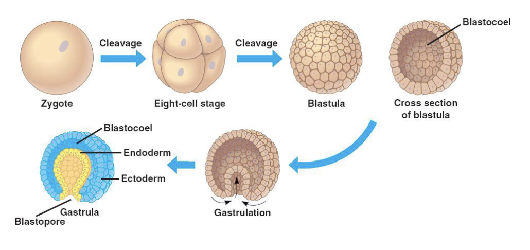
Blastula stage
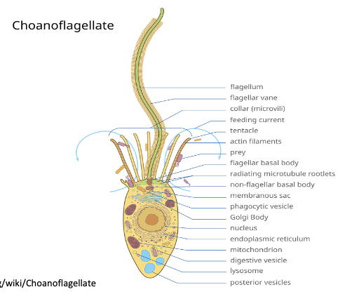
Choanoflagellata
Sister taxon of Metazoa
Single flagellum
Base of flagellum is surrounded by collar of actin filaments (microvilli)
Flagellar movement draws water through the collar, trapping edible particles (i.e. bacteria)
May be free-swimming or sessile.
Sessile forms tend to be colonial - connected with an ECM - likely resembles ancestral Porifera
Resemble choanocytes found in Porifera
Choanocytes
Porifera cell type, resemble Choanoflagellata.
Have the same general cell structure, Microvilli collar
Metazoan traits
Epithelial cells
Mesenchymal cells
Epithelial cells
Polarized
Arranged in parallel
Basement membrane
Joined by belt-form junctions

Mesenchymal cells
No particular alignment with other mesenchymal cells
Bear only spot-form junctions
Surrounded by ECM
The blastula
Ball of hollow cells
Comes from equal radial cleavage
2 main difficulties: Cannot feed (no mouth, cannot further develop because of ciliation (if cilia were shed, the embryo would sink).
Solution=gastrulation
Gastrulation
Fundamental feature of metazoans,
Cells on the interior can divide/ differentiate without compromising motility.
Five known clades of Metazoa
Porifera (sponges)
Placozoa (“flat animals”)
Cnidaria (jellyfish, corals, hydroids, etc)
Ctenophora (comb jellies)
Bilateria (everything else
Placozoa
“Ciliated plates”
Single known species
Up to 2 mm size
Global distribution in littoral of tropical and subtropical oceans
Development only observed up to 64 cells

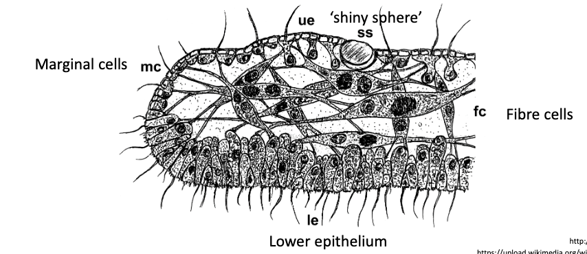
Placozoan anatomy
Simpler organization than any other living metazoan:
Sandwich organization: Upper and lower epithelium with loose network of fiber cells
No mouth, organ systems, nerve or muscle cells
No symmetry of any kind
Irregular body shape, constantly changes
Placozoan feeding
Moves on surfaces via ciliary creeping
Feeds by overlaying an algal cell and lysing it with extracellular digestive enzymes.
Grades of organization in metazoa
Non-epithelial (incomplete compartmentalization):
Porifera
Placozoa
Epithelial diploblasts:
Cnidaria
Epithelial triploblasts:
Bilateria
Choanocyte
Uniflagellate cell with a collar of microvilli.
Draws water through the collar
Retains particles larger than 0.2 micrometers (ex. bacteria)
Ingests particles via phagocytosis, transfers to Archaeocytes for digestion
Each choanocyte captures ~ 3-4 bacteria every day.
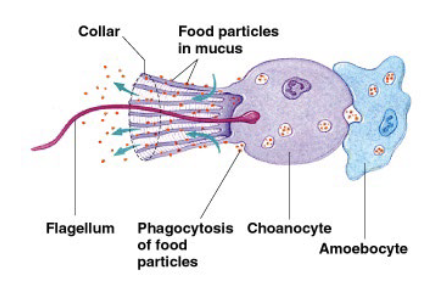
Archaeocytes/ Amoebocytes
Porifera cells that digest bacteria
Totipotent cells
Found in mesohyl
Porocytes
“Doughnut-shaped” cells
Form pores through which water is drawn into the sponge
Bridge the mesohyl
Spongocoel
Interior of the sponge
Pinacoderm
Outer layer of protective cells in sponges.
Not real epithelium
Mesohyl
Gelatinous layer between pinacoderm and choanocyte chambers
Contains archaeocytes
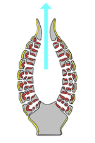
Spicules
Porifera structure and protection
Made of calcite, silicate, or protein
Distributed in mesohyl
Give shape to sponge body
Often characteristic to species
All Porifera cell types:
Choanocytes
Pinacocytes
Archaeocytes
Porocytes
Osculum
Opening of the spongocoel
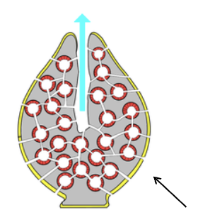
Asconoid Body Plan
Found in Porifera
Simplest body plan
Choanocytes arranged on spongocoel walls
Syconoid Body Plan
Folded inner wall
Increased surface area for choanocytes
Increased power of water flow
Allows for larger individuals
Leuconoid Body Plan
Folded folds create chambers:
Allows sponges to grow to 1m+ size
Vegetative Propogation (Sponges)
Budding, branching, fragmentation possible
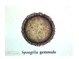
Freshwater sponges survive winter as reduction bodies called gemmules
Sexual Cycle (Sponges)
No discrete gonads
Ova and sperm develop from somatic cells
Most sponges are hermaphroditic
Sperm are released en masse into the water
Sperm are then phagocytosed by choanocytes of neighbouring individuals
Then de-differentiated into ameboid carrier cells
Migrate into mesohyl and fertilize oocytes

Sponge Development
Wide range of development
Cleavage is usually total and equal (no order/ size differentiation)
Late blastulae experience extensive cellular reorganizations
Homology of developmental processes in sponges with gastrulation and germ layer formation in other metazoans uncertain
Sponge life cycle
Small, ciliated pelagic larve
Metamorphosis into benthic adult
Lecithotrophic larvae (feed on maternally supplied yolk)
Lecithotrophic larvae
Larvae that feed on maternally supplied yolk
Planktotrophic larva
Rely on external food sources
Sponge symbionts
Wide variety of symbiotic microbes in the mesohyl
Up to 30% of the wet weight of sponges
Some (e.g. cyanobacteria) are mutualists
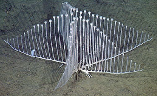
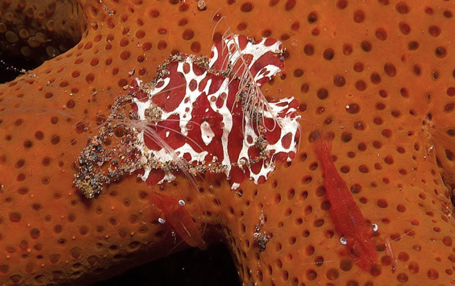
Carnivorous sponges
Demosponges in the family Cladhorizae can capture living prey (i.e. shrimp)
Use hooked spicules
Most live in deep water (below 1000m) or in caves
Some retain chambers and choanocytes
Others have lost their choanocytes
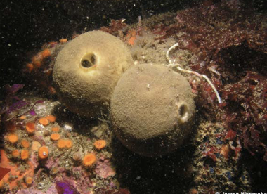
Calcarea (Calcispongiae)
Sponge clade
Sister group of Homoscleromorpha
3-4 radiate spicules made of calcium carbonate
Mostly small, drab, inconspicuous
Simple asconoid or syconoid body plans
~ 400 species
Homoscleromorpha
Porifera clade
Sister group of Calcarea
Reduced skeleton of uniform siliceous spicules (without axial filament)
Epithelium with basement membrane and specialized cell junctions
Possibly branched later than other Porifera

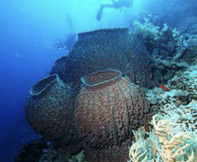
Demospongiae
Porifera clade
Sister group of Hexactinellida
Siliceous spicules organized around an axial filament (shared with Hexactinellida)
Spicules not with six rays
Spongin network often present
Mostly complex leuconoid body plan
Ocean and freshwater
Includes carnivorous sponges
All of the really big sponges
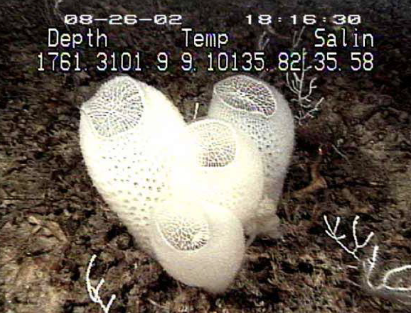
Hexactinellida
Porifera clade
Sister group of Demospongiae
Siliceous spicules organized around an axial filament (shared with Demospongiae)
Rigid skeleton of 6-pointed siliceous spicules
Syncytial network of soft body tissue
“Collar bodies” instead of “collar cells”
Deep-water forms
Most common in cold seas, especially the Antarctic
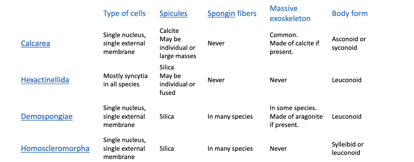
Porifera groups summary sheet
Classes of Cnidaria
Three classical classes
Anthozoa: Corals and anemones
Scyphozoa: Jellyfish
Hydrozoa
Two additional classes:
Staurozoa: Stalked jellyfish
Cubozoa: Box jellyfish
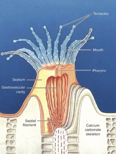
Anthozoa
Cnidarian class
Subclasses:
Hexacorrallia
Octocorrallia
Most corals and sea anemones
Polyp only - completely suppressed medusa
Includes reef-building corals with massive calcareous exoskeleton for zooids
Medusozoa
Cnidarian class
Sister group of Anthozoa
Contains 2 sister groups:
Staurozoa (stalked jellyfish), Scyphozoa (jellyfish), Cubozoa (box jellyfish)
Hydrozoa
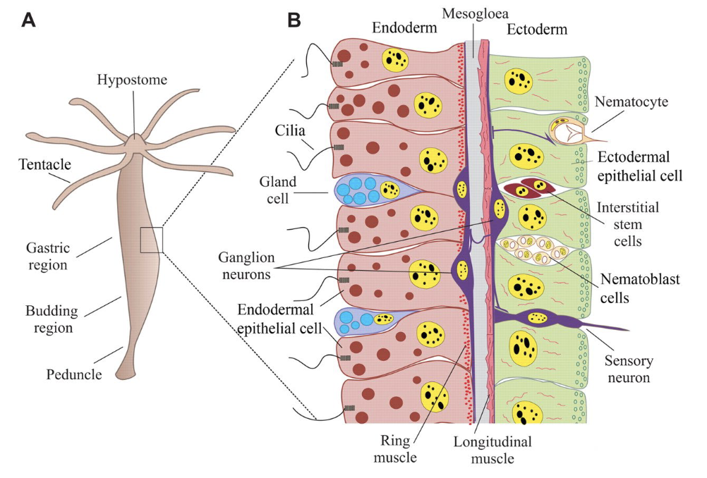
Cnidarian Characteristics
Diploblastic:
Ectoderm and Entoderm
Oral-aboral axis and radial symmetry
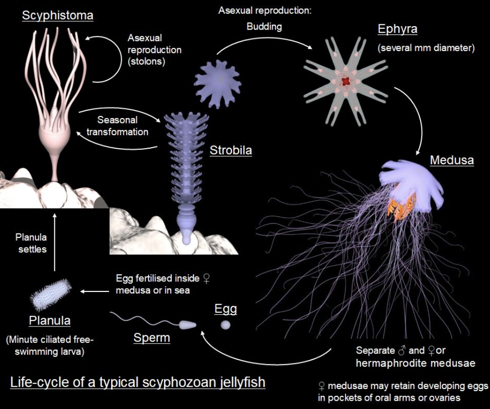
Life Cycle:
Ciliated Planula larvae
Polyp (asexual) and Medusa (sexual) - with variations and reductions
Solitary or colonial
Tentacles
Neurons → nerve net
Musculature
Simple sensory structures (more complex in some Cubozoa
Parasitic Myoxozoa have virtually none of these characteristics
Myoxozoa
Parasitic Cnidarians, have virtually no typical traits
Cnidarian germ layers
Ectoderm → Epidermis
Endoderm → Gastrodermis
Mesoglea between layers
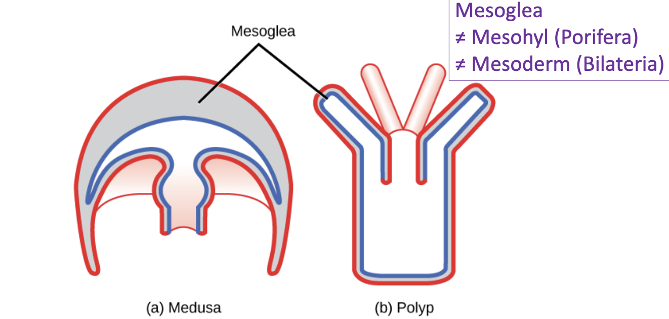
Mesoglea
Between epidermis and gastrodermis in Cnidarians

Zooid
An animal arising from another through budding or division, especially in each of the organisms of a colony.
Ex: The hydrozoan polyp
Hydrozoan development
Cleavage:
Equal and radial
Blastula → Gastrula → Planula
Ciliated Planula larvae
Cnidarian larvae
Ciliated ectoderm and solid endodermal mass
Cellular differentiation (nerve, muscle, sensory and gland cells, nematocysts)
Feeds in anthozoans, but not hydrozoans
Planula attaches at the aboral end and becomes a polyp, developing mouth and gastric cavity
Cnidarian colonies
Budding and persistent stolon lead to colonies
Differentiation leads to polymorphic colonies
Ex: Hydractinia
Hydractinia
A colonial hydrozoan growing on gastropod shells
Several kinds of morphologically/ functionally distinct zooids:
Gastrozooid
Gonozooid
Dactylozooid
Gastrozooid
Colonial zooid type found in Cnidarian Hydractinia
Feeding zooid with mouth, tentacles, and gastric cavity
Gonozooid
Colonial zooid type found in Cnidarian Hydractinia
Reproductive zooid that forms sexual medusa
Dactylozooid
Colonial zooid type found in Cnidarian Hydractinia
Protective zooid with nematocysts but no mouth
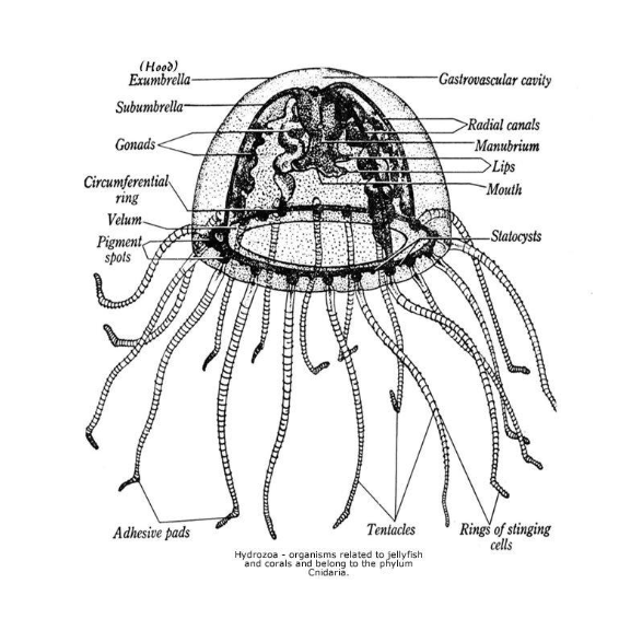
The Hydrozoan medusa
Motile zooid
Swims by contraction of muscular ring
Sexual zooid, bearing gonads - some budding occurs, but rarely
Gametes shed directly into seawater, fertilization is usually external
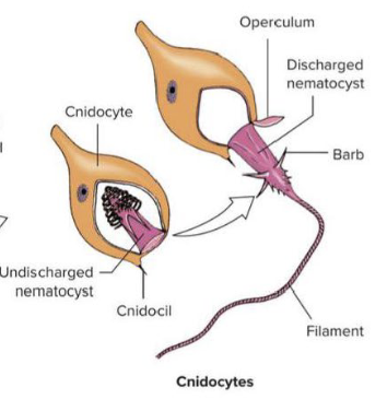
The cnidocyte
Perhaps the most complicated of all metazoan cell types
Discharge stimulated by chemical (prey body fluids) and mechanical (displacement of cnidocil) signals
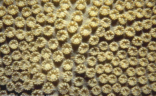
Coral zooids
Anthozoa (Cnidaria)
Form large non-polymorphic colonies
Nematocyst-armed tentacles
Usually rely on endosymbiotic zooxanthellae for most of their metabolism
Each zooid occupies a cup on the surface of the block of secreted calcium carbonate
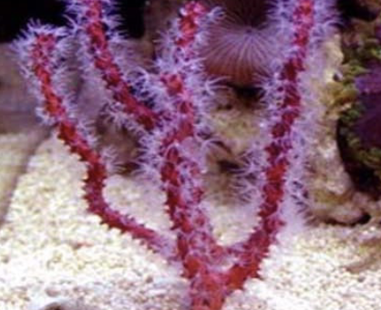
Gorgonacea
Anthozoa (Cnidaria)
Sea whips and sea fans
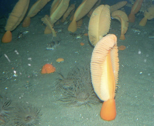
Pennatulacea
Anthozoa (Cnidaria)
Soft corals and sea pens
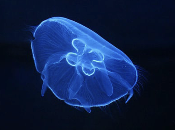
Scyphozoa
Traditional Cnidarian class
Jellyfish
Primarily predatory marine medusae
Highly reduced polyp phase
~ 200 species worldwide
Tetramerous symmetry
Bell with motile tentacles or drawn out into 4 oral arms
Capture prey with nematocysts on tentacles, arms, and bells
Mesoglea contains fibres and ameboid cells, often stiff
Swimming by jet propulsion
Sensory organs on periphery of bell - Rhopalia
Rhopalia
Scyphozoan sensory organ
Contains statocyst, ciliated sensory pit, and sometimes ocelli
Scyphozoan development
Solid or hollow ball develops into a polyp, the scyphistoma
Immature medusae (ephyrae) bud from polyp by horizontal fission (strobilation)
Cubozoa (Cubomedusae)
Box jellyfish- cuboidal bell with 4 flattened sides
Tentacle or bunch of tentacles at each corner
Rhopalia, containing large, complex, lens-bearing eye
Active predators of fish
Highly venemous
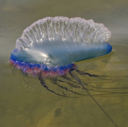
Hydrozoa
Most marine, colonial, with typical life cycle
Ex: Portuguese man-of-war
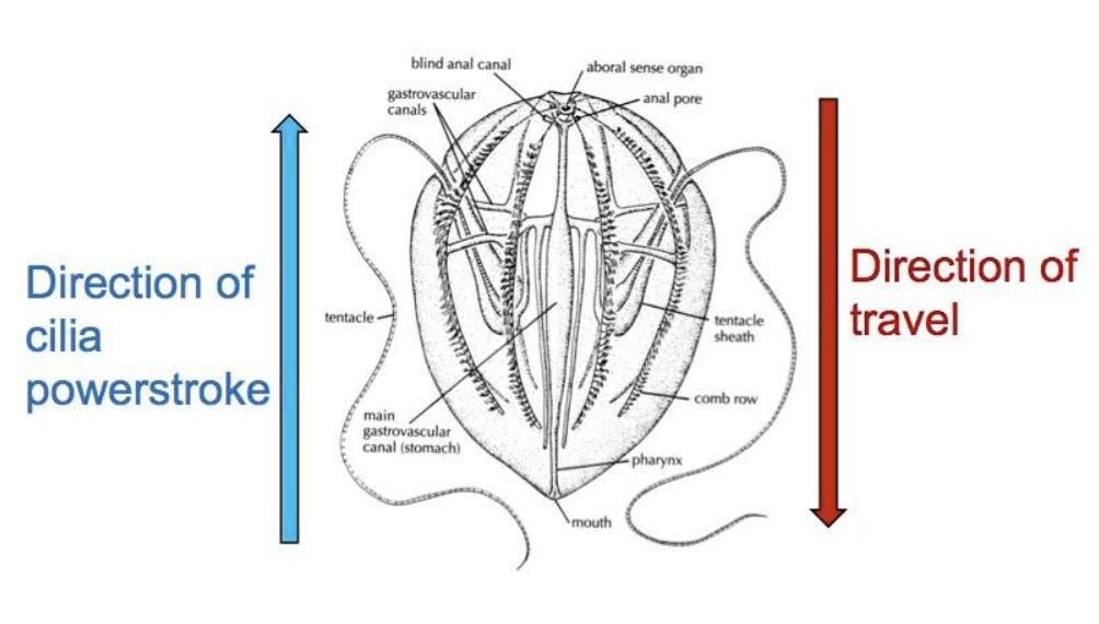
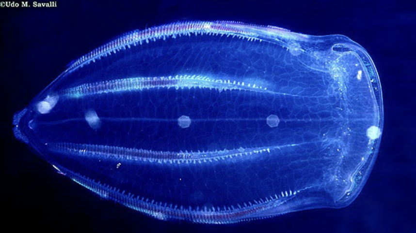
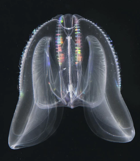
Ctenophora
Marine predators
200 species, mostly pelagic
>1 cm to 2 m
Shared with Cnidaria:
Diploblasts with Mesoglea
Oral-aboral axis with radial symmetry
Gelatinous body
Tentacles
Nerve and muscle cells
Unique traits
8 rows of ctenes (comb-like, ciliated plates)
Colloblasts (adhesive cells)
Statocyst-based sensory organ
Bioluminescence is common
Ctenophore general anatomy
Ectoderm + entoderm, separated by mesoglea
Muscle cells in mesoglea
Epidermal body wall with basement membrane
Mouth → Blind gut → anal pores
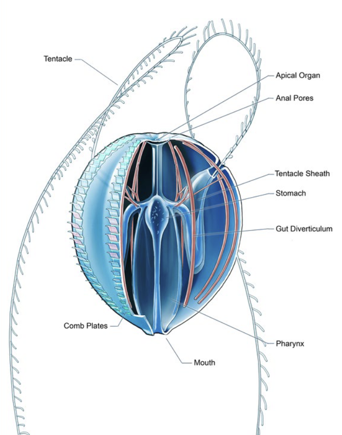
Comb rows (ctenes)
Comb = blade of fused cilia
Beat in sequence for propulsion
Ctenophores= larges animals propelled by cilia
Ctenophore tentacles
Paired
Capture prey with colloblasts
Different from cnidarian tentacles, may have evolved separately
Colloblasts
Adhesive cell used by Ctenophores to capture prey
Ctenophore nervous system
Neurons with synapses
Nerve net with some neural concentrations
Lack many bilaterian genes for neural developments and neurotransmission
Possibly evolved independently
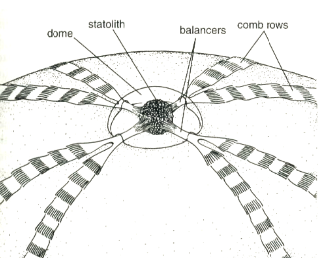
Apical organ
Complex statocyst-based organ at the aboral pole
Senses gravity and controls movement, including ciliary beating
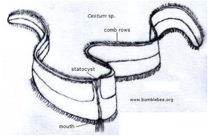
Flattened Cestida (“Venus girdle”
Ctenophore example
Benthic Platyctenida
Ctenophore
Settles of sea stars
Tentacles and colloblasts
Predatory Beroida
Ctenophore
8 comb rows
No tentacles
Mnemiopsis leidyi (“Sea Walnuts)
Invaded Black Sea in the 1980s
Introduced from North America through ballast water
Feeds on zooplankton and fish larvae
Disrupted whole ecosystem
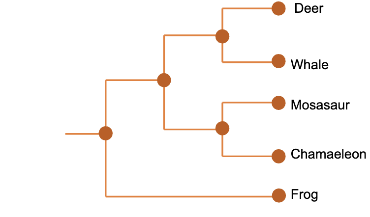
Current consensus view on Bilateria Phylogeny
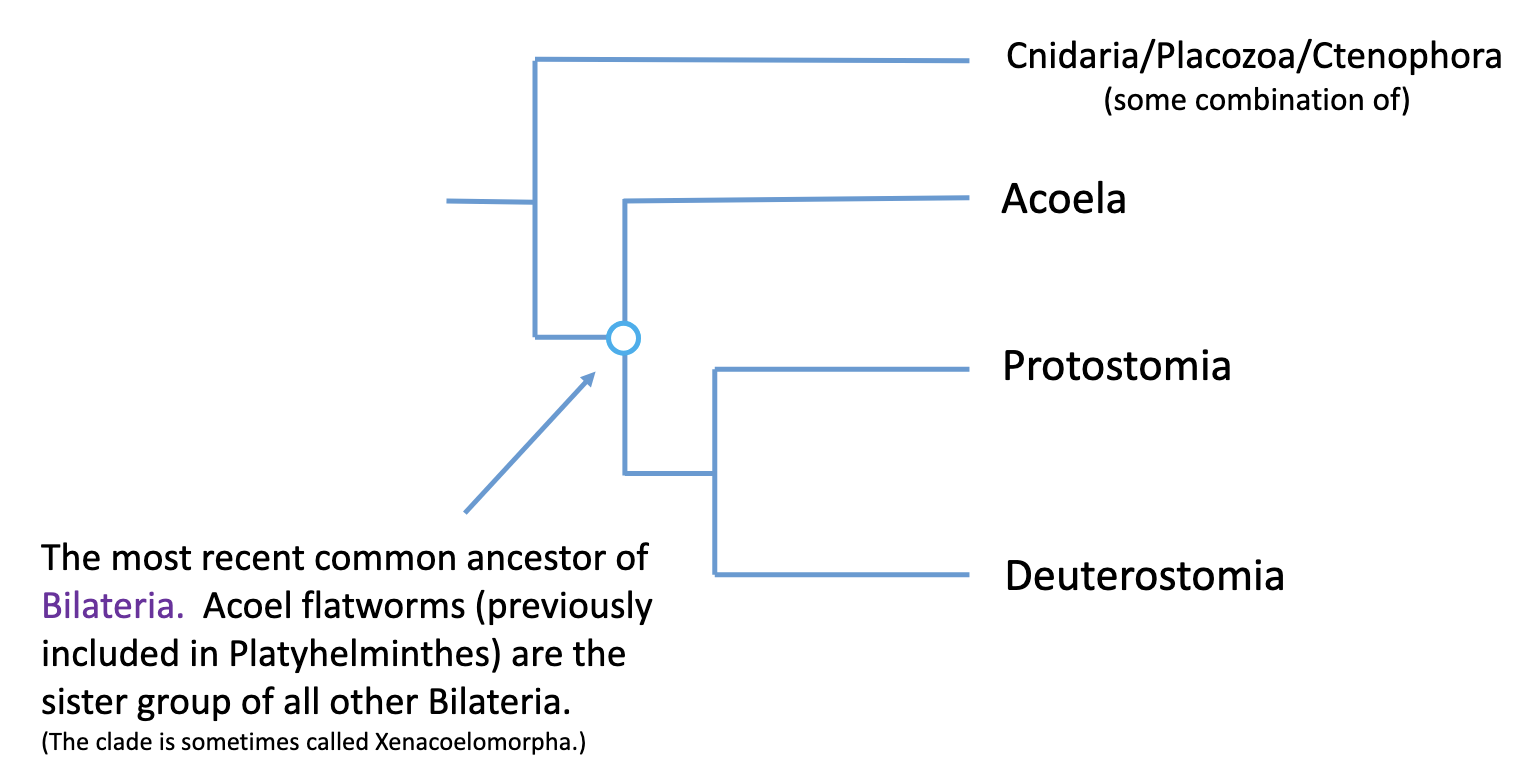
Bilateria
Bilateral symmetry at some life stage
Triploblastic
Mesoderm → Muscles
Mesoderm → Coelom
Bilateral symmetry
Two mirrored portions - left & right
Typically accompanied by cephalization
Three planes
Anterior - posterior
Dorsal - ventral
Medial - lateral
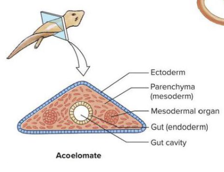
Acoelomates
e.g. Acoela, Platyhelminthes
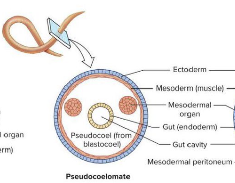
Pseudocoelomates
e.g. Nematoda
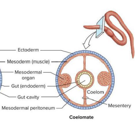
Coelomates
e.g. Annelida
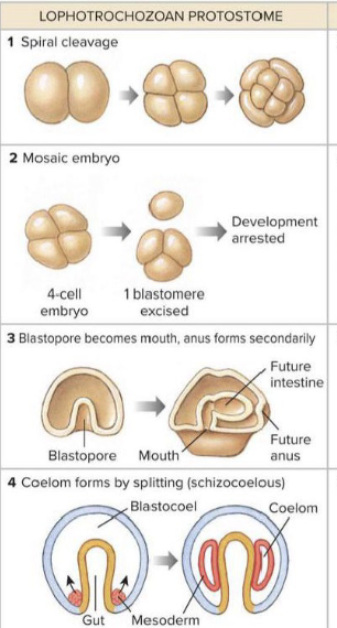
Protostome Developmental Tendency
Spiral Cleavage
Mosaic Embryo
Blastopore becomes mouth
Coelom forms by splitting (Schizocoelus)
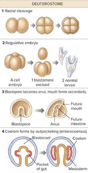
Deutorostome Developmental Tendency
Radial Cleavage
Regulative Embryo
Blastopore becomes anus
Coelom forms by outpocketing (enterocoelus)
Xenacoelomorpha
a.k.a Acoela
Acoel flatworms
Formerly grouped in Platyhelminthes
Sister group of all other Bilateria
3 internal sister clades
Marine worms
Examples:
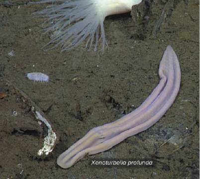
Xenoturbellida
Amphiscollops
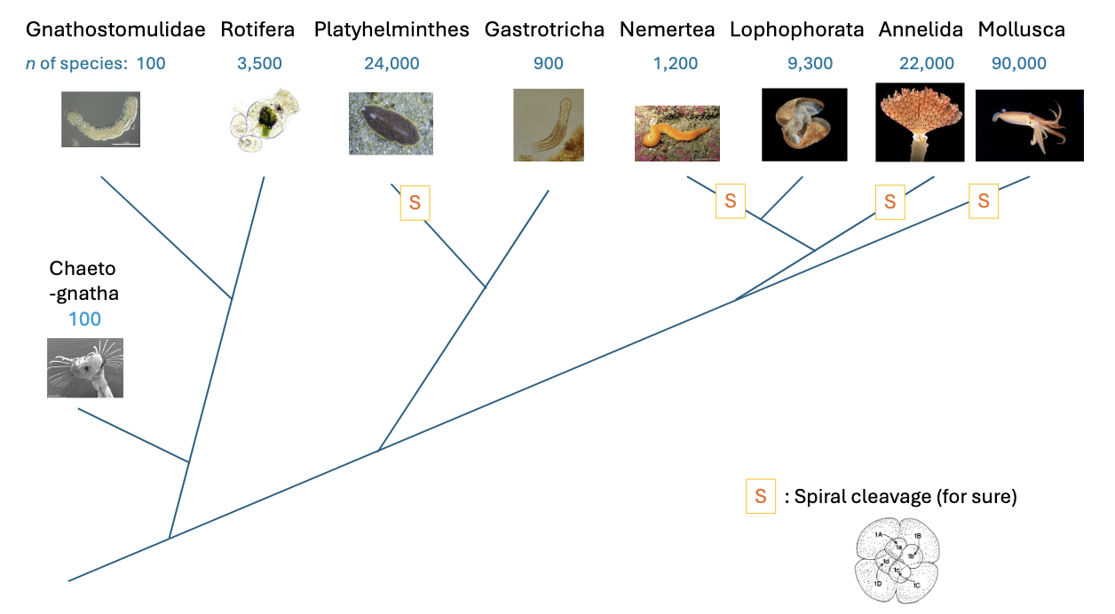
Spiralia
Protostome clade characterized by spiral cleavage
= Lophotrochozoa
Sister group of Ecdysozoa
Spiral Cleavage
3rd division is unequal:
4 macromeres
4 micromeres
Subsequent divisions also unequal
Result:
A hollow, ciliated blastula that develops into a prototroch larva
Current consensus phylogeny of Spiralia
Gnathifera
Gnathostomulidae and Rotifers
Sister group of Chaethognaths
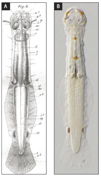
Chaetognatha
“Arrow worms”
Marine, ~100 species
Abundant planktonic ambush predators - up to 10% of plankton biomass
Torpedo-shaped, 1-12 cm
Trunk with “fins”
Mouth with grasping spines and “teeth”
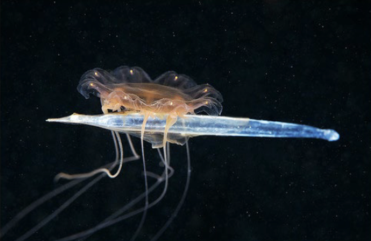
Chaetognath anatomy
Lateral fins for swimming
Chitinous spines for capturing prey
Body usually turgid with a well-developed coelom
Gnathostomulidae
Sister group of Rotifera, part of Gnathifera
Marine and brackish worms, up to 3 cm
Acoelomates
Complex jaws
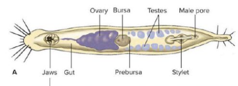
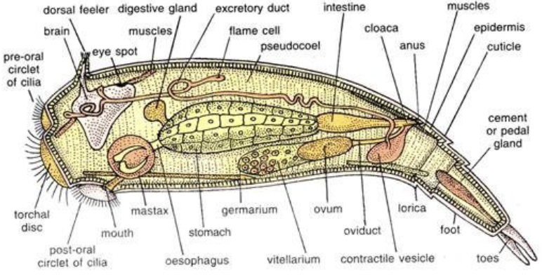
Rotifera
Mostly free-living, freshwater, microscopic; ~ 2000 species (plus ~1500 in Acanthocephala)
Corona of cilia (rotary or wheel organ)
Feeding apparatus with muscular mastax and trophi (“jaw”)
“Toes”
Pseudocoelomate
Eutely; syncytial epidermis
Cryptobiosis common
Reduction of male sex common
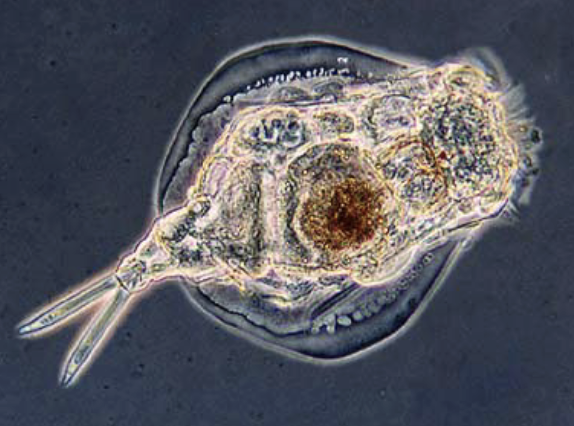
Rotifer anatomy
Body wall: Cuticle, epidermis, and subepidermal muscles
Syncytial epidermis with scattered nuclei
Spacious body cavity, pseudocoel
No respiratory or circulatory systems