Bio
1/113
There's no tags or description
Looks like no tags are added yet.
Name | Mastery | Learn | Test | Matching | Spaced | Call with Kai |
|---|
No analytics yet
Send a link to your students to track their progress
114 Terms
Mentalism
An immaterial mind (psyche) controls behavior
Aristotle: 384-322 B.C.E.
Inspired by story of Cupid and Psyche: thought Psyche was a good representation of human
mentalism - The Cardiac Hypothesis
He thought psyche produced behavior by communicating with heart which he believed controlled the body
Dualism
Two entities, an immaterial mind (psyche) and a material body control behavior
Descartes: 1596-1650
Thought that bodies were kinda like robots/body consisted of very complex reflexes
how dualism worked
First to contribute some importance to brain, specifically what we now call pineal gland
He thought ventricles were important and rest of brain was a cushion to protect pineal gland
Claimed his theory could explain both reflexive and voluntary movements
problem with dualism
how does an individual know if others have a mind or if there just an automaton
They came up with test: to have mind you need language and memory skills
- babies don’t have mind till about 7, people with disabilities may have troubles passing test, most animals can't pass test
- cant feel pain or distress if you can’t pass test so allows excuse for abuse and neglect
Materialism
the brain and nervous system control behavior
Body form, behavior and environments of animals
Darwin: 1809-1882/book: The Origin of Species
Theory all animals originate from single animal and evolution began to create diverse animals, bodies, behaviors
Theory of Natural Selection lead to materialism
Behavior and mind is result of brain and bodies that have evolved to suit our environments
No immaterial mind
How does the brain produce behavior? The Brain Theory
Donald O. Hebb: 1904-1985/Organisation of Behavior
When you produce behavior it activates group of cells
Neurons that fire together, wire together (connections between those neurons strengthen and you improve each time you do that behavior because connection continues to strengthen (you create neural circuit/memory))
Most modern theory
True or False: neurons, are similar in all animals with nervous system
true
Sea slugs
used to understand learning
zebra finches
used to study learning languages + dialects
fruit flies
used to study because easy to manipulate their genes: study chromosomes
extra chromosomes causes what?
down syndrome
cladograms
Graph that depicts predicted evolutionary relationships between organisms
Each branch point identifies a change
Each species in branch share new feature
neurons original purpose
allow organisms to move
Simple animals to complex animals
Neurons + muscles -> Nerve net -> Bilateral symmetry -> segmentation (clear top and bottom) -> Ganglia (knots of neurons) -> Spinal cord -> Brain
common relative to hominids
australopithecus
Why Did the Hominid Brain Get So Big?
Climate changes (adaptable brain and behavior)
Primate lifestyle (large social groups + diets)
Physiology (effective brain cooling systems (big brains produce lots of heat) and smaller jaws - because of change in diet)(smaller jaw = larger brain)
Altered maturation (neoteny and prolonged development)
Genetic mutations (SARGP2 responsible for determining number of neurons in cortex - appears to have duplicated at least 3 times)
Prolonged development time period
Neoteny
Neoteny
Juvenile features of the ancestors are retained in the mature form of the descendents (ex juvenile apes)
is the central or the peripheral nervous incased in bone?
the central nervous system
Structures near top of brain
dorsal / superior
Structures near midline of brain
medial
Structures on side of brain
lateral
Structures near bottom of brain
ventral / inferior
Structures near front of brain
anterior / rostral
Structures near back of brain
posterior / caudal
Coronal section
a cut from top to bottom / produces a frontal view of brain
Horizontal section
creates dorsal view / cut front to back
Sagittal section
cut front to back on vertical axis / creates medial view
Frontal view
white ventricles
dorsal view
black ventricles
bilateral
two of something / one per side
unilateral
one thing
contralateral
opposite side
Ipsilateral
same side
proximal
close to
distal
far from
meninges
membranes
dura mater is soft - true or false
false its hard
Arachnoid membrane
looks like spider
pia mater is soft - true or false
true
another name for sub arachnoid space
cerebral spinal fluid
what is cerebral spinal fluids purpose
removes toxins from brain and back into blood stream and is made by the choroid plexus
where are ventricles located and how many are there
four ventricles
in the middle of the brain and filled with fluid
the right and left ventricles are…
bilateral
cerebral spinal fluid is made by…
the choroid plexus
what are ventricles lined with?
glial cells
what prevents the brain from crushing itself?
cerebral spinal fluid
hydrocephalus (what & treatment)
born with condition / build up of fluid in ventricles that can't escape / pushes brain tissue into skull and causes brain to die
Surgery insert shut into base of skull to release fluid from ventricle into abdomen (blood stream)
brain organization (original three areas → new 5 areas)
original three:
forebrain
midbrain
hindbrain
new five:
telencephalon
diencephalon
mesencephalon
mesencephalon
mylencephalon
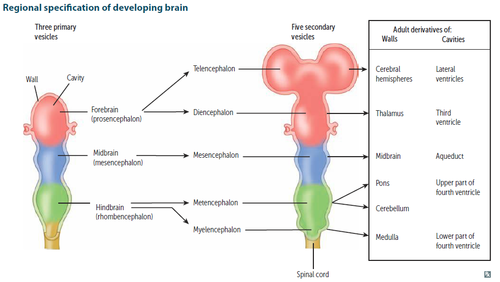
what allows nervous system to develop?
neural tubes
parts of forebrain
Telencephalon
Diencephalon
telencephalon - neocortex / general info
Six layers of cells / Thin (2-4mm)
Layers number I-VI in roman numerals
gyrus / gyri = outer bump
Sulcus / sulci = inner dips
Deep sulci = fisher
Grey matter: neuron cell bodies of neurons (no wrap) (absorb die in tests)/sends axons with messages = white matter
White matter: axons wrapped in glia sending signals
Corpus callosum = allows hemispheres to communicate
2 sheets of neocortex (one in each side of brains)
Neocortex is cerebral cortex (generally)
Grey matter
neuron cell bodies of neurons (no wrap) (absorb die in tests)/sends axons with messages = white matter
White matter: axons wrapped in glia sending signals
axons wrapped in glia sending signals
Corpus callosum
allows hemispheres to communicate
telencephalon - neocortex / structure info
Four lobes: frontal (motor), parietal (somatosensory), occipital (visual), temporal (auditory)
Named by bones structure above it
Between frontal and parietal = central sulcus (splits brain in half)
Lateral fissure/sylvian fissure = separates temporal lobe from frontal and parietal
neocortex - Brodmann's Area 312
post central gyrus (its a bump)
primary somatosensory cortex
broadmanns areas
Each brodmann's area has different shape or structured cells (neurons)
Structure determines function = each area has a different function
Dendrites receive info for neurons
Ex. more dendrites = more info coming in
Became most used map because it was good at predicting functions of neocortex
Sensory perceptions are created by neocortex (responsible for making you feel like yourself)
neocortex - broadmanns area 4
the primary motor cortex = darkest shades in brain pic on slides is the primary part of that brain
Darkest areas are most closely connected to inputs and outputs from the body (info arrives there first)
Lighter areas called association areas - further from inputs and outputs / responsible for processing higher/more complex details of info
Primary motor cortex = send info to spinal cord (does not receive much input its responsible for sending inputs)
neocortex - braodmanns area 17
Primary visual cortex also called striate cortex
what’s broadmanns area 41?
primary auditory cortex
allocortex
3-4 layers of cells
Also called limbic system/boarder system
Cingulate cortex:
Bilateral structure
Produces emotional expression and behaviors
Anterior cingulate cortex is overactive in major depressive disorder
allocortex - hippocampus
Bilateral structure
Latin for seahorse (due to shape)
Important for explicit memory / memories you can talk about / episodic or personal memories (very specific memories not habits/implicit memories)
Involved in retrieval of memories as well as making new memories
Involved in social navigation
Forms a cognitive map of environment
Personal memories are put on cognitive map to prevent bad things from rehappening which is why it is housed in the same structure
allocortex - amygdala
Responsible for processing emotions especially fear
Important to attributing value/intensity to emotional stimulus
Ptsd is overactivity of amygdala
Basal Ganglia (base knot) pathways
Direct pathway: responsible for the excitation of the muscles/movement
Indirect pathway: inhibits unwanted movements
Basal Ganglia (base knot)
Bilateral
Motor behavior - connected to motor cortex: they produce voluntary movements
Motor cortex is responsible for initially organizing/sequencing your movement
Basal ganglia observes the sequence to do something (ex. Brush teeth) / the more you do it the stronger the connection become until it is habit - once its a habit the neocortex is not involved in the action only the basal ganglia is
Stores procedural memories
Caudate, Putamen, Globus pallidus, Nucleus accumbens = basal ganglia
Diseases involved with basal ganglia = tourettes, huntington's chorea, parkinsons
huntington's chorea: deterioration of caudate, putamen, globus pallidus
caudate, putamen and globus pallidus = the striatum
forebrain - diencephalon (the rooms)
Bilateral structure
Epithalamus (epi means above)
Thalamus
Hypothalamus (below)
diencephalon - Epithalamus (epi means above)
Seasonal rhythms (pineal gland - waking and sleeping behavior)
Pineal gland releases melatonin
Regulates Circadian rhythm
diencephalon - thalamus
Cortical relay system/station: redirects and filters information
Cortical relay system integrated info entering and leaving cerebral cortex
View slide 9 - lecture 2 (pulvinar and lateral geniculate body)
Contains many nuclei
diencephalon - hypothalamus
Maintain body homeostasis via regulatory behaviors
Physiological symptoms maintained
Many nuclei compete for control of the pituitary gland
Pituitary gland: master endocrine gland/master hormone producer/releaser
Hypothalamus regulates: drinking, eating, temp regulation, sleep salt regulation, sexual behavior, parental behavior
midbrain (location & structure)
below hypothalamus
mesencephalon (tectum + tegmentum)
mesencephalon - Tegmentum (floor)
Periaqueductal gray matter: species typical behaviors (ex. Cat stalking bird, sexual, hunting, fighting behaviors behaviors) / motor circuits & pain relief circuits (opioid receptors)
Reticular formation: goes through mid and hindbrain
Red nucleus: older motor pathway (basal ganglia to spinal cord)
Substantia nigra: produces dopamine and projects to basal ganglia
Ventral tegmental area (NuAcc): produces dopamine and projects to nucleus accumbens (basal ganglia) / high activity is associated with drug addiction
mesencephalon - tectum
four bumps - sensory / older systems
Superior colliculi (top bumps) - visual map
Inferior colliculi (bottom bumps) - auditory map
Only visual and auditory systems on reptiles and amphibians
The colliculi allow you to respond to loud sound or sudden movement to keep you safe / evolutionary old visual orienting system
substantia nigra
Cells make dopamine
When not making dopamine (no pigment) - parkinsons
Less dopamine - less able to initiate movement
Hypokinesia
Bradykinesia - slowness of movements
Akinesia - difficulty initiating voluntary movements
Rigidity - increased muscle tone
Dopamine is not about pleasure its about motivation (wanting to seek reward) / initiating and motivating movements
Awakenings / Oliver Sacks: gave patients L-Dopa
Certain stimuli elicited movements = certain motor circuits intact just difficulties activating them
They are conscious just cant move
L-Dopa allows them to move but eventually it stops working - before it completely stops working it creates weird side effects (choreic movements)
Visual motion may help initiate movements
Bradykinesia
slowness of movements
Akinesia
difficulty initiating voluntary
Rigidity
increased muscle tone
reticular formation
Arousal (ex. Waking up)
Moruzzi and Magoun - cats / electrode in neck
Coma patients - small subset
Narcolepsy
hindbrain
Metencephalon:
Pons (bridge)
Cerebellum (little brain)
Myelencephalon
Medulla oblongata
hindbrain - metencephalon / Cerebellum (little brain)
Dorsal structure (motor but mostly sensory)
Behind fourth ventricle
12 lobes, cerebellum cortex, cells that form gray and white matter
Purkinje cells: receives lots of sensory input (gray matter) - uses info to refine your motor behavior (smooth movements)
Cerebellar Ataxia - clunky, jerky movements (look drunk when walking)
More skilled motor behaviors = larger cerebellum
hindbrain - metencephalon / Pons (bridge)
Mostly made up of nerve fibers that connect cerebellum and neocortex
Helping regulate unconscious actions (breathing, swallowing, bladder control…)
Ventral portion of brain stem (motor control)
hindbrain - mylencephalon / Medulla oblongata
Bottom of brainstem
unconscious behaviors
Older than pons = less sophisticated behaviors
nervous system division
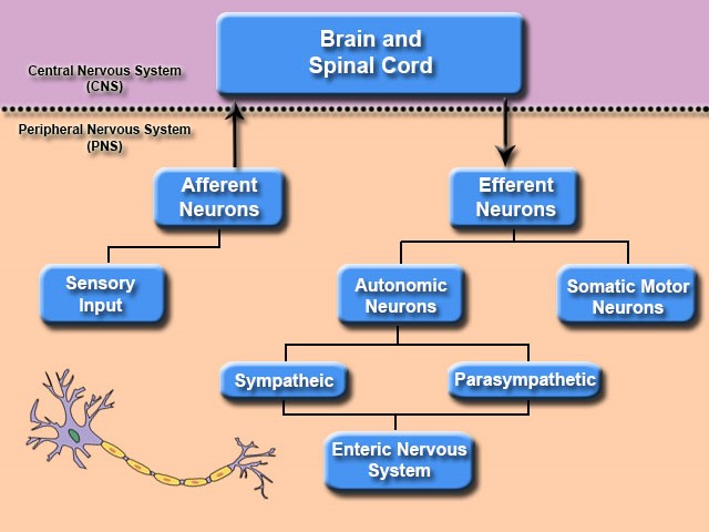
autonomic nervous system -
Involuntary movements and functions of organs (beating of heart, pupil movement …)
Unconscious control (unless using mindfulness)
Divided into 2 sections sympathetic and parasympathetic
autonomic nervous system - enteric
Responsible for gut
Gut lining = plexus
Gut extends through esophagus, stomach, small intestine, and colon
Millions of neurons embedded in it to control bowel motility, nutrient absorption, enzyme secretion
Little input from brain (unless regarding emotions in relation to stomach issues)
Processes signals in gut (microbiome = our individual gut bacteria) that can influence physiological and psychological processes
Microbiome in gut produces majority of our serotonin
Lots of gut brain feedback
autonomic nervous system - parasympathetic
Stores energy reserves
Rest and digest system
Long term survival
Neurons originate in brain stem and sacral area (2 neuron system)
Opposite function of sns
Preganglionic neuron is long and synapse with second postganglionic neuron right before final destination/organ or in target organ
autonomic nervous system - sympathetic
Activate energy stores in body
Fight or flight - because energy stores are released (increase heart and breathing rate)(energy sent to skeletal muscles)
Shuts down activity in unnecessary organ systems (ex. Immune and digestive system)
Thoracic and lumbar are of spinal cord - neurons sent in body synapse in body then travel to right part of body
First neuron is preganglionic neuron (closer to spinal cord) then postganglionic neuron travels to final destination / organ
If active long term could cause physical/neurological damage
Camillo Golgi (1843 - 1925)
The reticular theory (rete = net): a web or net of tubes and fluid flowed through tubes and that's how information was transferred
Developed dies for staining body tissues
Golgi stain on brain showed image of tree like images
Golgi stain made of silver absorbed by tissue
Ramony Cajal (1852 - 1934)
Used golgi stain and saw what he believed were individual cells that communicate with one another
Developed the neuron doctrine = brain made of individual cells (now called neurons)
Golgi and Cajal shared nobel prized in 1906 (they hated each other)
the neurone doctrine
The neuron: main info processing unit of brain
The synapse: individual units communicate with each other at specific points (now called synapse)
Connection specificity: in well working brain pattern/rhyme for neuron communication / neurons form larger info processing circuits
Dynamic polarization: info flow through neuron in particular direction
Somatic Nervous system
Voluntary controls of spinal muscles; 1 neuron (generality stimulatory effect)
cranial nerves
head, neck, and face
spinal nerves
originate in spinal cords
voluntary controls skeletal muscles – both motor neurons (spinal cord to nerves), sensory neurons (originate in skin, muscles, joints → project sensory input back to the spinal cord)
segmentation
Repetition of parts
Doral (Posterior) nerve
sensory
Ventral (Anterior) nerve
motor
spinal cord segmentation
Evolutionary conserved system
Divided into four main segments (cervical, thoracic, lumbar, sacral, coccygeal (tailbone))
Each Segment Receives and sends information to specific dermatome or specific section of body
Segments determined by genes → Hox genes (master genes for building bodies)→ transcription factors (protein that combines other genes in DNA that turn on and turn off other genes responsible for growing legs/arms, ect.)→ basically found in all animals with segmentation
Dermatomes: Segment of spinal cord linked to a specific segment of body
Rostral: near top
Caudal: near bottom
Dermatomes
Segment of spinal cord linked to a specific segment of body
Localized spinal cord injuries
Distinguishing real and hysterical symptoms
Guiding the reattachment of limbs
The Bell-Magendie Law
Dorsal relays sensory info from the body to brain
Ventral relays motor info from the brain to body
Afferent: incoming; Carry incoming information
Efferent: exciting; Carry information away from body
where are Cell bodies of motor neurone located?
the centre of the spinal cord