4.4 - Circulation
 Call Kai
 Learn
Learn Practice Test
 Spaced Repetition
 Match
Match Flashcards
 Knowt Play
1/38

There's no tags or description
Looks like no tags are added yet.
Name | Mastery | Learn | Test | Matching | Spaced | Call with Kai | Chat |
|---|
No analytics yet
Send a link to your students to track their progress
39 Terms
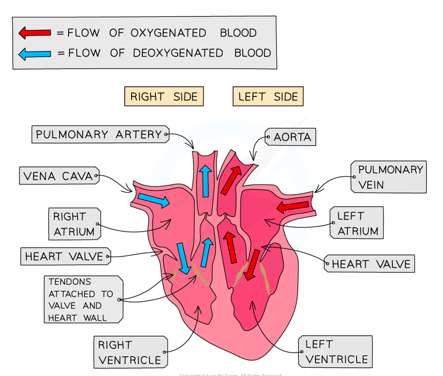
What type of circulatory system do humans have?
Double with a 4-chambered heart:
right side pumps deoxygenated blood to lungs only
left side pumps oxygenated blood to rest of the body
What are the similarities & differences between single & double circulatory systems?
Similarities:
both contain fluid medium, heart & vessels/valve
Differences:
in single, the blood passes through heart once per cycle, whereas in double, the blood passes through heart twice
in single, oxygenated & deoxygenated blood are not separated, whereas in double, they are separated
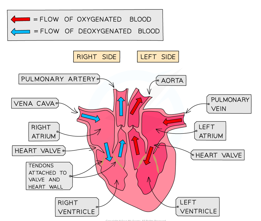
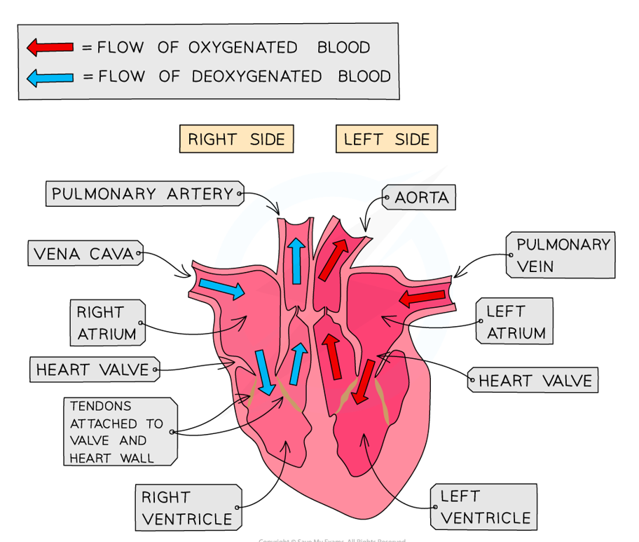
What is the structure of the heart?
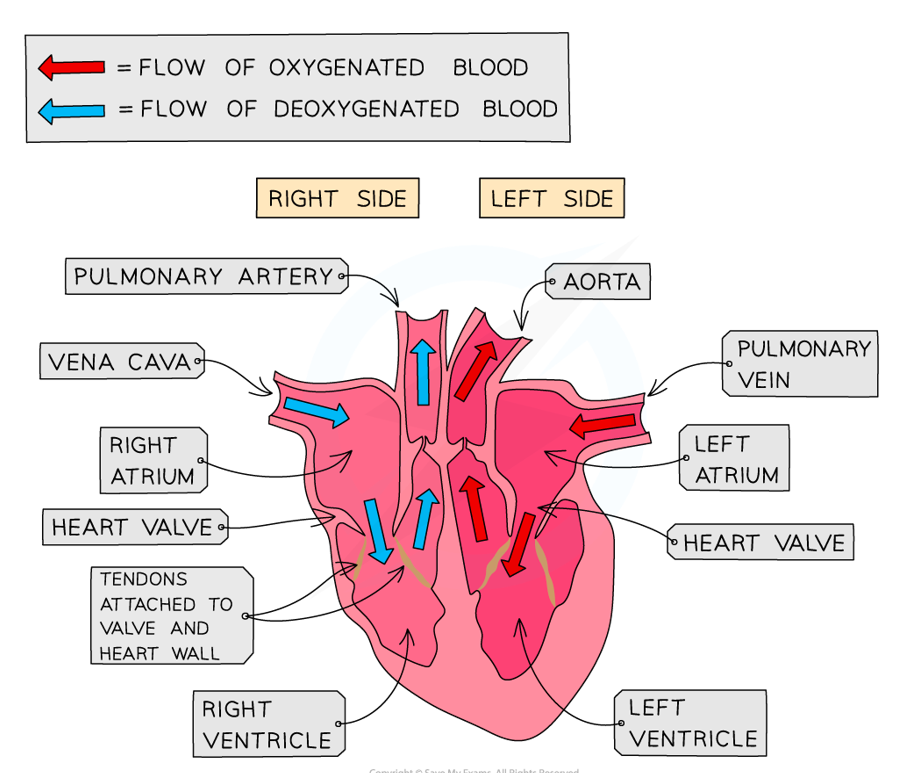
Four chambers: two-thin walled atria on top (receive blood) & two thick-walled ventricles underneath (pump blood)
Veins carry blood into atria & arteries carry blood away from ventricles
Between the atria & ventricles are atrio-ventricular valves (prevent back-flow of blood)
left valve (bicuspid) has 2 flaps, whilst right valve (tricuspid) has 3 flaps
Arteries have two semi-lunar valves (pulmonary & aortic)
What separates the two halves of the heart & why is the right ventricle thinner than the left?
Septum:
right ventricle pumps blood only to the lungs at low pressure, so it needs less muscle
left ventricle pumps blood to the whole body at high pressure, so it has a thicker wall
What is the heart made of?
Cardiac muscle, composed of myocytes:
when myocytes receive an electrical impulse they contract together → causes heartbeat
they require a lot of oxygen, so are fed by numerous capillaries from two coronary arteries
blood returns via the coronary sinus (drains directly into right atrium)
What is the purpose of blood vessels in the body?
To deliver blood to capillary beds, where substances are exchanged between cells & the blood
What is the structure & function of arteries?
Function:
carry blood away from the heart to every tissue in the body
Structure:
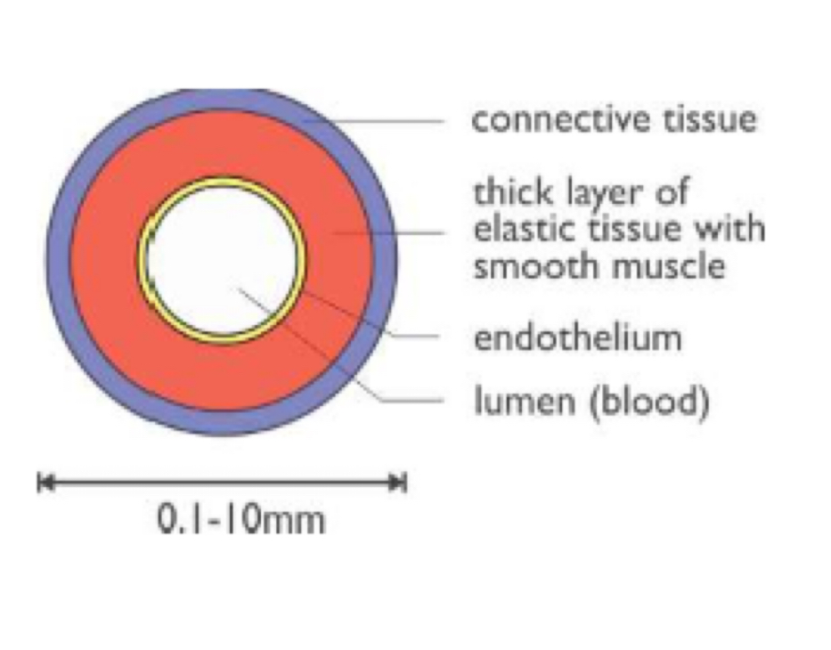
thick walls (made of 3 layers):
outer layer → connective tissue
middle layer → smooth muscle & elastic tissue (allows expansion during systole & withstands high blood pressure)
inner layer → endothelium with elastic basement membrane
small lumen
no valves (except in heart)
blood is at high pressure & usually oxygenated (except in pulmonary circulation)
What is the structure & function of arterioles?
Function:
carry blood from arteries to one capillary bed
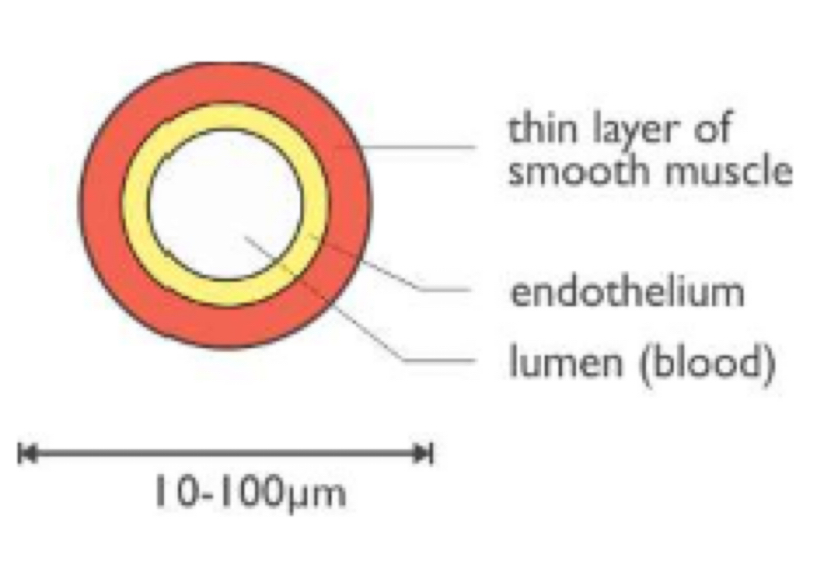
Structure:
thick walls with smooth muscle & endothelium to control blood flow to capillary bed
small lumen
no valves
blood pressure falls & is usually oxygenated (except in pulmonary circulation)
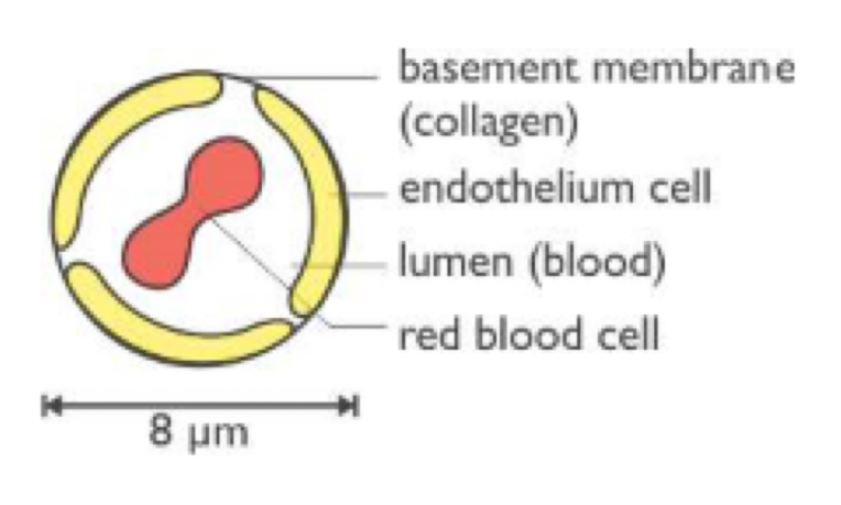
What is the structure & function of capillaries?
Function:
allows exchange of materials between the blood & the tissues
Structure:
very thin & permeable walls → one endothelium cell with fenestrations (gaps)
small lumen → blood cells must distort to pass through
no valves
blood pressure falls & changes from oxygenated to deoxygenated (except in pulmonary circulation)
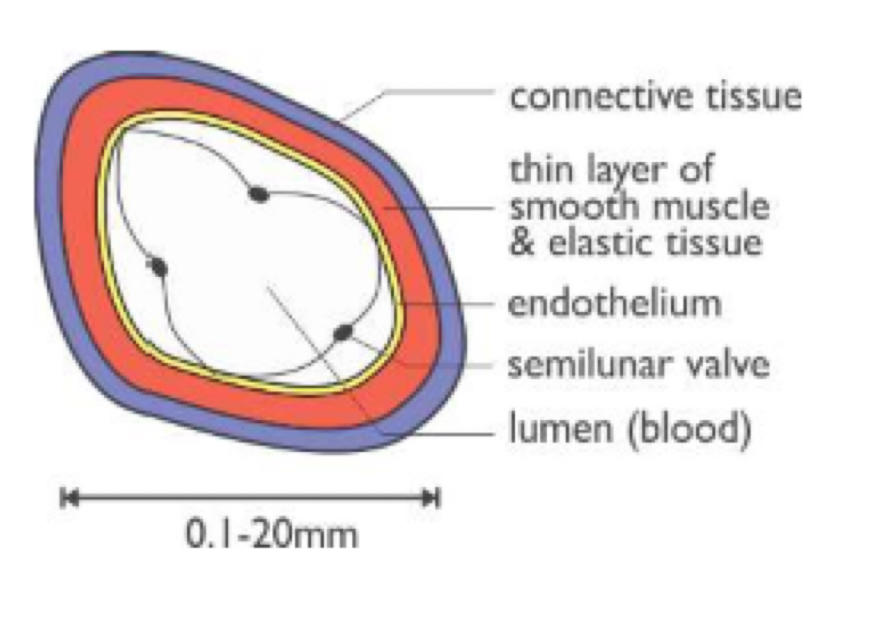
What is the structure & function of veins?
Function:
carry blood from every tissue in the body to the heart
Structure:
thin walls → consists of connective tissue, layer of smooth muscle/elastic tissue & endothelium
large lumen → reduces resistance to flow
many semi-lunar valves → prevents back-flow of blood
blood is at low pressure & usually deoxygenated (except in pulmonary circulation)
What are the advantages of a double, separate circuit in mammals over a single, linear circuit in fish?
Oxygenated blood reaches the respiring tissues undiluted by deoxygenated blood (two halves of the heart are separated by septum)
Oxygenated blood is delivered at high pressure to all body tissues (blood is returned to the heart from the lungs)
Cardiac muscles are myogenic. What does this mean?
They can contract on their own, without needing nerve impulses:
contractions are initiated within the heart by the SAN (pacemaker) in the right atrium
What are the three stages of the cardiac cycle?
Diastole
Atrial systole
Ventricular systole
What happens during diastole?
Atria & ventricles are relaxed
Blood enters the atria via the pulmonary vein & vena cava
Atria fill with blood, which increases the pressure
When the pressure in the atria exceeds the pressure in the ventricles, the atrioventricular valves open, allowing blood to flow into the ventricles
The blood pressure in the ventricles is lower than the pulmonary artery & aorta, so the semi-lunar valves shut (‘dub’ sound)
What happens during atrial systole?
The muscle walls of both atria contract simultaneously
This forces all remaining blood to flow into the ventricles below
Throughout this stage, the ventricles are relaxed
What happens during ventricular systole?
After a short delay (to allow the ventricles to fill with blood), both ventricles contract simultaneously
The blood pressure in the ventricles increases, so the atrioventricular valves shut (‘lub’ sound) → prevents the back-flow of blood into the atria
The semi-lunar valves are forced open, as the blood pressure is higher in the ventricles than the aorta & pulmonary artery. This forces blood into the aorta & pulmonary artery
The ventricles have thick muscular walls, so can contract with great force & pump blood at high pressure
Why does the impulse need to be delayed?
If the impulse spread straight from the atria into the ventricles, there would not be enough time for atrial systole to complete
Describe the sequence of stimulation that occurs in a mammalian heart in one cardiac cycle
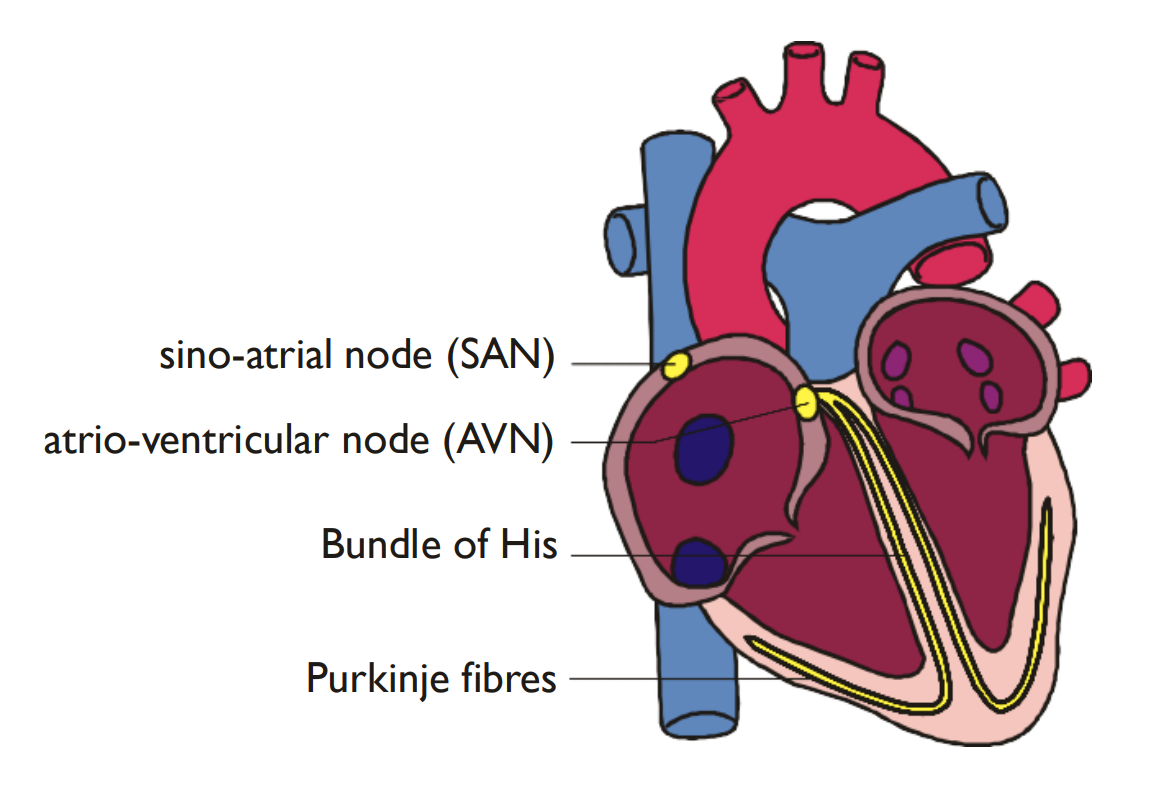
The sinoatrial node (SAN) acts as a pacemaker
The impulse travels through the heart muscle
The atrioventricular node (AVN) delays the transmission of the impulse
The impulse travels down the Bundle of His & spreads through the purkyne fibres
The ventricles contracts from the apex upwards
What is an electrocardiogram (ECG) ?
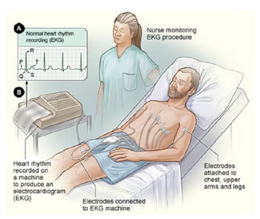
Used to measure rhythms of the heart by producing a record of electrical activity:
rhythm is caused by spread of a wave of electrical activity (depolarisation) through specialised tissue within the heart
causes tiny electrical changes on the surface of the skin that can be measured with 12 electrodes
What are the patterns of peaks/troughs seen in each cardiac cycle on an ECG?
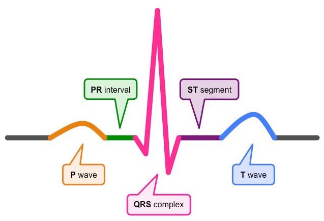
PQRST:
P = start of atrial systole (atrial depolarisation)
Q = Purkyne fibre excitation
R = start of ventricle systole (ventricular depolarisation)
S = ventricles fully contracted
T = ventricle relaxation (ventricular repolarisation)
What does the PR segment on an ECG trace show?
Represents the delay at the AVN
What are the 5 different cardiac rhythm diagnoses?
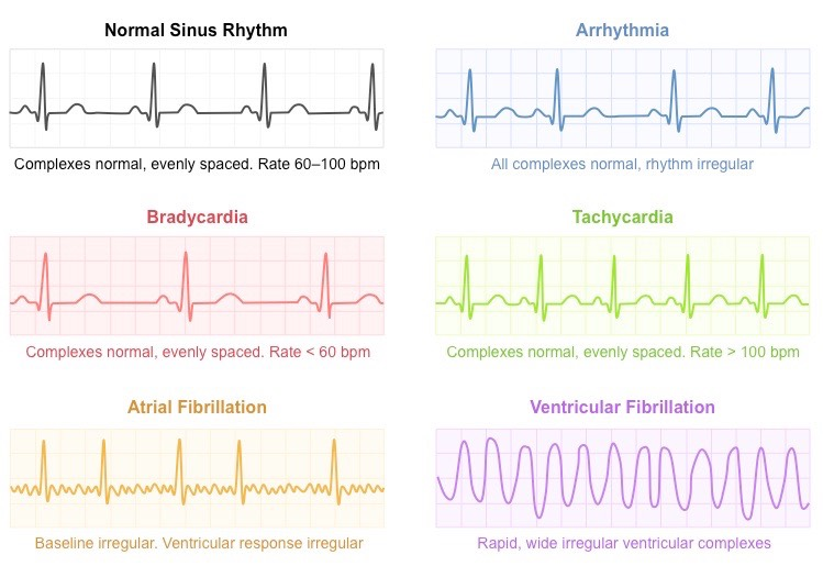
Arrythmia: normal complexes & irregular rhythm
Bradycardia: normal complexes, evenly spaced rhythm & rate < 60 bpm
Tachycardia: evenly spaced complexes & rate > 100 bpm
Atrial fibrillation: irregular baseline & clear ventricular response
Ventricular fibrillation: rapid, wide irregular ventricular complexes
What is cardiac output & stroke volume?
Cardiac output: the total volume of blood pumped by the heart per minute
Stroke volume: the volume of blood pumped out of the heart with each beat
What is the equation to calculate cardiac output?
Cardiac output = heart rate x stroke volume
What is the equation to calculate heart rate?
Heart rate (beats per minute) = 60 / cycle time (s)
Why does the cardiac output increase dramatically when the body exercises?
Oxygen & glucose can get to the muscles faster
Carbon dioxide, lactate & heat can be carried away from the muscles faster
What is blood & what is its function?
Complex mixture of solutes & cells suspended in plasma. Its functions include:
transport of nutrients & waste
forms tissue fluid
defends against foreign bodies
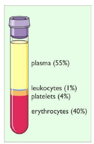
What are the main components of blood?
Plasma
Erythrocytes
Leukocytes
Thrombocytes
What is plasma?
Pale yellow-coloured aqueous solution. It transports:
nutrients (e.g. glucose/amino acids)
waste (e.g. urea/lactic acid)
hormones & heat
proteins (e.g. albumins/antibodies)
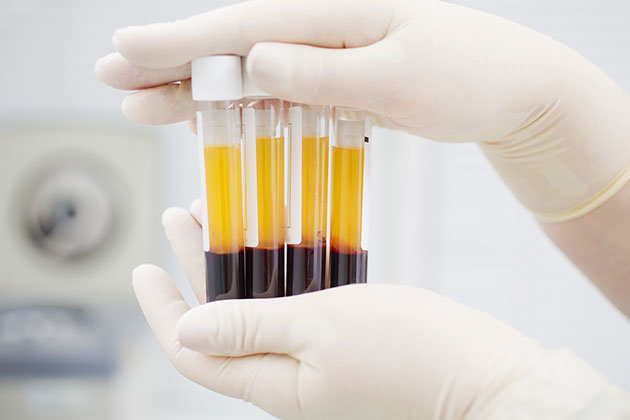
What are erythrocytes?
Red blood cells:
contain haemoglobin & made in the bone marrow
large surface area to volume ratio due to biconcave disc shape → oxygen can diffuse quickly into cells & bind quickly to haemoglobin
lack organelles, so there is more room for haemoglobin
their size & shape mean they can squeeze through capillaries & transport oxygen extremely close to cells
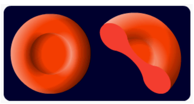
What are leukocytes?
White blood cells:
all have a nucleus, are spherical/irregular in shape & most are larger than erythrocytes
part of the immune system, killing pathogens in the blood & tissue fluid
two types: granulocytes & agranulocytes
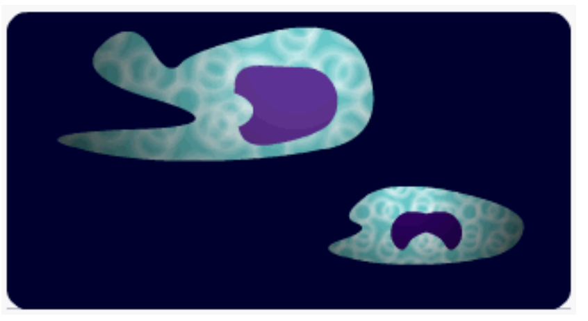
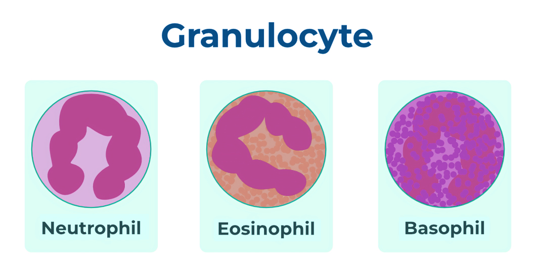
What are granulocytes?
Have visible granules & lobed nuclei
Three types (all part of non-specific immune system):
neutrophils → phagocytic & contain lysosomes to break down ingested bacteria
eosinophils → phagocytic & important in response against parasitic infection, allergic responses & inflammation
basophils → produce histamines involved in inflammation & allergic reactions
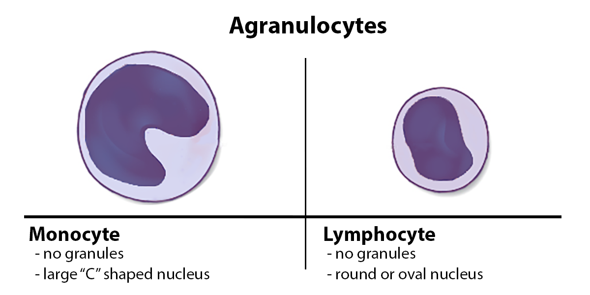
What are agranulocytes?
Have clear cytoplasm without granules & large, unlobed nuclei
Three types:
monocytes → phagocytic against bacteria & antibody coated viruses + part of non-specific immune system
macrophages (matured monocytes) → engulf & digest pathogens & present antigens to activate the immune response
lymphocytes → produces antibodies & involved in specific immune response
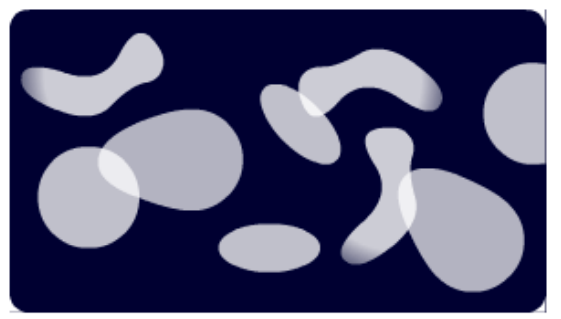
What are thromboctyes?
Platelets (cell fragments without nuclei):
formed by fragmentation of large cells in bone marrow
responsible for blood clotting
Why does blood need to clot?
Prevents blood loss
Prevents entry of harmful bacteria & infection
Provides a framework for repair
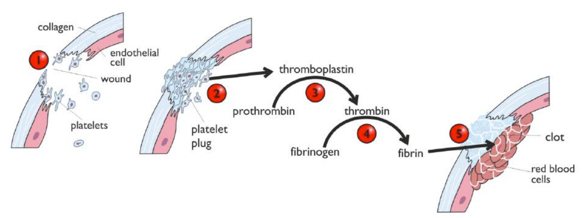
What is the process of the blood clotting cascade?
Damage to tissue exposes platelets to collagen
Platelets rush to damaged area & form platelet plug (serotonin also released → causes smooth muscle of blood vessel to contract)
Platelet plug triggers release of thromboplastin, which catalyses the conversion of prothrombin to thrombin
Thrombin catalyses the conversion of fibrinogen (soluble) to fibrin (insoluble)
Fibrin fibres form a mesh net that traps erythrocytes & more thrombocytes, resulting in a clot
What causes atherosclerosis?
Build-up of fibrous plaque (atheroma):
endothelium becomes damaged which can cause a blood clot
cells, salts, cholesterol & other substances build up & harden, forming a plaque that narrows the artery
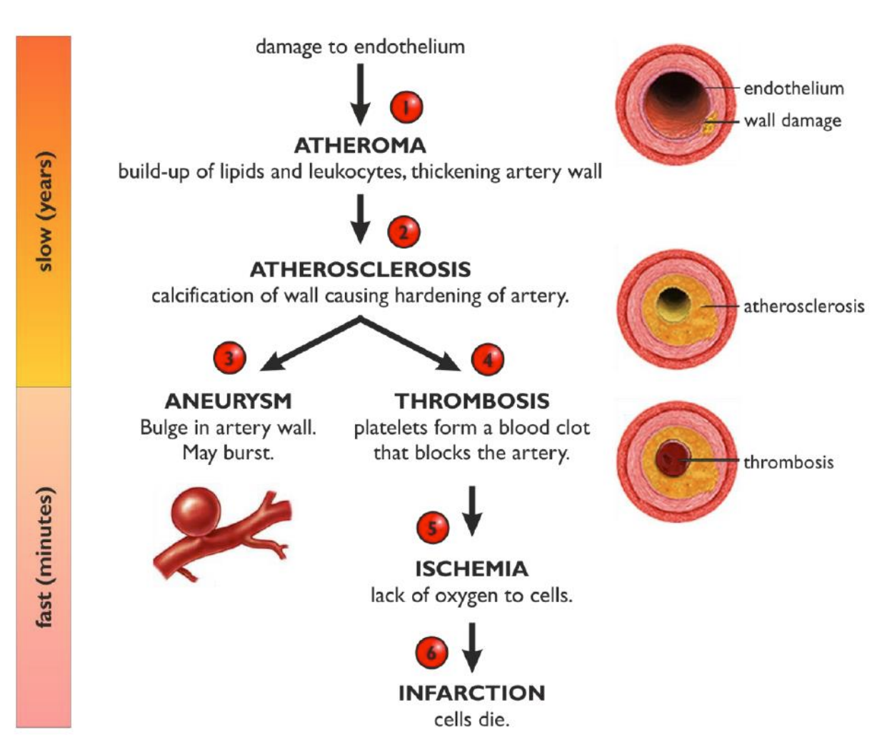
How does atherosclerosis affect health?
Increases risk of cardiovascular diseases (e.g. myocardial infarction (heart attack) stroke & angina)
What are examples of factors that increase risk of atherosclerosis?
Age
Genetics
Smoking/alcohol
Lack of exercise/obesity
Cholesterol levels