BV 1 Binocular and Spatial Vision
 Call Kai
 Learn
Learn Practice Test
 Spaced Repetition
 Match
Match Flashcards
 Knowt Play
1/66

There's no tags or description
Looks like no tags are added yet.
Name | Mastery | Learn | Test | Matching | Spaced | Call with Kai | Chat |
|---|
No analytics yet
Send a link to your students to track their progress
67 Terms
What are the two types of two-eyed vertebrate vision, and how do they differ?
Utrocular vision: Eyes function independently.
Ambiocular vision (true binocular vision): Eyes work together in a coordinated manner.
Binocular vision: Coordinated behavior of both eyes for the perception of space.
What are the key characteristics of utrocular vision?
Independent eye movements
Complete crossing (decussation) of optic nerves
Typical of lower vertebrates
What are the key characteristics of ambiocular (binocular) vision?
Coordinated eye movements
Follows Hering's Law of Equal Innervation (equal neural input to yoked eye muscles)
Partial decussation of optic nerve fibers
Characteristic of mammals
Are frontal eyes required for ambiocular (binocular) vision?
No. Frontal eye placement facilitates ambiocular vision by increasing visual field overlap, but it is not strictly required for binocular vision to occur.
What is ocular laterality, and how is it measured?
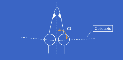
Ocular laterality is measured by ω (omega).
ω = angle between the optic axis (with the eye centered in the orbit) and the body's midline.
It quantifies how laterally positioned/oriented the eyes are relative to the head.
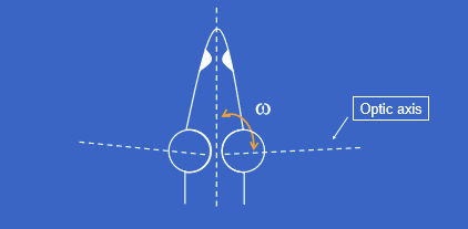
How does ocular laterality (ω) differ between prey and predator animals?
Large ω (greater ocular laterality): Eyes positioned more laterally → wider panoramic field of view (common in prey animals).
Small ω (less ocular laterality): Eyes positioned more frontally → greater binocular overlap and depth perception (common in predators).
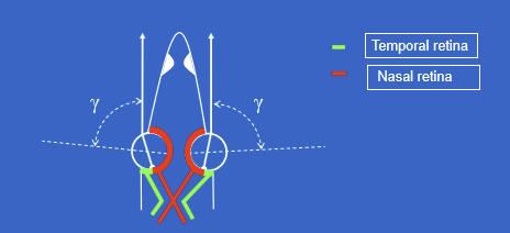
What is the angle γ (gamma), and what is its significance in ambiocular vision?
γ (gamma) = angle between the line of fixation and the optic axis.
In ambiocular vision, γ creates a nasal-temporal division of the retina.
This retinal organization contributes to proper binocular visual processing.
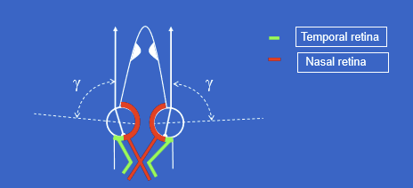
How are the nasal and temporal retinas related to binocular (ambiocular) vision?
The retina is divided into:
Nasal retina (medial)
Temporal retina (lateral)
This division allows the visual fields of both eyes to be integrated and supports binocular visual processing.
What is stereopsis, and why is it the major advantage of ambiocular vision?
Stereopsis = perception of depth from binocular vision.
It is the characteristic sensory advantage of ambiocular vision.
Allows accurate judgment of relative distances and 3D spatial relationships.
What mechanisms make stereopsis possible?
Stereopsis requires:
Coordinated eye movements
Retinal correspondence (matching retinal points from the two eyes represent the same point in space)
Besides stereopsis, what other evolutionary advantage may frontal eyes provide?
Improved visual acuity
Frontal eyes have a smaller angle γ (gamma).
A smaller gamma angle is associated with better optical performance.
Some theories suggest improved acuity may have evolved first, with stereopsis developing as a secondary benefit.
How is visual direction determined in utrocular vision?
Each eye assigns visual direction independently.
The left and right eyes maintain separate oculocentric directions.
Because there is no shared visual direction, the two eyes do not automatically combine their perceptions into a single spatial percept.
How does visual direction differ between utrocular and ambiocular vision?
Utrocular vision: Each eye has its own independent visual direction system.
Ambiocular vision: The two eyes share a common visual direction system.
What is Hering's Law of Identical Visual Directions?
Corresponding retinal points in the two eyes are assigned the same visual direction.
The two foveas share a common oculocentric direction.
This allows the brain to combine information from both eyes into a unified visual percept.
Why can visual confusion occur in ambiocular vision?
Since the foveas are assigned the same visual direction, different objects stimulating corresponding retinal points may be perceived as occupying the same location in space.
This creates visual confusion unless fixation is properly coordinated.
What is bifoveal fixation, and why is it important?
Bifoveal fixation occurs when both foveas are directed at the same target.
It is necessary to prevent confusion and maintain single binocular vision.
Proper bifoveal fixation ensures corresponding retinal points receive compatible images.
What condition must be met for an object to be seen as single during ambiocular vision?
The retinal angles must be equal: βR = βL
When an object's image falls on corresponding retinal locations in both eyes, the object is perceived as single rather than double.
What are the major functional advantages of binocular vision over monocular vision in humans?
Improved depth perception (stereopsis)
Differences in brightness perception
Differences in light adaptation
Improved light detection threshold
How do monocular and binocular vision compare in depth perception?
Binocular vision provides far superior depth perception through stereopsis.
At a viewing distance of 1 meter, the threshold for detecting depth differences is much smaller with binocular vision than monocular vision.
Binocular vision can detect much finer differences in target separation.
Why does binocular vision provide better depth perception than monocular vision?
Each eye views an object from a slightly different angle.
The brain compares these retinal disparities.
This process creates stereopsis, allowing precise perception of relative depth and distance.
How does brightness perception differ between monocular and binocular viewing?
Viewing with both eyes does not normally make an object appear twice as bright.
The visual system effectively combines information from both eyes without simply adding the luminance values.
How are luminance signals combined during binocular vision?
The visual system tends to process the luminance from the two eyes as an average, not a sum.
How does dark adaptation occur in the two eyes?
The eyes dark adapt independently of one another.
Dark adaptation in one eye does not automatically transfer to the other eye.
How does binocular vision affect light detection threshold?
Two eyes improve the probability of detecting a very dim stimulus.
Combining information from both eyes increases the likelihood that at least one eye detects the target.
What is the probability summation principle for binocular light detection?
q=1−(1−p)^n
Where:
q = probability of detection
p = probability of detection by one eye
n = number of eyes/independent detectors
For two eyes at threshold: q=1−(1−0.5)² = 0.75
So the probability of detecting the stimulus increases from 50% to 75%.
What are the three types of visual direction?
Ocular (eye) direction (α)
Oculocentric (relative) direction (β)
Egocentric (absolute) direction (χ)
Differentiate ocular (α), oculocentric (β), and egocentric (χ) directions.
Ocular direction (α): Direction of an object relative to an individual eye.
Oculocentric direction (β): Direction relative to the eye's visual reference system.
Egocentric direction (χ): Direction of an object relative to the observer/body as a whole.
Mnemonic:
α = eye
β = between retinal/ocular references
χ = self (ego)
What sign convention is used for visual directions?
Right and up = positive (+)
Left and down = negative (−)
Right & Rise = Positive
Left & Lower = Negative
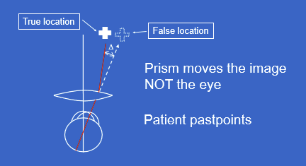
How do plus (+) spectacle lenses affect visual direction and eye movements?
Plus lenses create a prismatic effect that shifts the image.
The image moves, not the eye.
The eye must rotate more than normal to align with the object.
Patients tend to past-point (point beyond the true target location).
Why do patients wearing plus lenses often past-point?
The prism effect displaces the image to a false location.
The brain interprets the displaced image as real.
Increased eye rotation causes reaching movements that overshoot the true target.
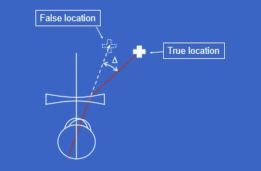
How do minus (−) spectacle lenses affect visual direction and eye movements?
The image is shifted in the opposite direction compared with plus lenses.
The eye requires less rotation than normal to fixate the target.
Perceived location differs from true location.
What is meant by "the prism moves the image, not the eye"?
Prismatic effects alter the apparent location of objects.
The object's retinal image is displaced.
The ocular motor system subsequently adjusts eye position to look at the displaced image.
How do patients adapt to new spectacle prescriptions?
The nervous system recalibrates limb proprioception and motor responses to match altered visual input.
With time, reaching and pointing become accurate despite the optical distortion.
What happens when a patient frequently switches between spectacles and contact lenses?
The brain can develop separate sensorimotor adaptations ("matrices") for each optical system.
Patients may rapidly switch between these learned calibrations.
Why may patients with traumatic brain injury (TBI) struggle with adapting to new optical corrections?
Visual adaptation after TBI is often impaired.
Changes that normally become automatic may remain disorienting.
Patients may have difficulty recalibrating visual and proprioceptive systems.
Why can switching from glasses to contact lenses be particularly disorienting in some patients?
Glasses create prismatic/image displacement effects that contact lenses largely do not.
The brain must switch between different learned sensorimotor calibrations.
This process may be especially difficult after TBI.
Why are bifocals often difficult for patients with poor visual adaptation?
Different lens zones produce different optical effects.
Patients must adapt to changing visual directions and image locations.
Those with impaired adaptation (e.g., TBI) may find bifocals particularly disorienting.
What are the two major theories explaining binocular ocular direction?
Dominant Eye Theory
Cyclopean Theory
What is the Dominant Eye Theory of binocular ocular direction?
The perceived visual direction comes from the dominant eye.
Binocular direction is assigned as either:
αL (left eye direction), or
αR (right eye direction)
What is the Cyclopean Theory of binocular ocular direction?
Visual direction is the average of the two eyes' directions.
The brain behaves as if vision originates from a single central eye ("cyclopean eye").
αB= (αL+αR)/2
where:
αB = binocular direction
αL = left eye direction
αR = right eye direction
According to Cyclopean Theory, where is binocular visual direction located?
At the bisector of the angle of convergence.
Perception is referenced to an imaginary central viewing position between the two eyes.
What evidence supports the Cyclopean Theory over the Dominant Eye Theory?
Placing a minus lens or prism before only one eye changes perceived egocentric direction.
If only the dominant eye determined direction, altering the nondominant eye should have little effect.
Because changing one eye affects overall directional perception, binocular direction appears to be a combination of information from both eyes.
What is the egocenter?
The egocenter is the body's reference point for egocentric direction.
It serves as the perceived origin from which directions in space are judged.
Where is the egocenter located according to the Dominant Eye Theory?
At the dominant eye.
Since directional judgments are assumed to come from one dominant eye, the reference point is placed there.
Where is the egocenter located according to the Cyclopean Theory?
Approximately midway between the two eyes.
Consistent with the concept of an imaginary central "Cyclopean eye."
How can the egocenter be experimentally determined?
Have a person fixate targets in space.
Mark perceived locations along the lines between the targets and the observer.
The intersection of these perceived directional lines estimates the egocenter.
What is the experimentally determined location of the egocenter?
Located between the eyes.
Approximately 10 cm behind the plane of the eyes.
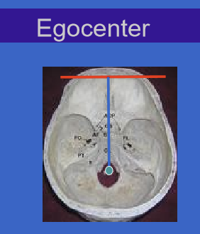
What is the relationship between the egocenter and the dens (odontoid process)?
The dens of C2 (axis) is often considered anatomically close to the body's rotational reference point.
The egocenter is thought to lie near the body's midline and central axis of rotation.
This supports the concept of a central reference point rather than one located at either eye.
What are corresponding retinal points?
Corresponding retinal points are points, one in each retina, that share the same oculocentric direction.
When stimulated simultaneously, they are perceived as representing the same location in space.
Definition: βL=βR
What condition defines retinal correspondence?
Corresponding retinal points occur when: βL=βR
where:
βL = oculocentric direction in the left eye
βR = oculocentric direction in the right eye
What is Hering's Window, and what does it demonstrate?
Hering's Window demonstrates retinal correspondence and Hering's Law of Identical Visual Directions.
When fixation occurs at a point in a window, images may stimulate corresponding retinal points despite representing different objects.
This can produce visual confusion because different objects are perceived in the same direction.
Why do confusion and diplopia occur simultaneously in Hering's Window?
Confusion
Different objects are assigned the same visual direction.
Diplopia
Images do not fall on corresponding retinal points in both eyes.
Separate images are perceived in addition to the confusion.
What is required for normal binocular vision?
Bifoveal fixation
Retinal correspondence
Equal oculocentric directions
During normal binocular vision, what happens to non-fixated objects?
Images of non-fixated objects fall on another pair of corresponding retinal points.
Because those points share the same visual direction, objects are perceived in their correct spatial locations.
What happens to visual direction in a right esotropia?
The right eye deviates inward.
The fixated object is not imaged on the right fovea.
Correspondence relationships become abnormal.
Perceived locations of objects become incorrect.
Result: Both confusion and diplopia may occur.
How does right esotropia disrupt retinal correspondence?
The deviating eye no longer places the fixation target on its fovea.
βL and βR no longer match appropriately.
Objects that should fall on corresponding points stimulate noncorresponding retinal locations.
Consequence: Loss of normal binocular single vision.
What is Anomalous Retinal Correspondence (ARC)?
Correspondence is shifted so that the image of the fixation target falls on newly corresponding retinal points.
The objects appear in their correct locations despite the eye turn.
Result: Single binocular perception is preserved.
What is harmonious ARC?
A type of ARC in which the anomalous correspondence completely compensates for the ocular deviation.
The fixation target is superimposed correctly for both eyes.
Perceived spatial locations are normal.
How does ARC affect confusion and diplopia?
ARC reassigns corresponding retinal points.
This restores compatible visual directions.
Confusion and diplopia are prevented.
What retinal change occurs in ARC of a right esotropia?
Corresponding points in the right eye become shifted nasally.
The fovea of the fixing eye corresponds to a nonfoveal retinal location in the deviating eye.
What would vision look like if there were no retinal correspondence (utrocular vision)?
Each eye would localize objects independently. There would be no shared binocular directional system.
Why can utrocular vision be confused with harmonious ARC?
In both situations, objects may appear correctly localized.
However:
ARC: binocular sensory adaptation with correspondence.
Utrocular vision: no correspondence; each eye acts independently.
Which retinal points are the primary corresponding points in a normal visual system?
The two foveae are corresponding retinal points.
Both foveae share the same oculocentric direction.
What is retinal (absolute) disparity?
Retinal disparity results when an object stimulates noncorresponding retinal points in the two eyes.
It represents the difference in oculocentric direction between the two retinal images.
Retinal disparity = βL−βR
How is retinal disparity calculated?
Retinal disparity = βL−βR
where:
βL = oculocentric direction in the left eye
βR = oculocentric direction in the right eye
The magnitude of disparity equals the angular separation of the retinal images in the two eyes.
What is the relationship between corresponding retinal points and retinal disparity?
Corresponding retinal points have identical oculocentric directions: βL=βR
Therefore: Retinal disparity = 0
What does a retinal disparity of zero indicate?
The object's images fall on corresponding retinal points.
The object lies on the fixation surface (horopter conceptually).
Single binocular vision is produced without disparity cues.