Lower Respiratory System
1/17
There's no tags or description
Looks like no tags are added yet.
Name | Mastery | Learn | Test | Matching | Spaced |
|---|
No study sessions yet.
18 Terms
What are the 2 main components of the respiratory system?
Upper respiratory system
Lower respiratory system
What are the organs in the lower respiratory system?
Bronchus
Bronchioles #a700ff
Lungs #ff00fe
Bronchus
Begins at
Forming
What special bronchus is present in pigs and ruminants
Function
What do the principal bronchi divide into
Function
What are segmental bronchi
Location
Flow chart of bronchus organisation
Walls of bronchi are supported by
Begins at: Bifurcation of trachea
Forming: Right and left principal bronchus
What special bronchus is present in pigs and ruminants: Tracheal bronchus
Function: Supplies the cranial lobe of the right lung
What do the principal bronchi divide into: Lobar bronchi
Function: Each supply a lung lobe
What are segmental bronchi: Tertiary bronchi
Location: Within lung lobes and branch from lobar bronchi
Flow chart of bronchus organisation: Trachea —> Primary bronchi —> Lobar Bronchi —> Segmental Bronchi
Walls of bronchi are supported by: Cartilaginous plates
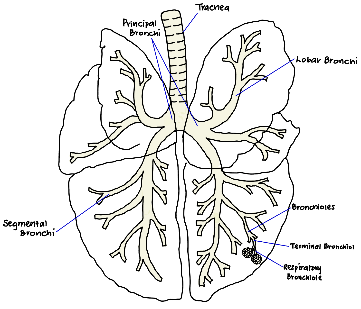
Bronchioles #a700ff
What structural change defines bronchiole
Bronchioles branch into
Bronchioles terminate in clusters of
Where does gas exchange
What structural change defines bronchiole:
When airways are less than 1 mm in diameter
And lack cartilage
Bronchioles branch into:
Terminal bronchioles
Respiratory bronchioles
Alveolar ducts
Bronchioles terminate in clusters of: Alveolar sacs and pulmonary alveoli
Where does gas exchange: Alveoli
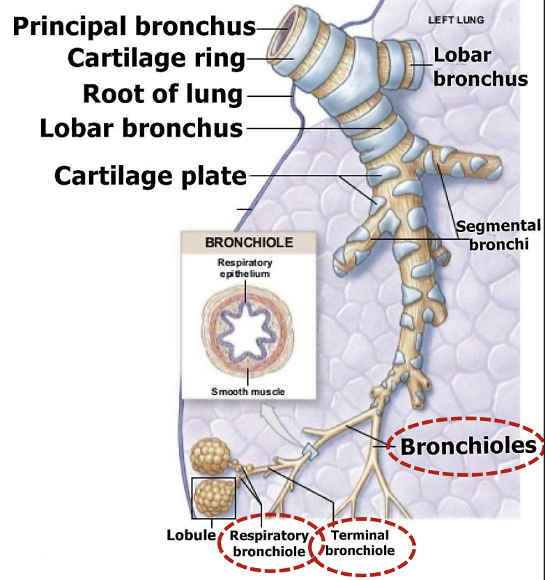
The respiratory zone includes?
Respiratory bronchioles
Alveolar ducts
Alveoli
Lungs #ff00fe
Consist of
Which side is larger
Location
Normal texture
Allows it to
Fresh color is
Shaped as
What are the parts
Location
Curved lateral surface is called
Flattened surface is called
Faces the
Surface that lies against the convex surface of diaphragm is called
Consist of: Left and right lung
Which side is larger: Right
Location: Occupies greater part of thoracic cavity
Normal texture: Sponge
Allows it to: Be flexible and regains shape quickly after being compressed
Fresh color is: Bright pink
Shaped as: Cone
What are the parts
Apex
Location: Lies in thoracic inlet
Base
Location: Lies adjacent to diaphragm
Convex lateral surface
Curved lateral surface is called: Coastal surface
Flattened surface is called: Medial surface
Faces: Mediastinum
Surface that lies against the convex surface of diaphragm is called: Diaphragmatic surface
Lungs #ff00fe
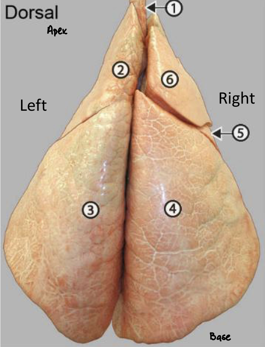
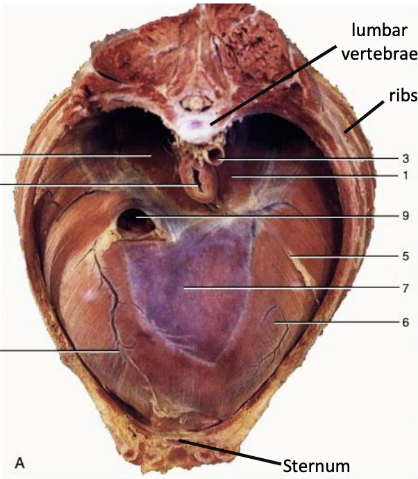
Label 1-6
1: Trachea
2: Left cranial lobe
3: Left caudal lobe
4: Right caudal lobe
5: Middle lobe
6: Right cranial lobe
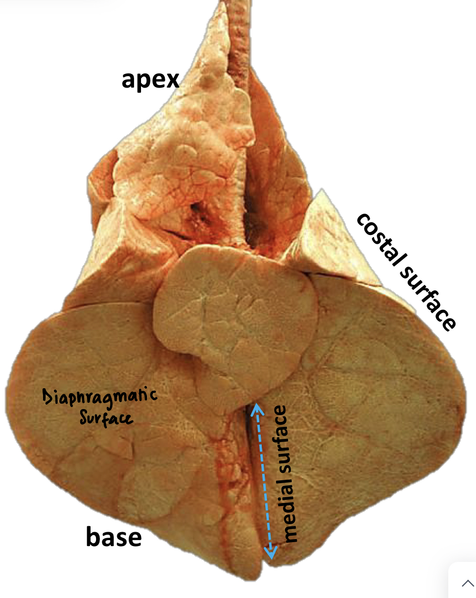
Lungs: Surfaces
Therefore how many surfaces does the lung have?
The lungs have 3 surfaces:
Coastal surface
Medial surface
Diaphragmatic surface
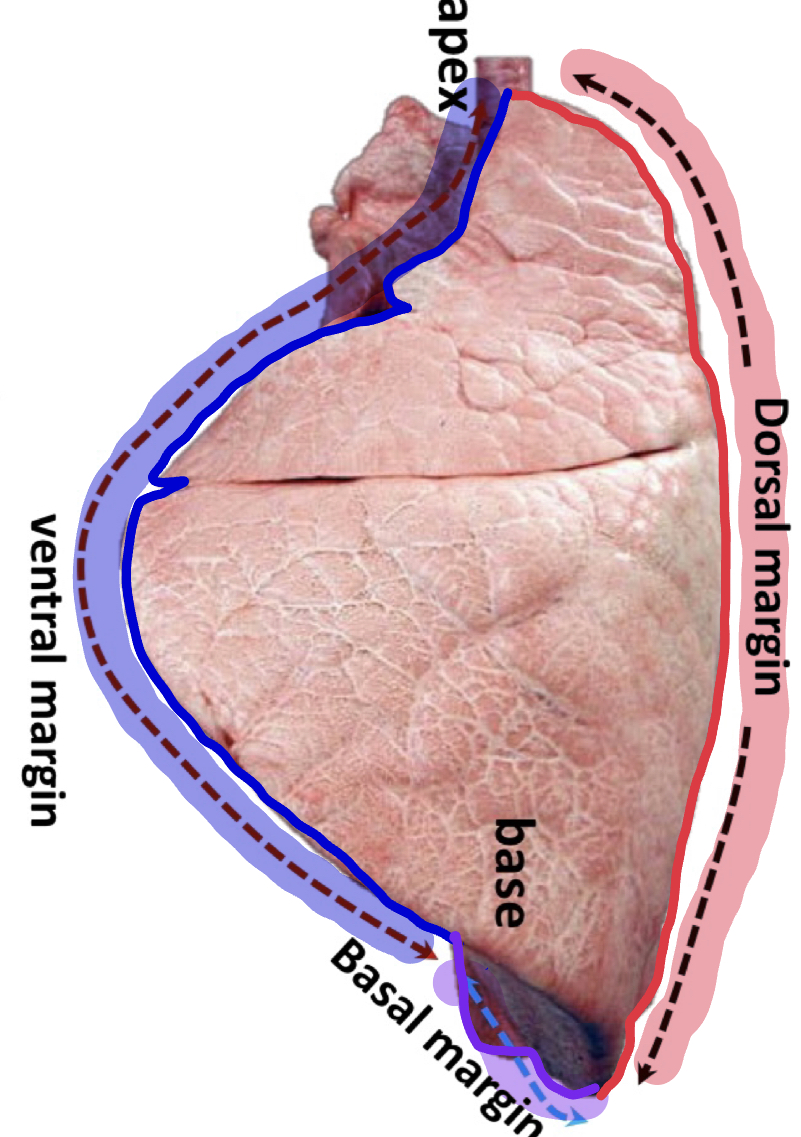
Lungs: Margins
Where is the dorsal margin
Where is the ventral margin
Where is the basal margin
Continuous with
Positioned along
Dorsal margin: Along vertebral (spine) part of lung
(posterior border)
Ventral margin: Along sternum (front) part of chest
(anterior border)
Basal margin: Along the caudal (bottom) part of lung
Continuous with: Ventral margin
Positioned along: Diaphragm
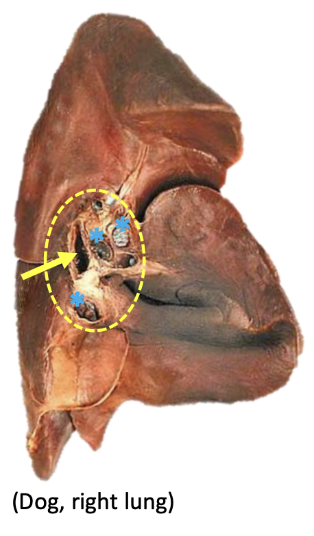
Lungs: Hilus #ff00fe
What is the hilus
Location
What is the root of the lung
What is the hilus:
Area where:
Principal bronchus
Pulmonary artery and vein
Lymphatic
Nerves
enter and leave the lung
Location: On the medial surface
What is the root of the lung: Bundle of the above structures that pass through the hilus
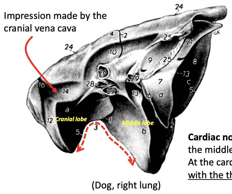
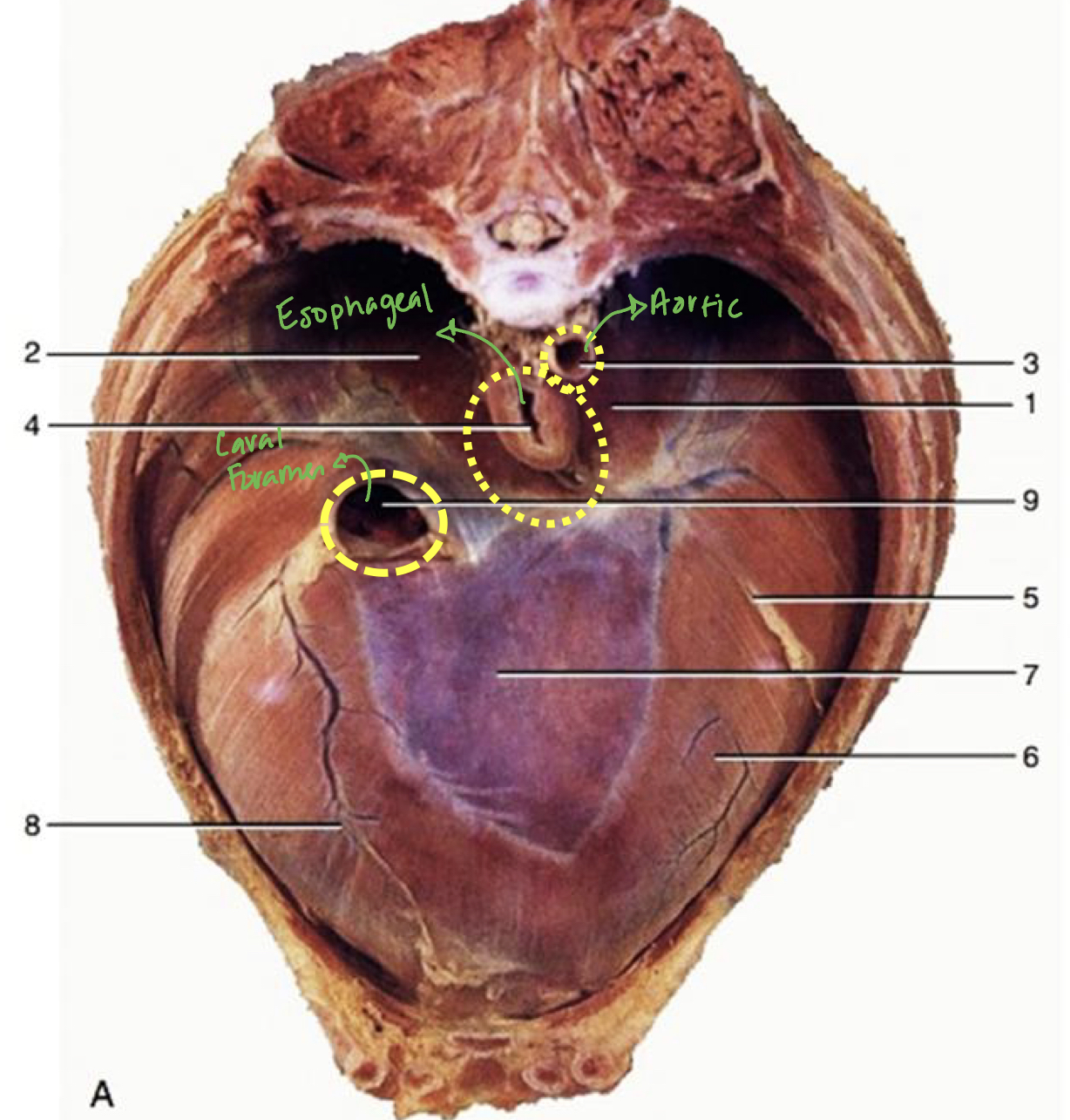
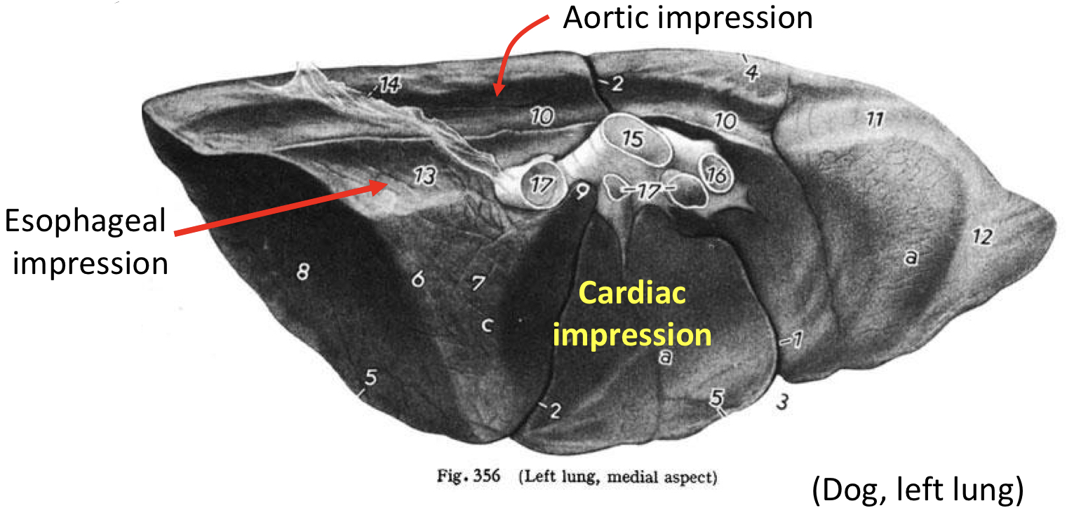
Lungs: Impressions #ff00fe
Where is the cardiac impression located
What are the 2 other most prominent impressions
Location
On which lung can the impression of the cranial vena cava can be seen
What is the cardiac notch
Where is the cardiac impression located: On the medial surface between the 3rd and 6th ribs
What are the 2 other most prominent impressions: Aortic and esophageal impressions
Location: Dorsal (above) to the cardiac impression
On which lung can the impression of the cranial vena cava can be seen: Right lung
Cardiac notch: Gap between the middle and cranial lobes of the right lung where the heart makes contact with the thoracic wall
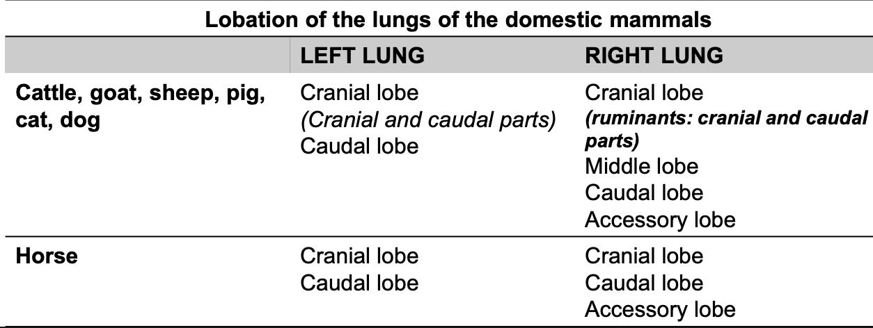
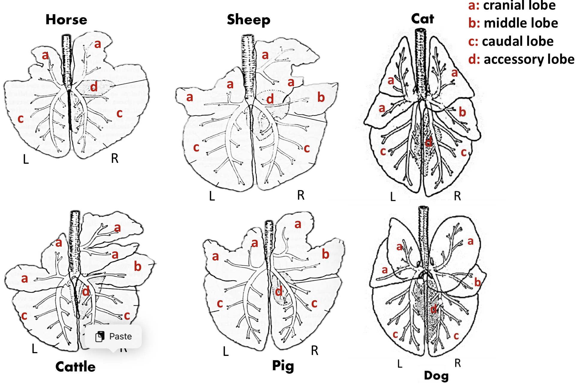
Lungs: Lobes #ff00fe
Defined by
In which species are the lobes not divided by deep fissues
In which species is the cranial lobe divided into cranial and caudal parts
(look at table)
Defined by: By the presence of the lobar bronchi (secondary bronchi)
In which species are the lobes not divided by deep fissures: Horse
In which species is the cranial lobe divided into cranial and caudal parts: Ruminants
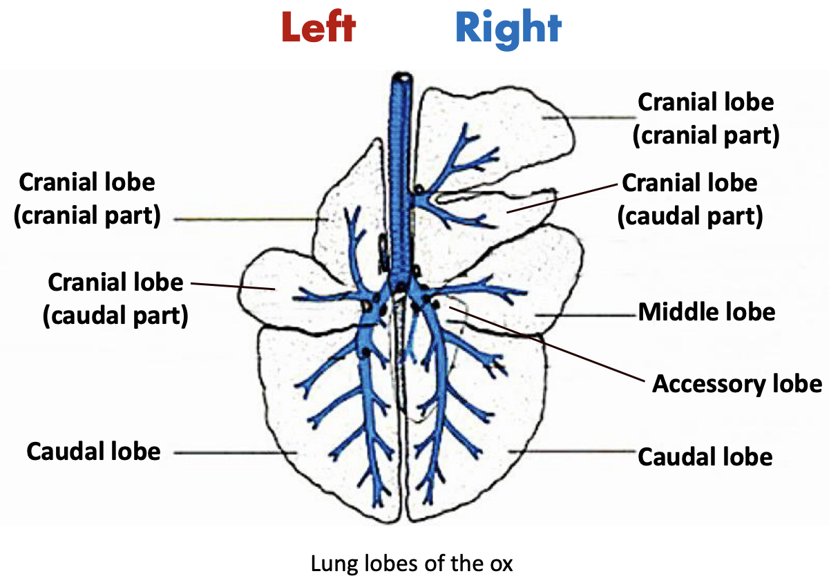
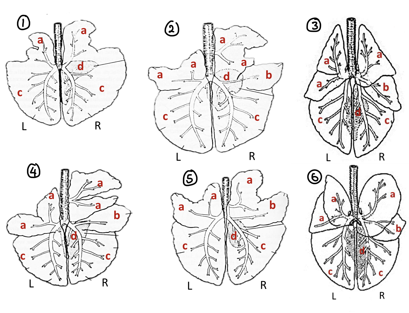
Lungs: Label Lobes #ff00fe
Label the species 1-6
Label the lobes a-d
Species:
1 = Horse
2 = Sheep
3 = Cat
4 = Cattle
5 = Pig
6 = Dog
Lobes:
a = Cranial lobe
b = Middle lob
c = Caudal lobe
d = Accessory lobe
Lungs: Blood Supply, Lymphatic Drainage #ff00fe
Lung receives blood flow by
Function
Blood supply comprises of what 2 vessels
Lung receives blood flow by:
Bronchial Circulation
Function: Provides oxygenated blood and nutrition to the bronchi, lungs, large blood vessels, lymph nodes and visceral pleura
Pulmonary Circulation
Function: Movement of blood from heart —> lungs for oxygenation then back to heart again
Blood supply comprises of what 2 vessels:
Bronchial vessels (nutritional blood supply)
Bronchial artery and vein
Pulmonary vessels (functional blood supply)
Pulmonary artery and vein
Lungs: Innervation #ff00fe
What provides autonomic and sensory innervation
Autonomic
Sympathetic
What is the effect of sympathetic stimulation on bronchial smooth muscle
How does sympathetic stimulation affect pulmonary vessels
What is the overall result of sympathetic innervation
Parasympathetic
What is the effect of parasympathetic stimulation on bronchial smooth muscle
What is the function of increased gland secretion during parasympathetic stimulation
What effect does parasympathetic innervation have on pulmonary blood flow
What is the overall result of parasympathetic innervation
Sensory
What structures send sensory information to brainstem
What kind of signals are carried
What provides autonomic and sensory innervation: Pulmonary nerve plexus
Autonomic
Sympathetic
What is the effect of sympathetic stimulation on bronchial smooth muscle: Causes bronchodilation (relaxes bronchial smooth muscle), increases airflow
How does sympathetic stimulation affect pulmonary vessels: Causes vasoconstriction which redirects blood flow
What is the overall result of sympathetic innervation: ↑ breathing rate and ↑ oxygen delivery
Parasympathetic
What is the effect of parasympathetic stimulation on bronchial smooth muscle: Causes bronchoconstriction (contracts bronchial smooth muscle) which decreases airflow
What is the function of increased gland secretion during parasympathetic stimulation: Produce mucus to moisten airways and trap dust/particles
What effect does parasympathetic innervation have on pulmonary blood flow: Causes vasodilation which increases blood flow to lungs
What is the overall result of parasympathetic innervation: ↓ Breathing rate in a low-demand state
Sensory:
What structures send sensory information to brainstem:
Larynx
Trachea
Bronchi
Vessels
Stretch receptors
What kind of signals are carried: Pain and reflex signals to brainstem
Diaphragm
Separates
Shape
Consist of
Muscular periphery is divided into portions arise from
(Check labeled diagram)
Separates: Thoracic and abdominal cavities
Shape: Dome-shaped, convex
Consist of:
Central tendon (tendinous center)
Muscular periphery
Muscular periphery is divided into portions arise from:
Lumbar vertebrae
Sternum
Caudal ribs

Diaphragm: Peripheral Muscles
Lumbar portion
AKA
Consist of
Arises from
Coastal part
AKA
Arises from
Attaches in what direction
Sternal part
AKA
Arises from
Location
Lumbar portion:
AKA: Pars lumbalis
Consist of: Left and right crura
Arises from: Ventral border of L4 and body of L3
Coastal part:
AKA: Pars costalis
Arises from: Inner surfaces of the ribs and costal cartilages
Attaches in what direction: Oblique direction to the 13th through 7th rib
Sternal part:
AKA: Pars sternalis
Arises from: Dorsal surface of sternum
Location: Runs dorsally to meet tendon
Diaphragm: Openings
What are the 3 openings
Contains
Location
Which opening doesn’t allow movement
Why
What are the 3 openings:
Aortic Hiatus
Contains:
Aorta
Azygous vein
Thoracic duct (lymphatic vessels)
Location: Most dorsal
Esophageal Hiatus
Contains:
Esophagus
Dorsal and ventral vagal nerves
Vessels
Caval Foramen
Contains: Caudal vena cava
Location: Dorsal to tendinous center of diaphragm
Which opening doesn’t allow movement: Caval foramen
Why: Tendon fused with adventitia of vessels