Anatomy Practical 1
1/208
There's no tags or description
Looks like no tags are added yet.
Name | Mastery | Learn | Test | Matching | Spaced |
|---|
No study sessions yet.
209 Terms
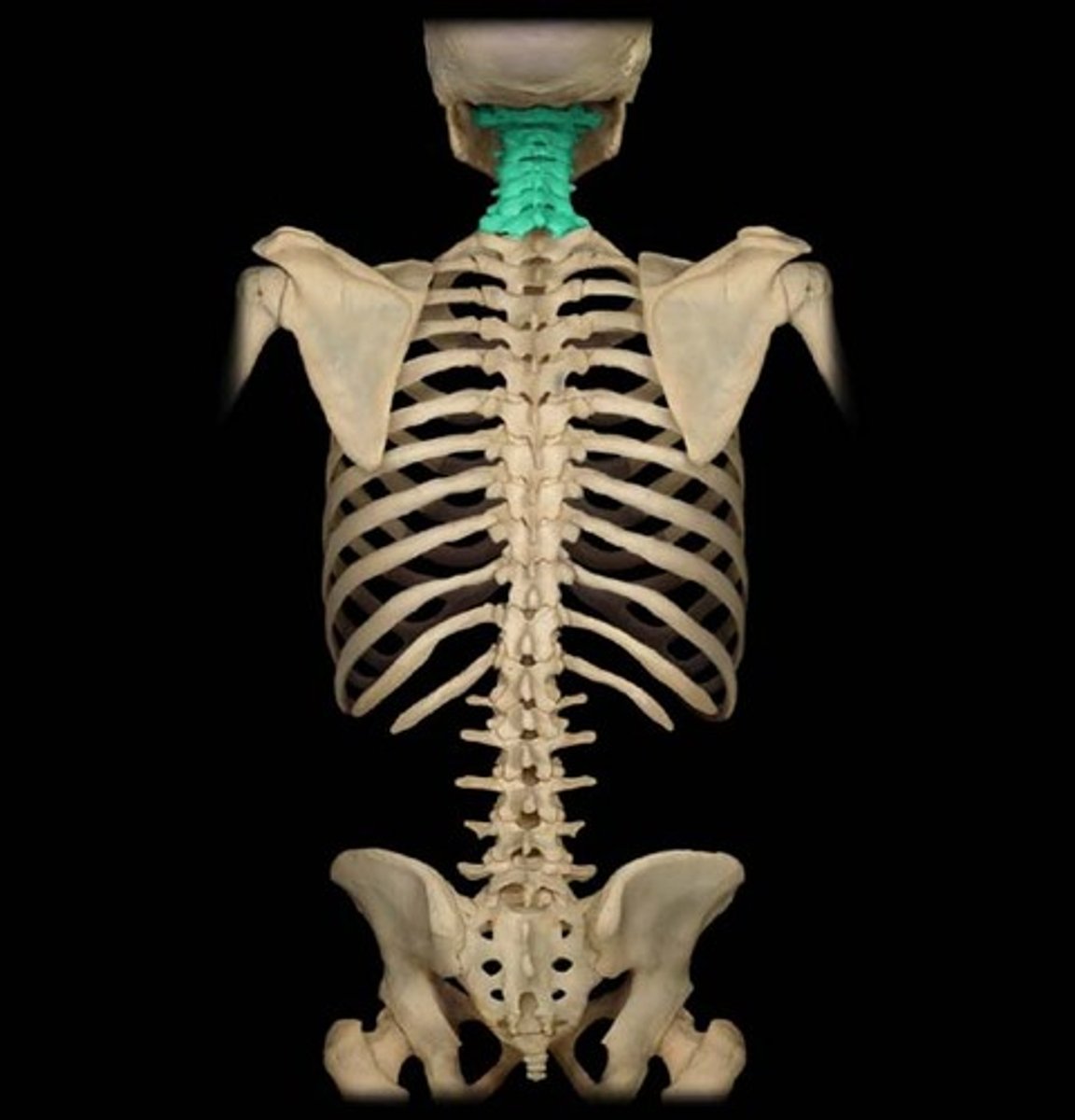
Cervical vertebrae (C1-C7)
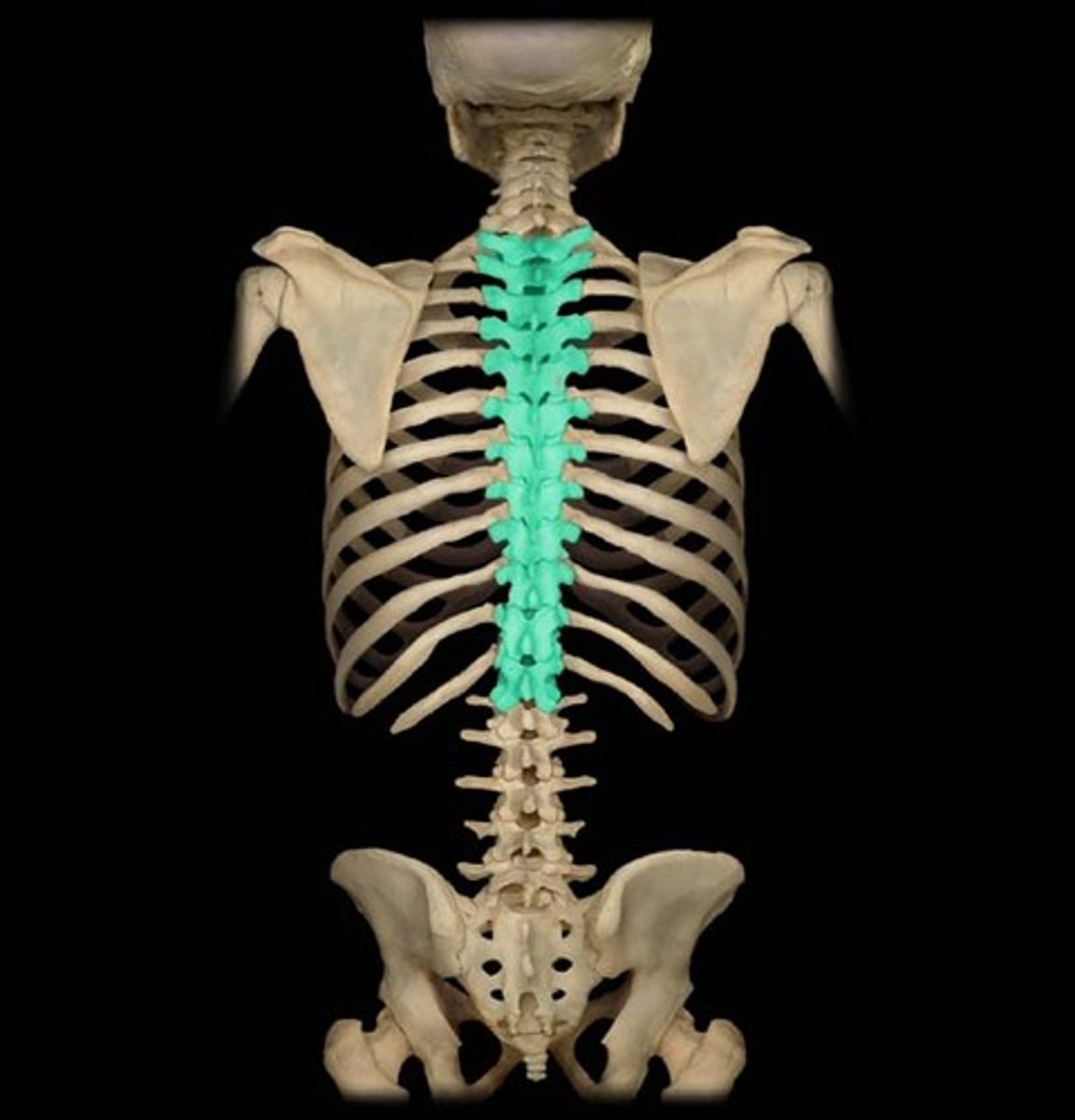
Thoracic vertebrae (T1-T12)
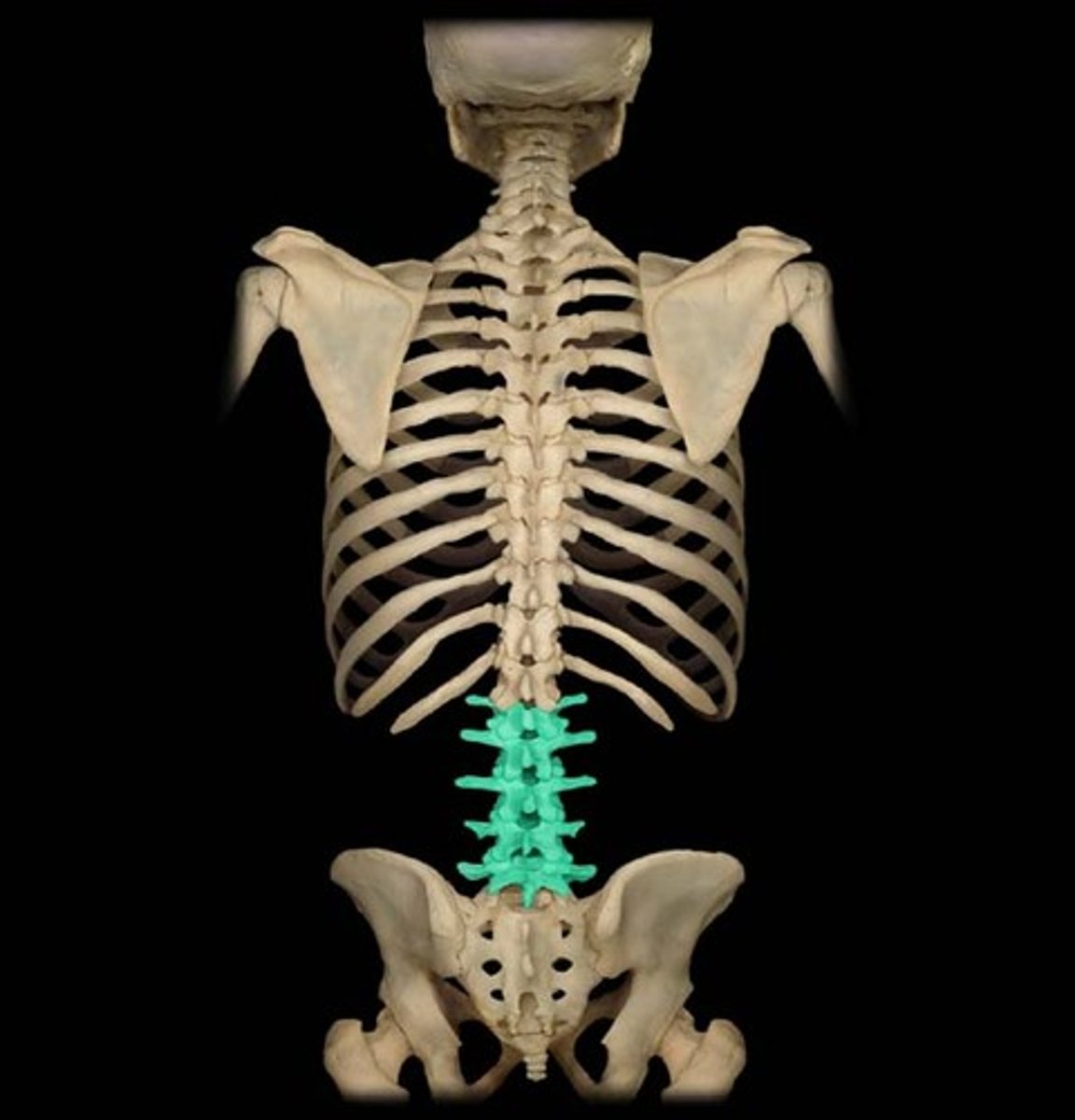
Lumbar Vertebrae (L1-L5)
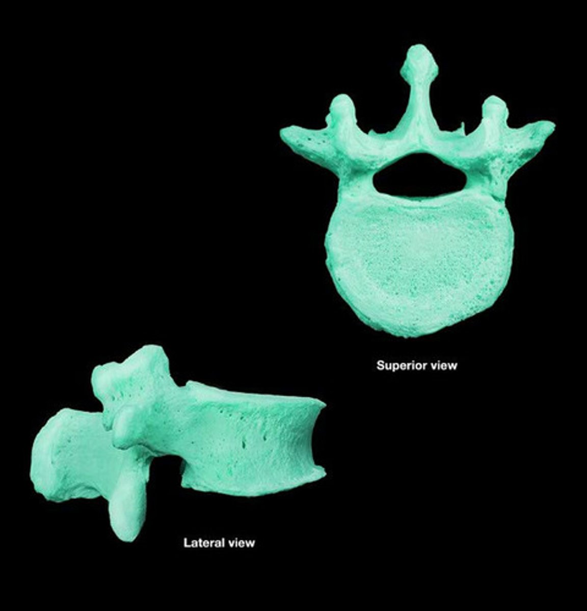
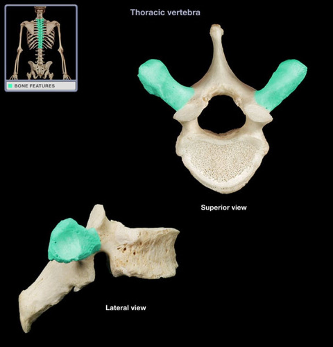
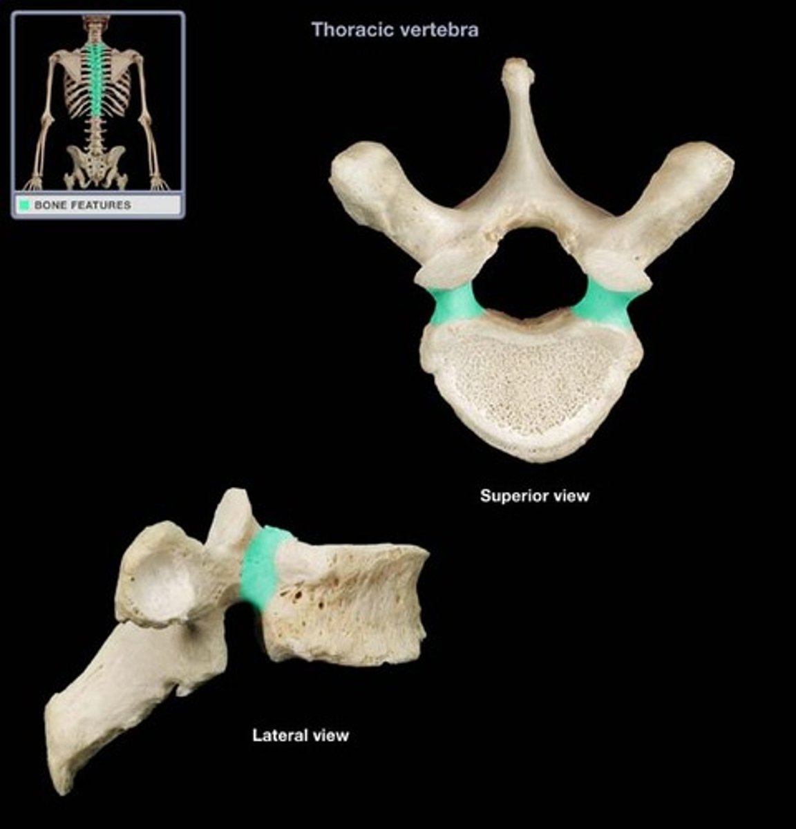
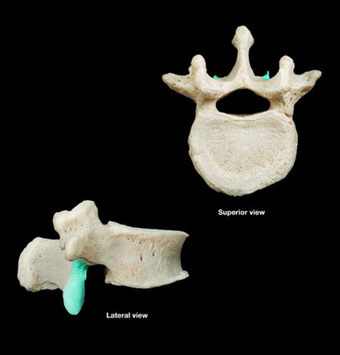
thoracic
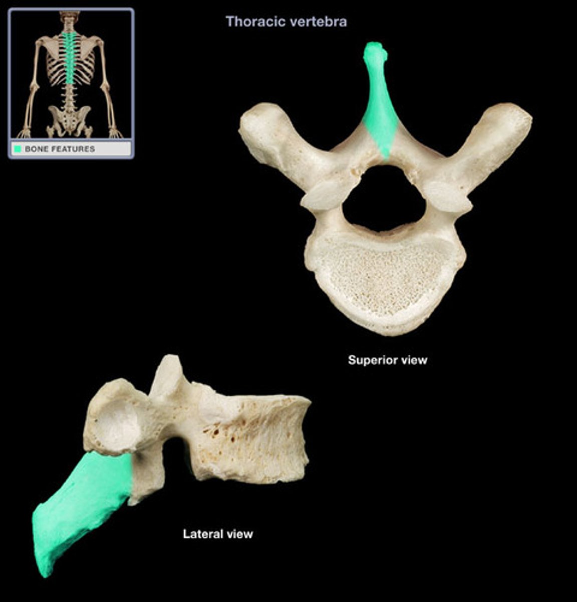
- long inferiorly positioned spinous process
- has costal facets
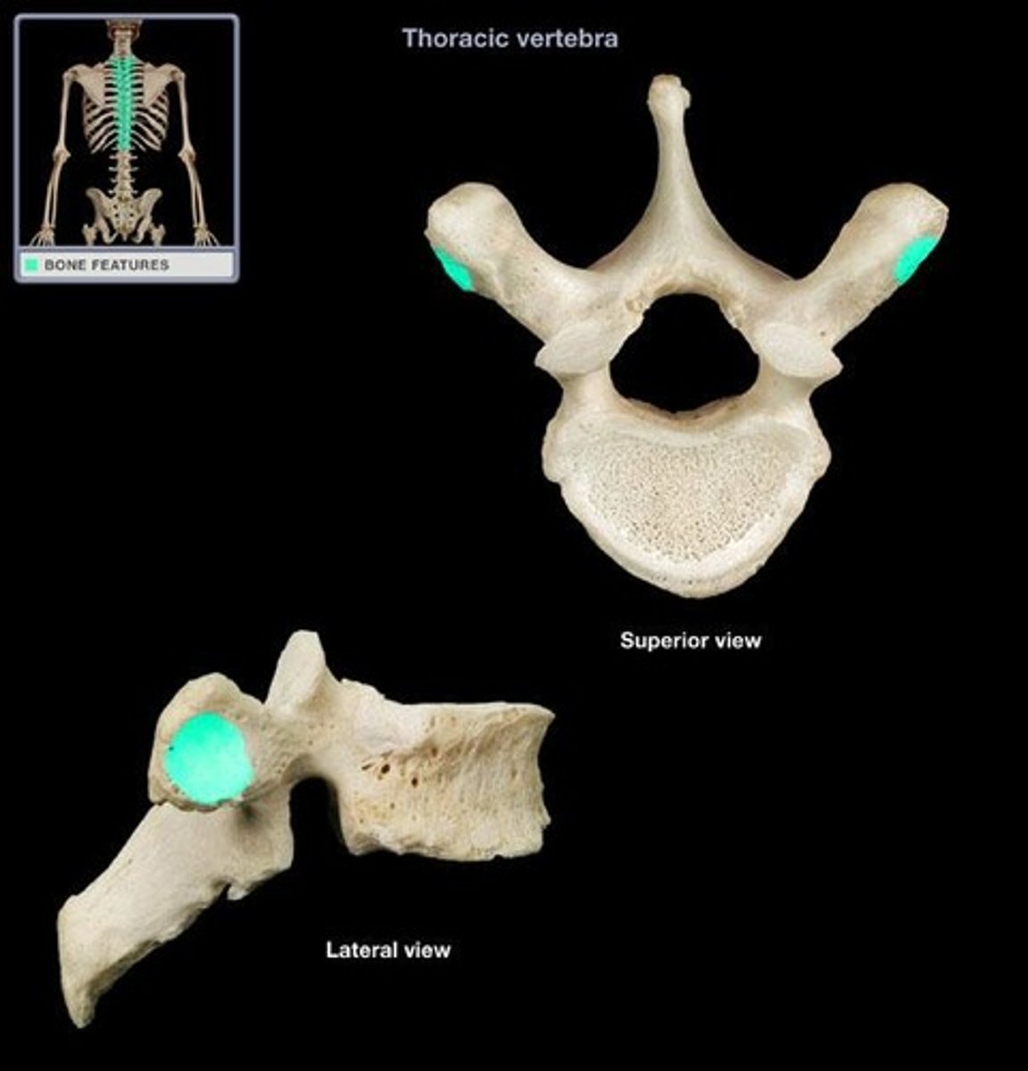
- long transverse processes
What type of vertebra is this?
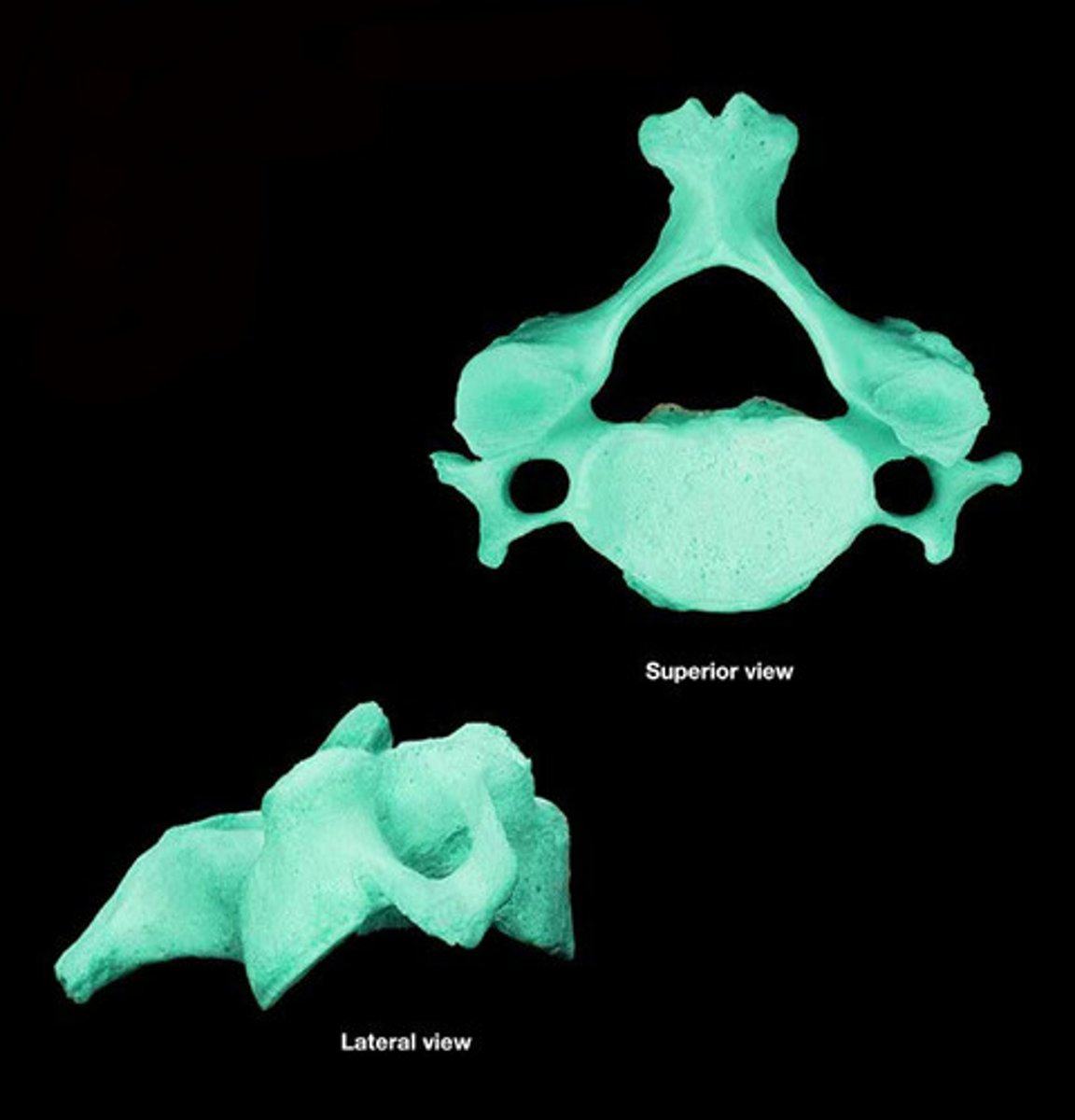
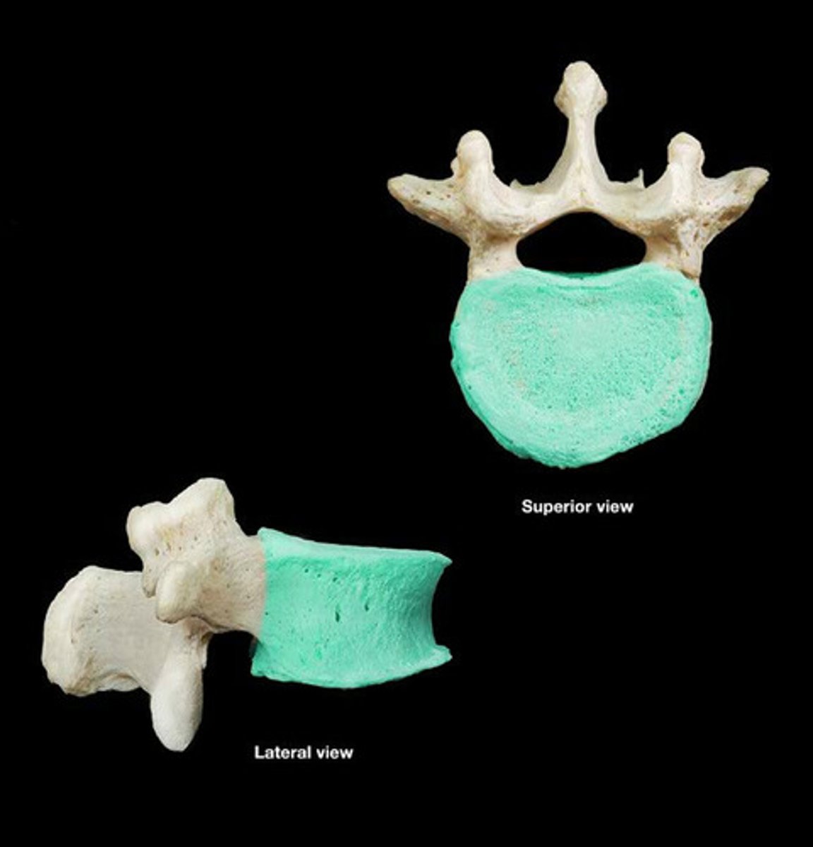
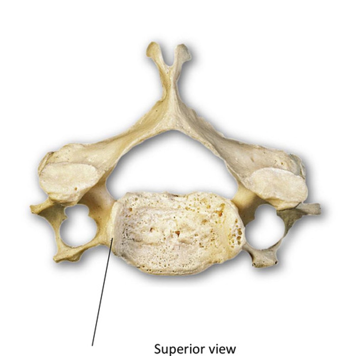
lumbar
- large vertebra body
- short, wide spinous process
What type of vertebra is this?
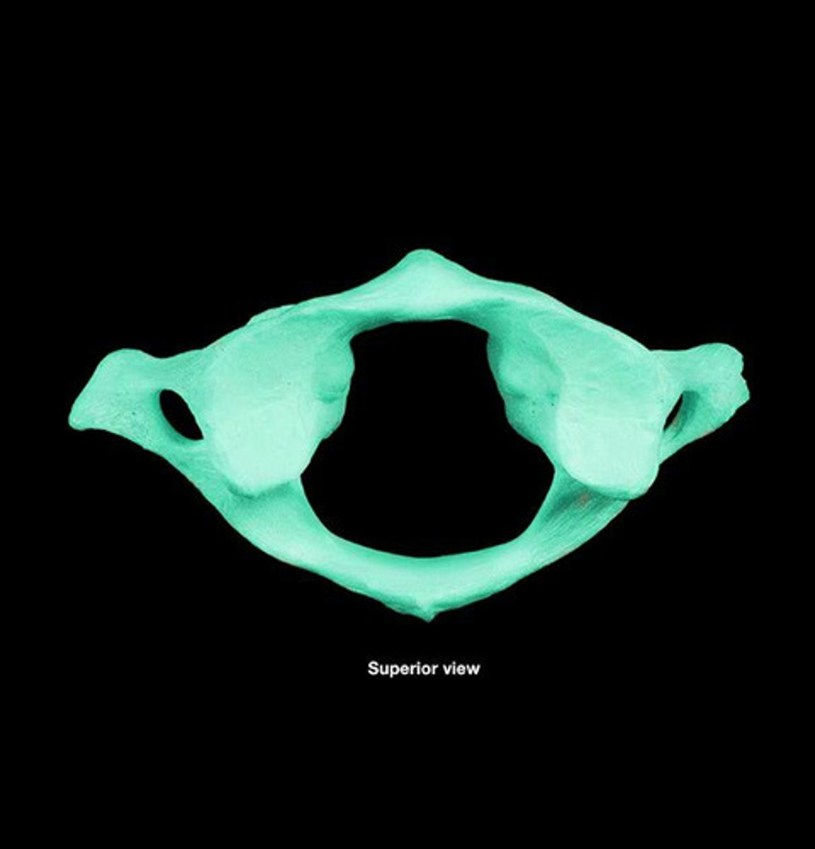
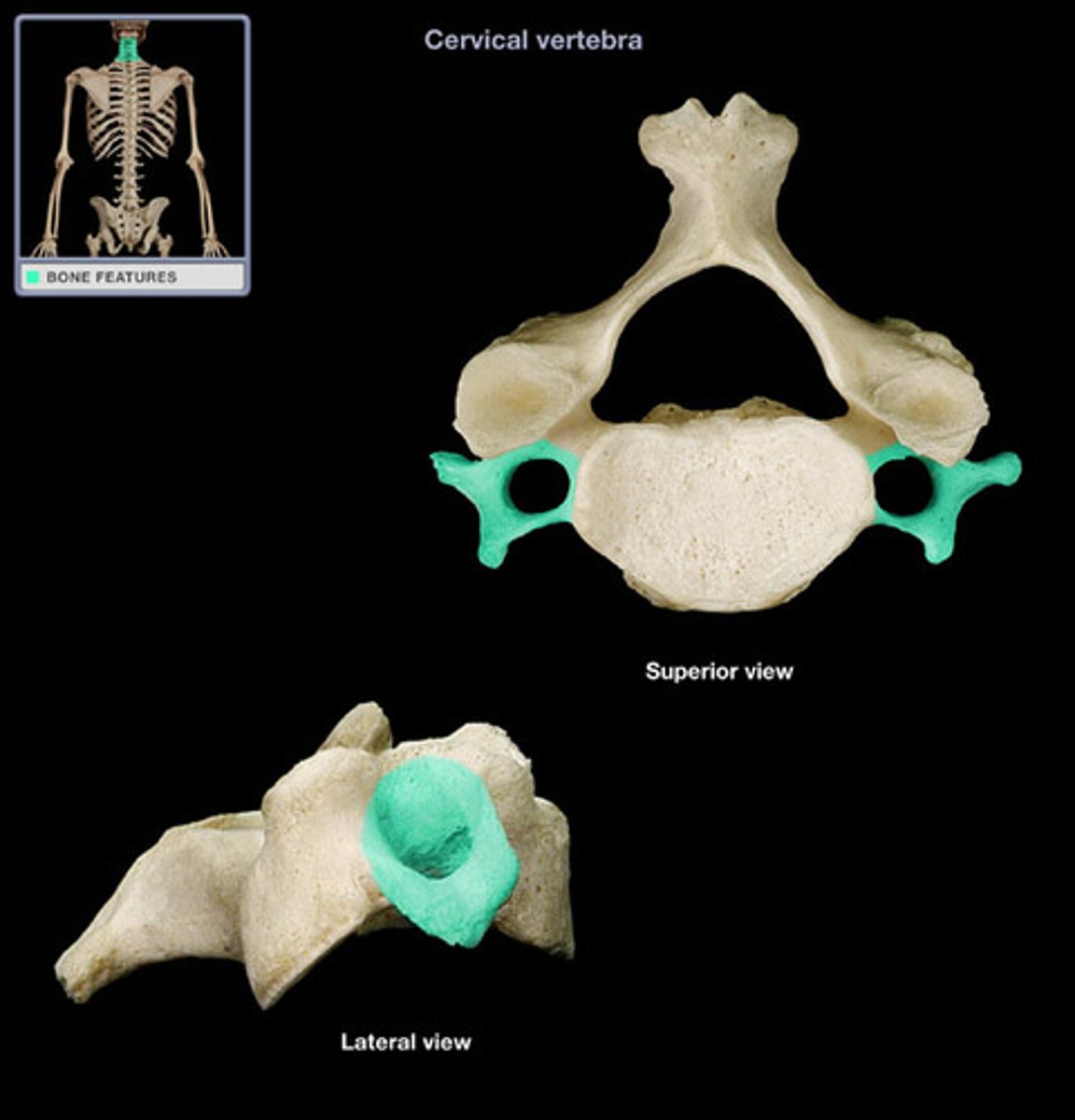
cervical
- has transverse foramen
- bifid spinous process
What type of vertebra is this?
Atlas (C1)
- no vertebral body or spinous process
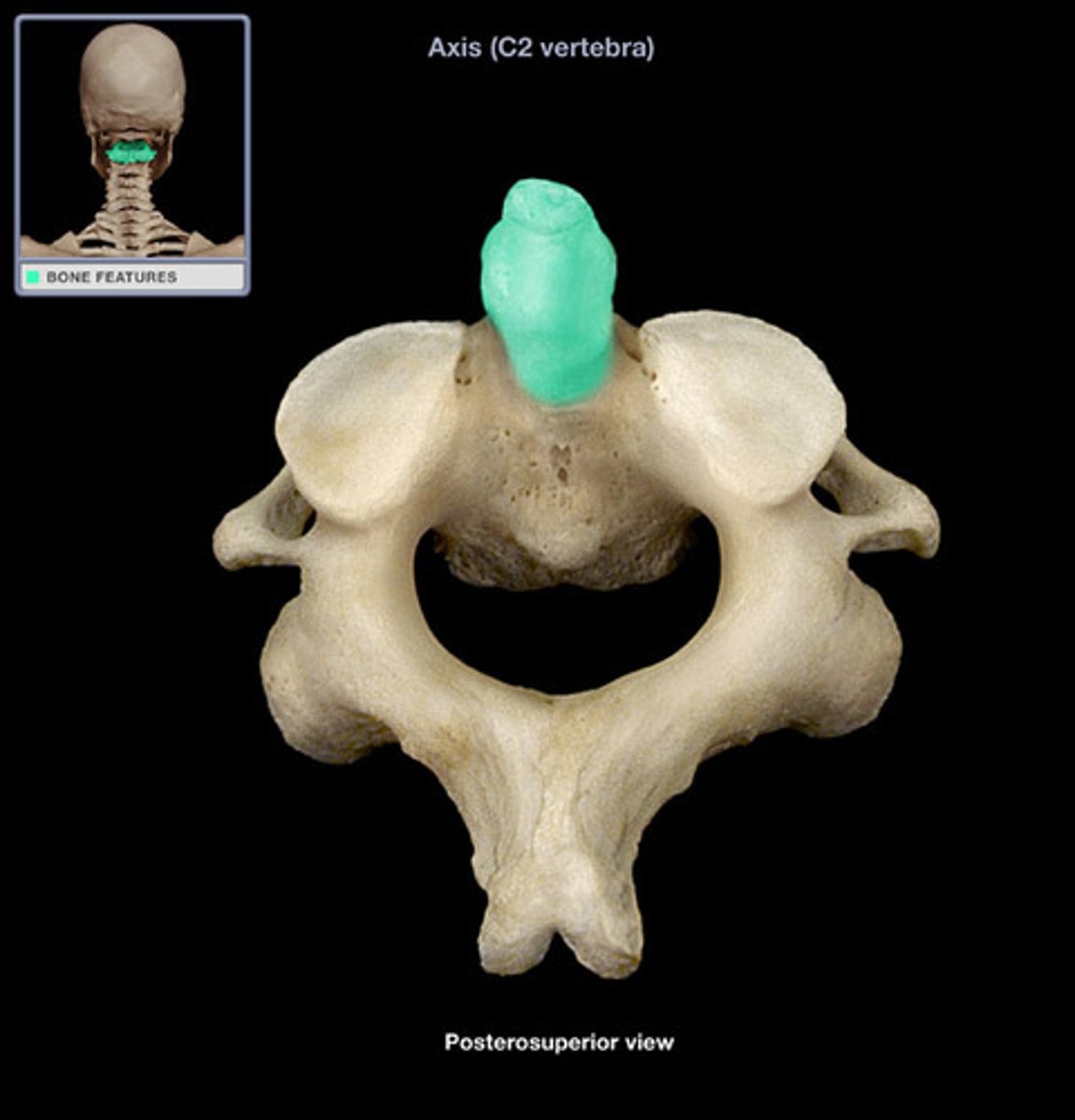
odontoid process (dens) on Axis (C2)
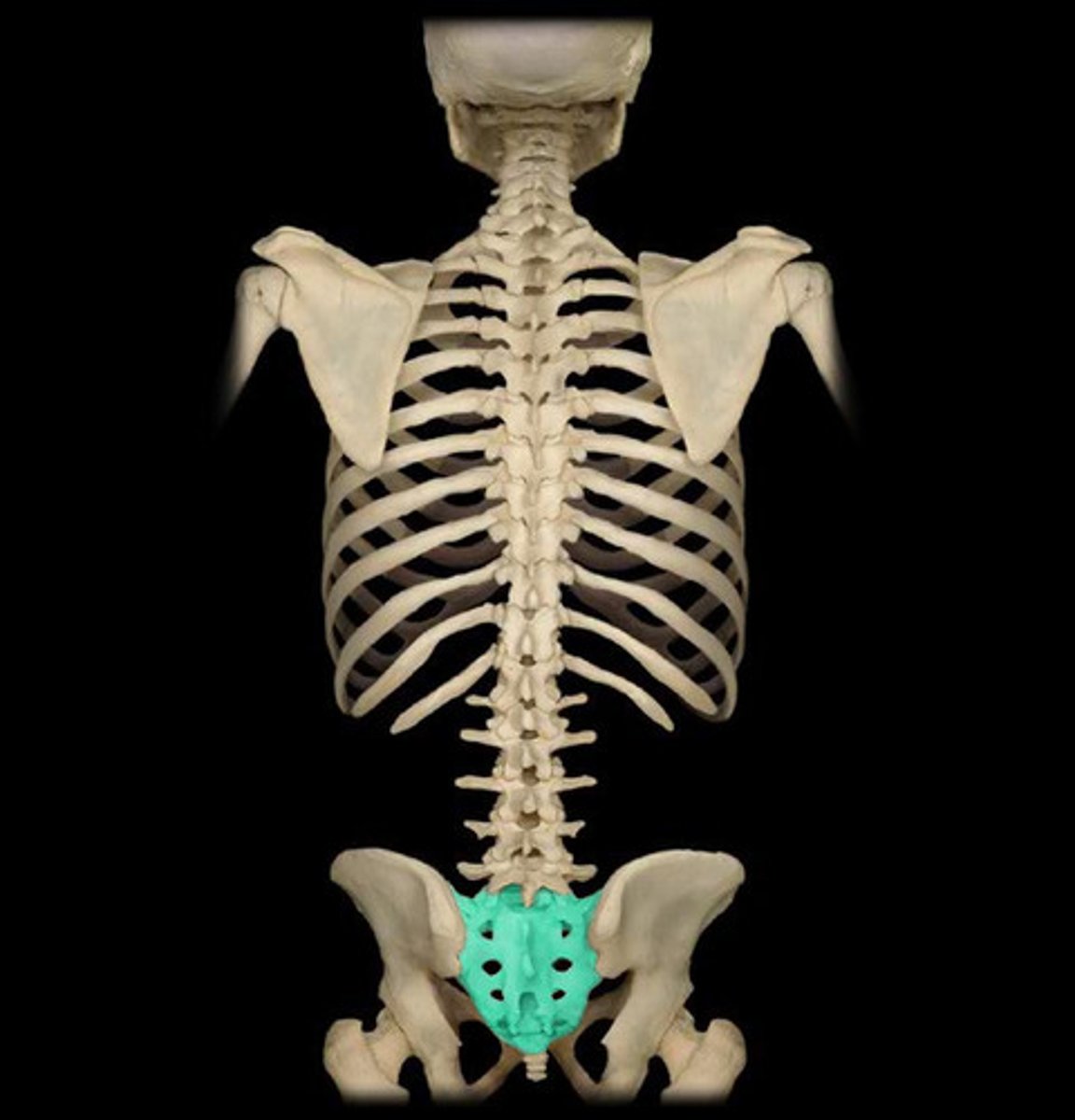
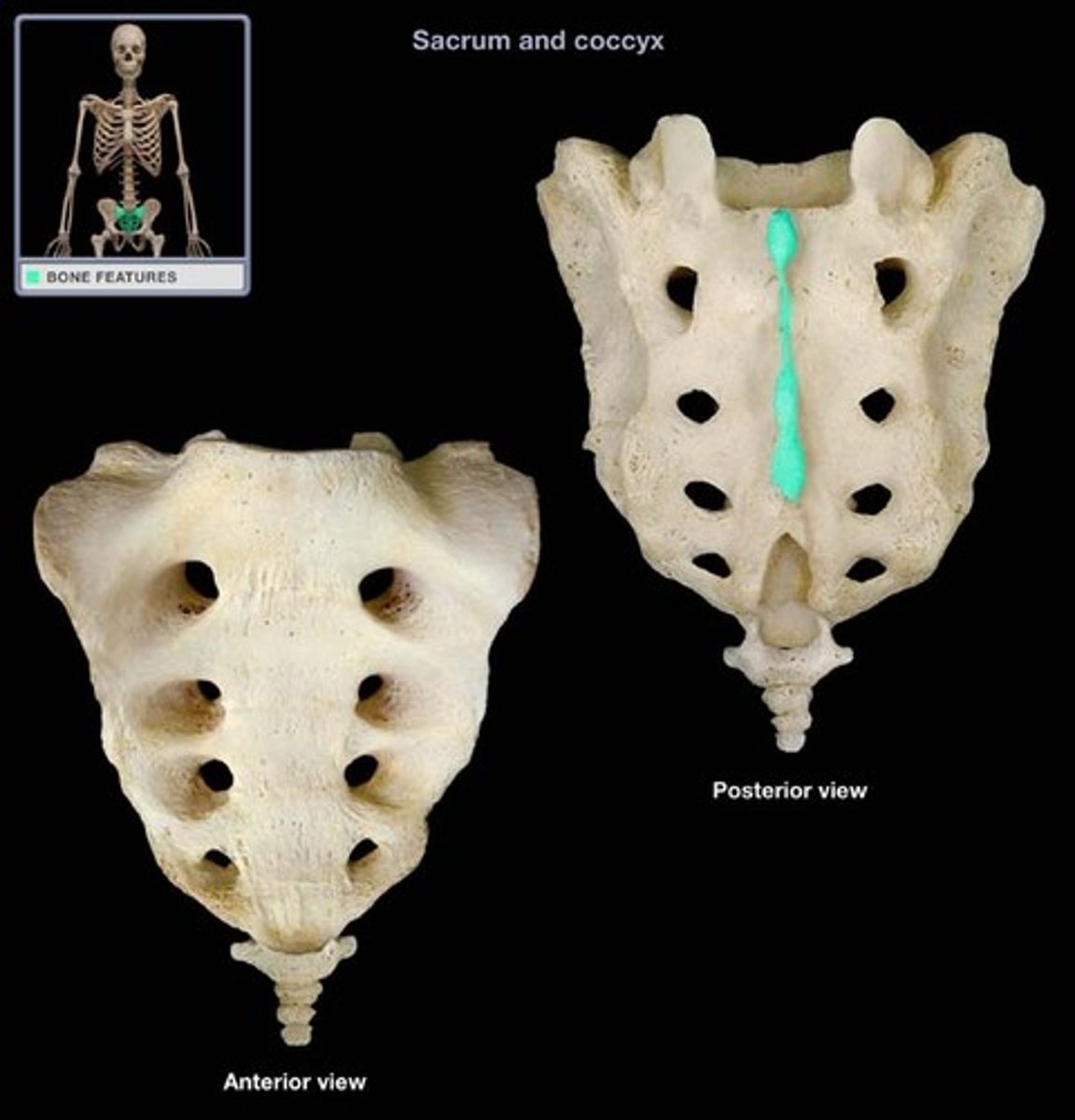
sacrum (5 fused vertebrae)
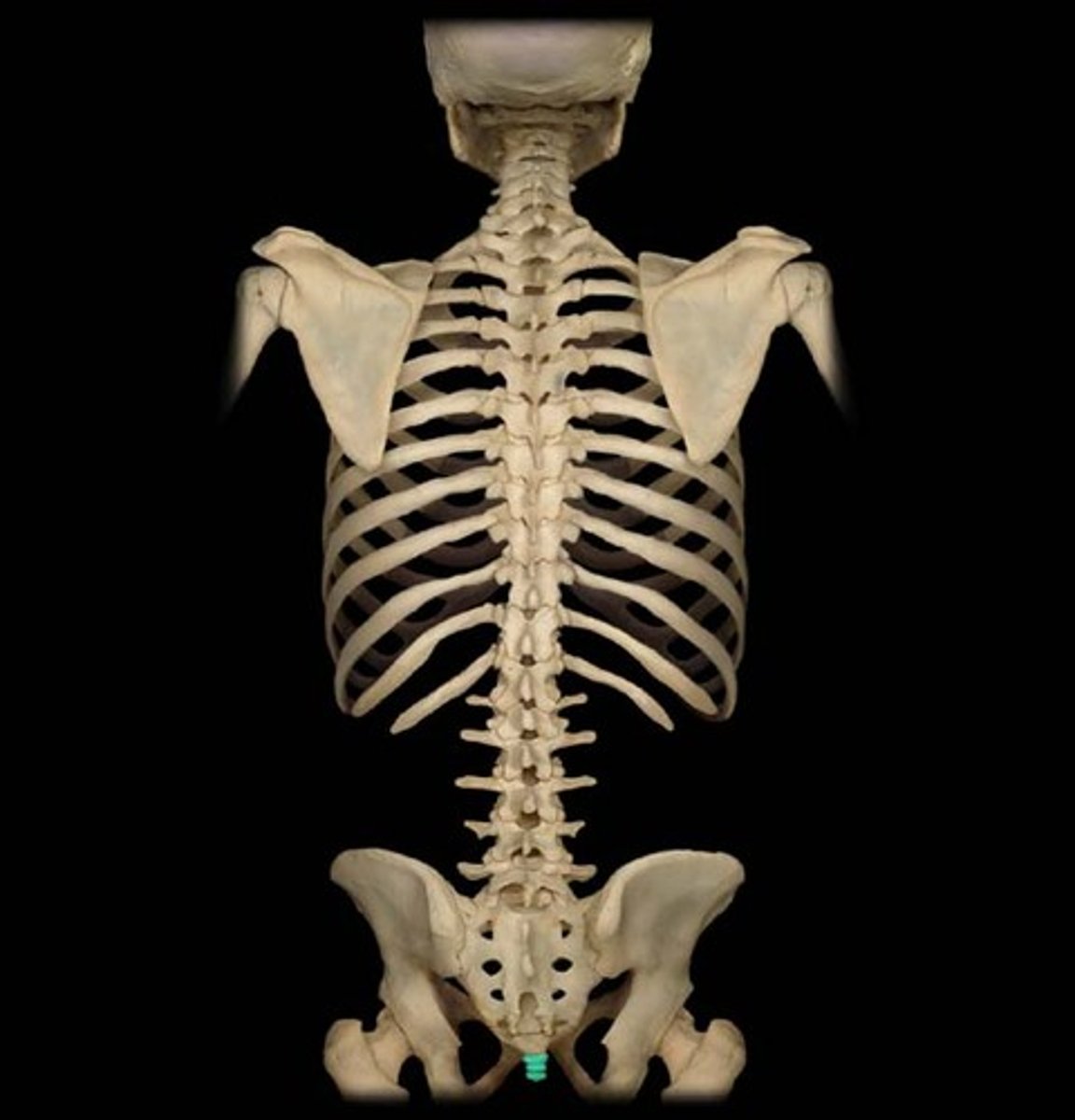
Coccyx (3-5 fused vertebrae)
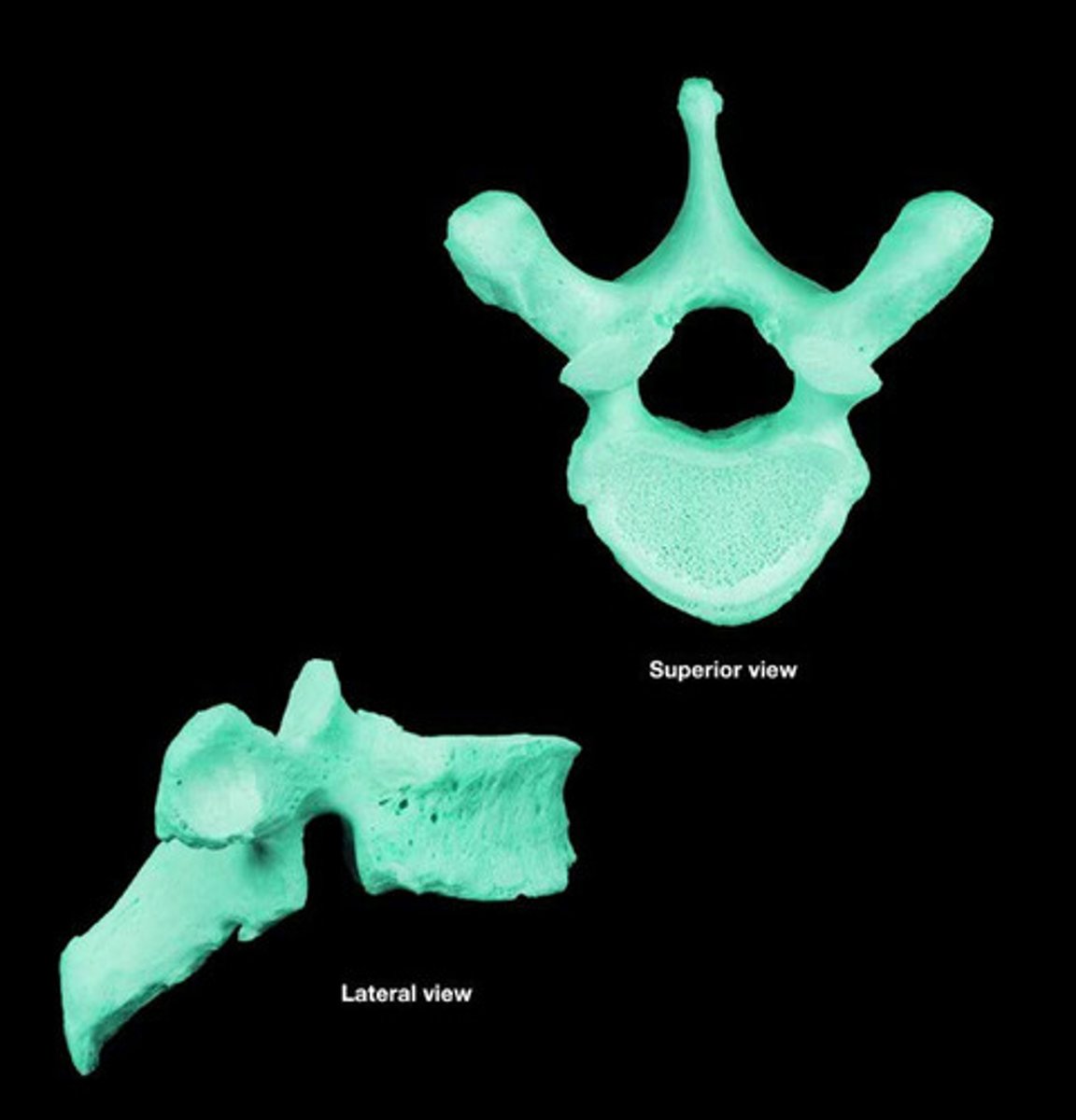
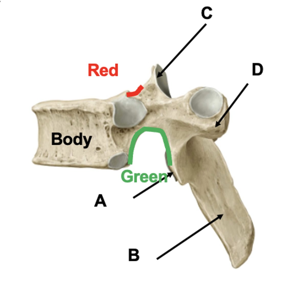
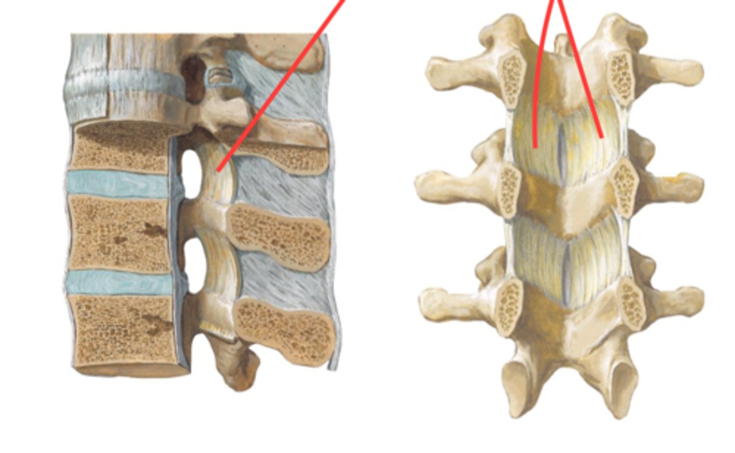
vertebral body
vertebral arch (formed by pedicles and laminae)
transverse processes
spinous process

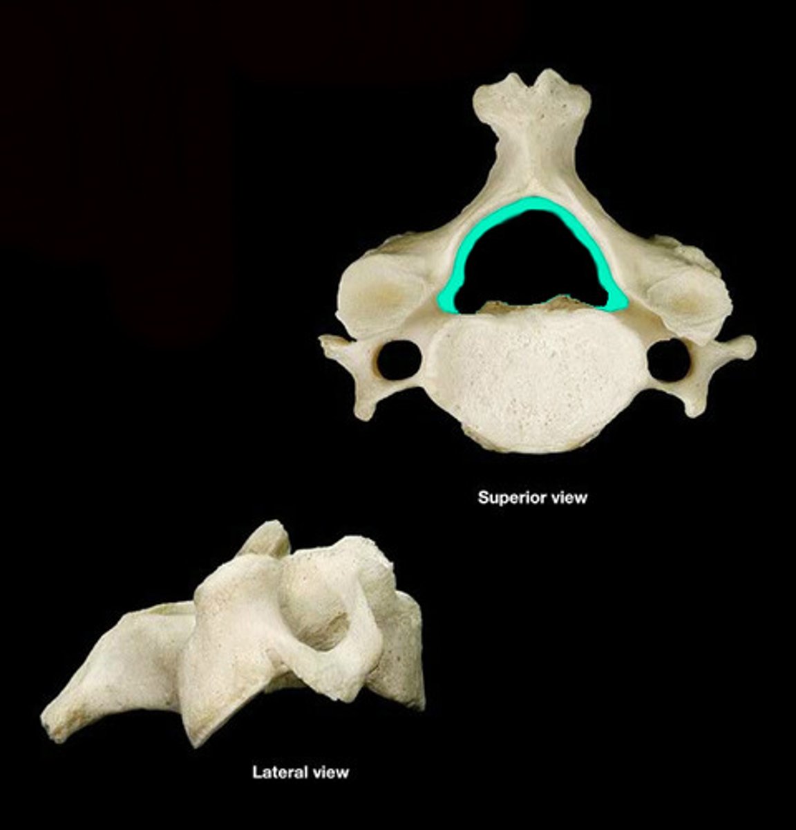
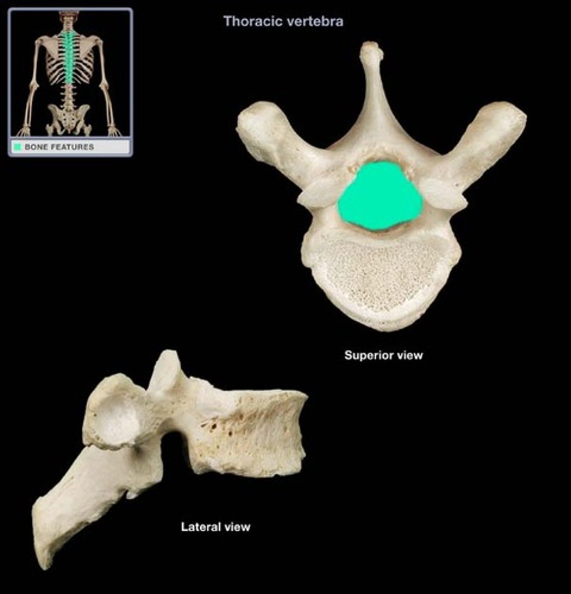
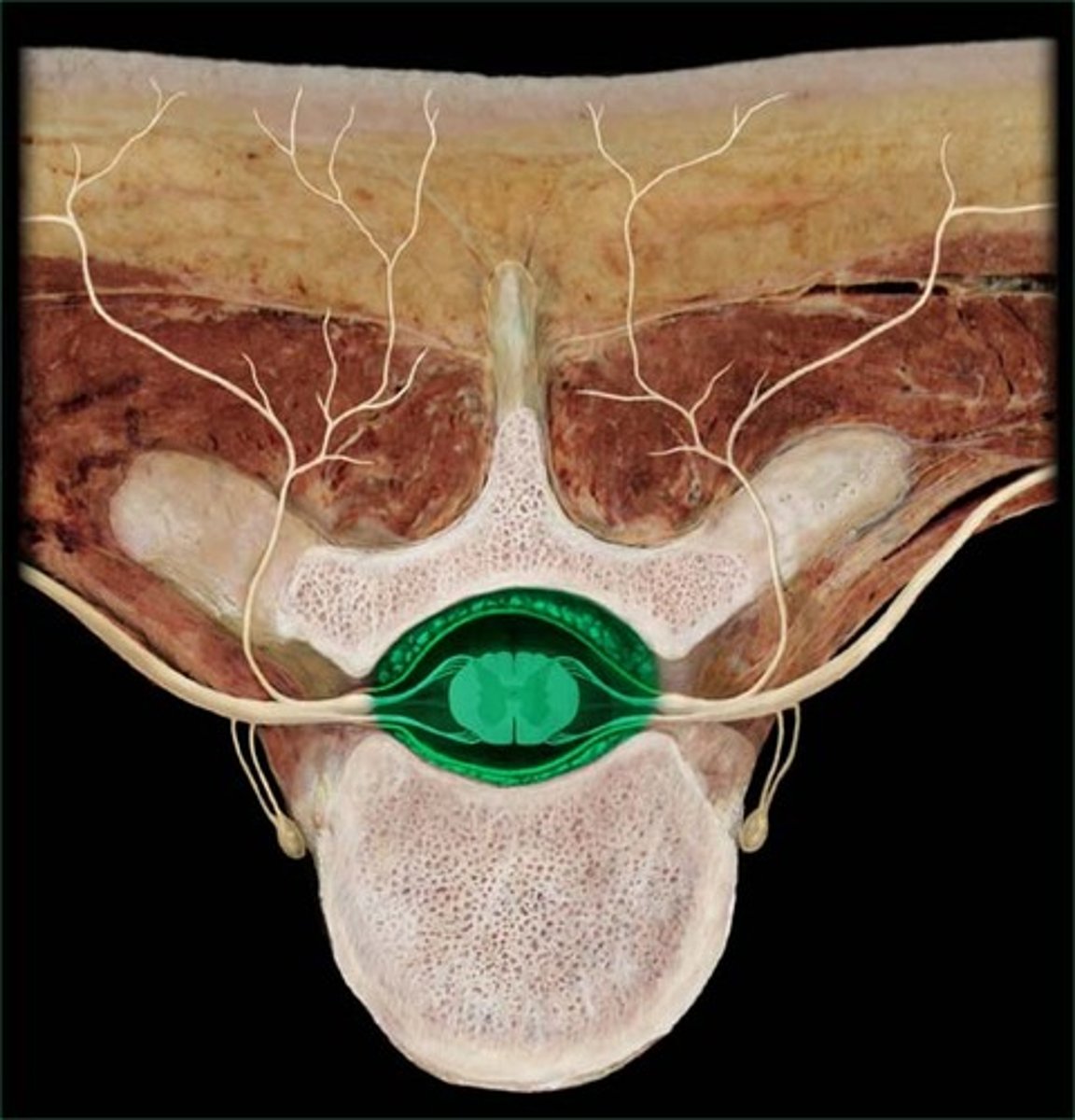
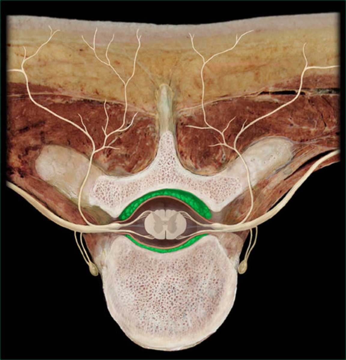
vertebral neural foramen
- holds the spinal cord
transverse foramen (cervical vertebrae only)
- holds vertebral arteries
Vertebra prominens (C7)
- spinous process is NOT forked
- transverse foramen so still cervical
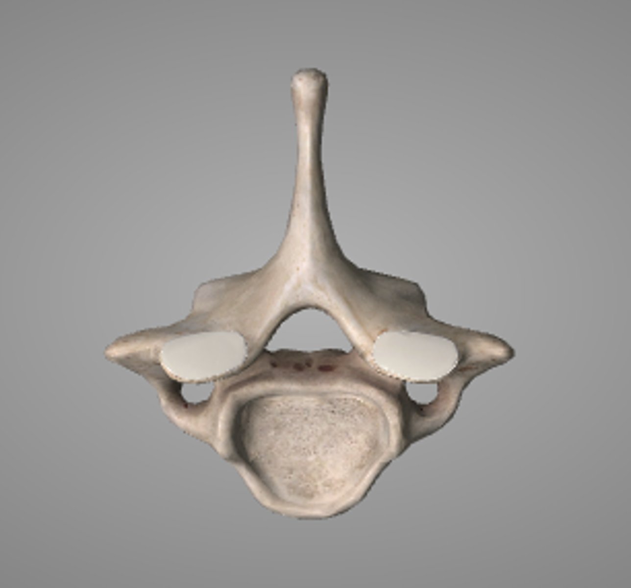
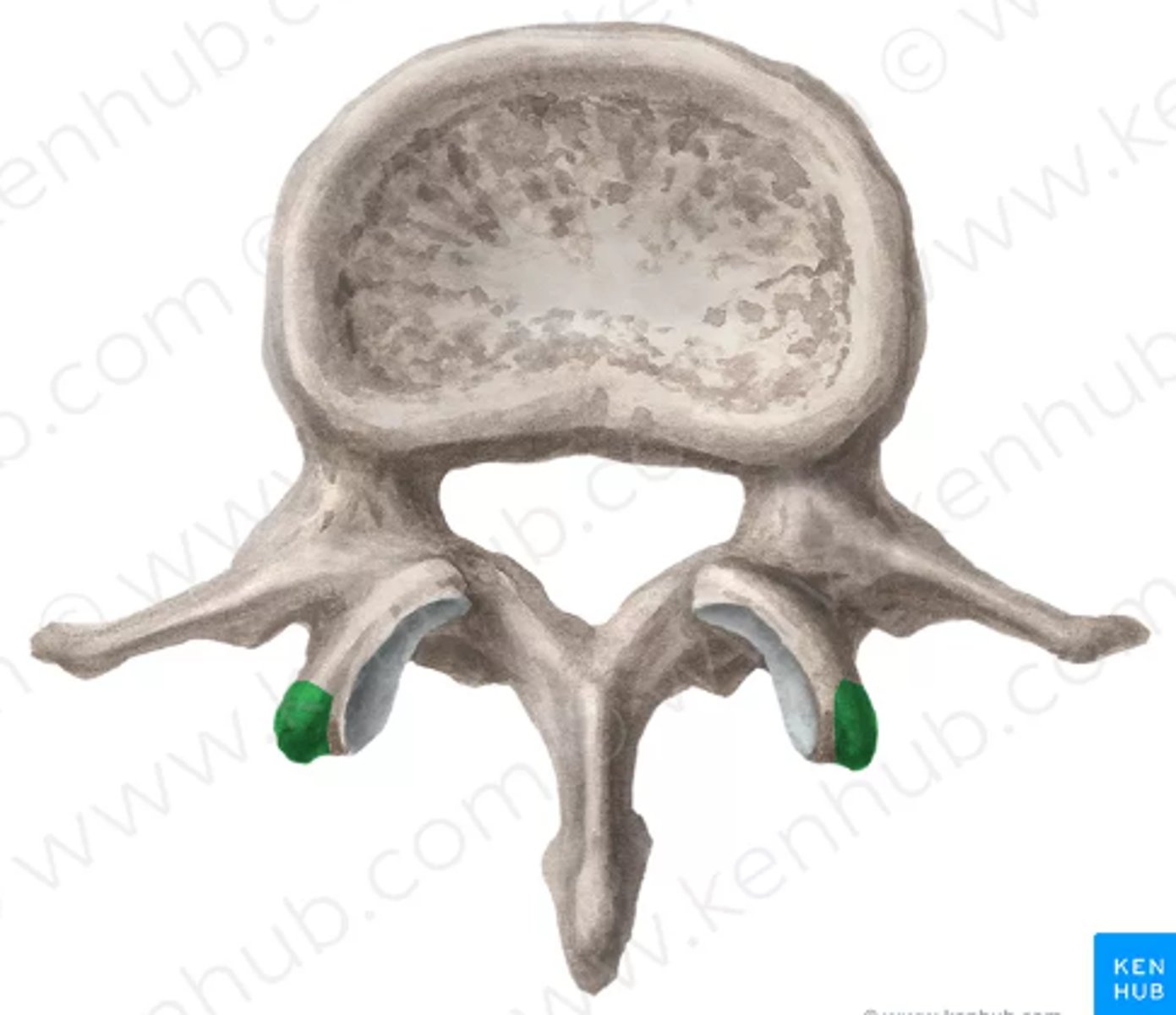
superior and inferior costal facets (thoracic)
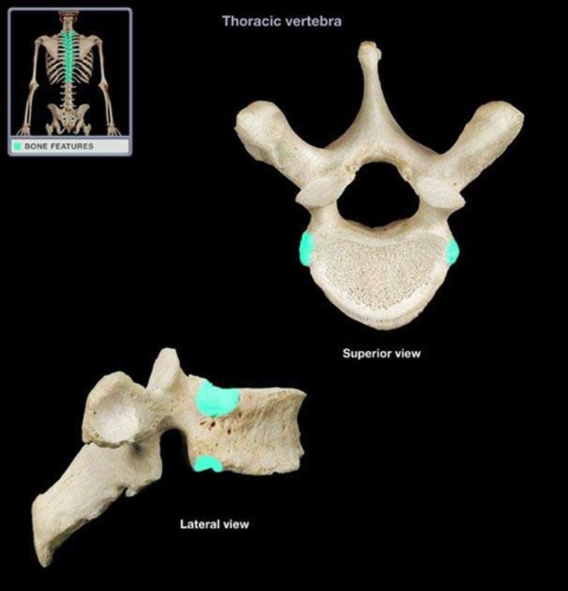
Transverse costal facet (T1-T10 only)
- articulates the tubercles of ribs
Spinous process
Lamina
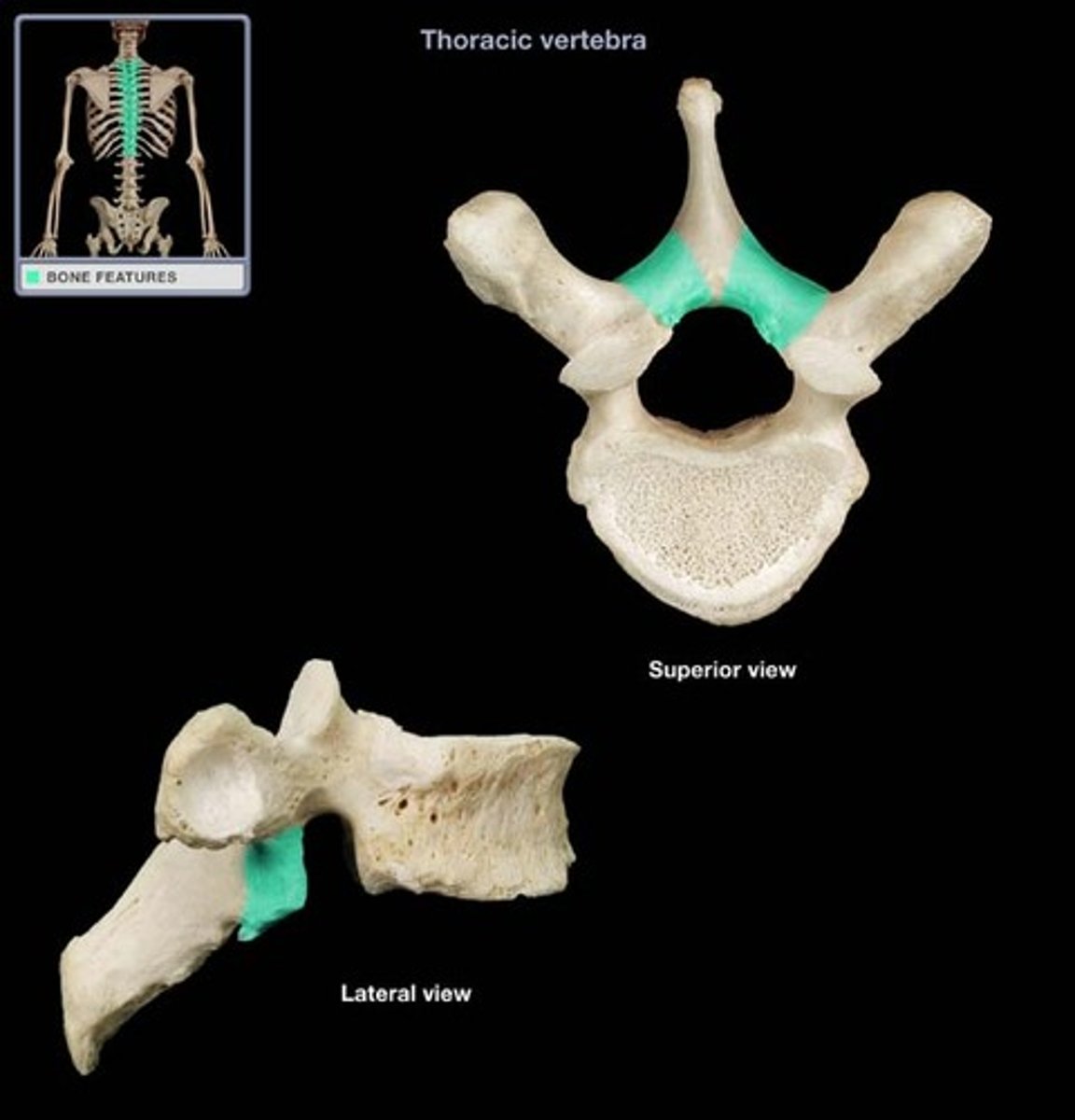
pedicle
red: superior vertebral notch
green: inferior vertebral notch
What is red and green?
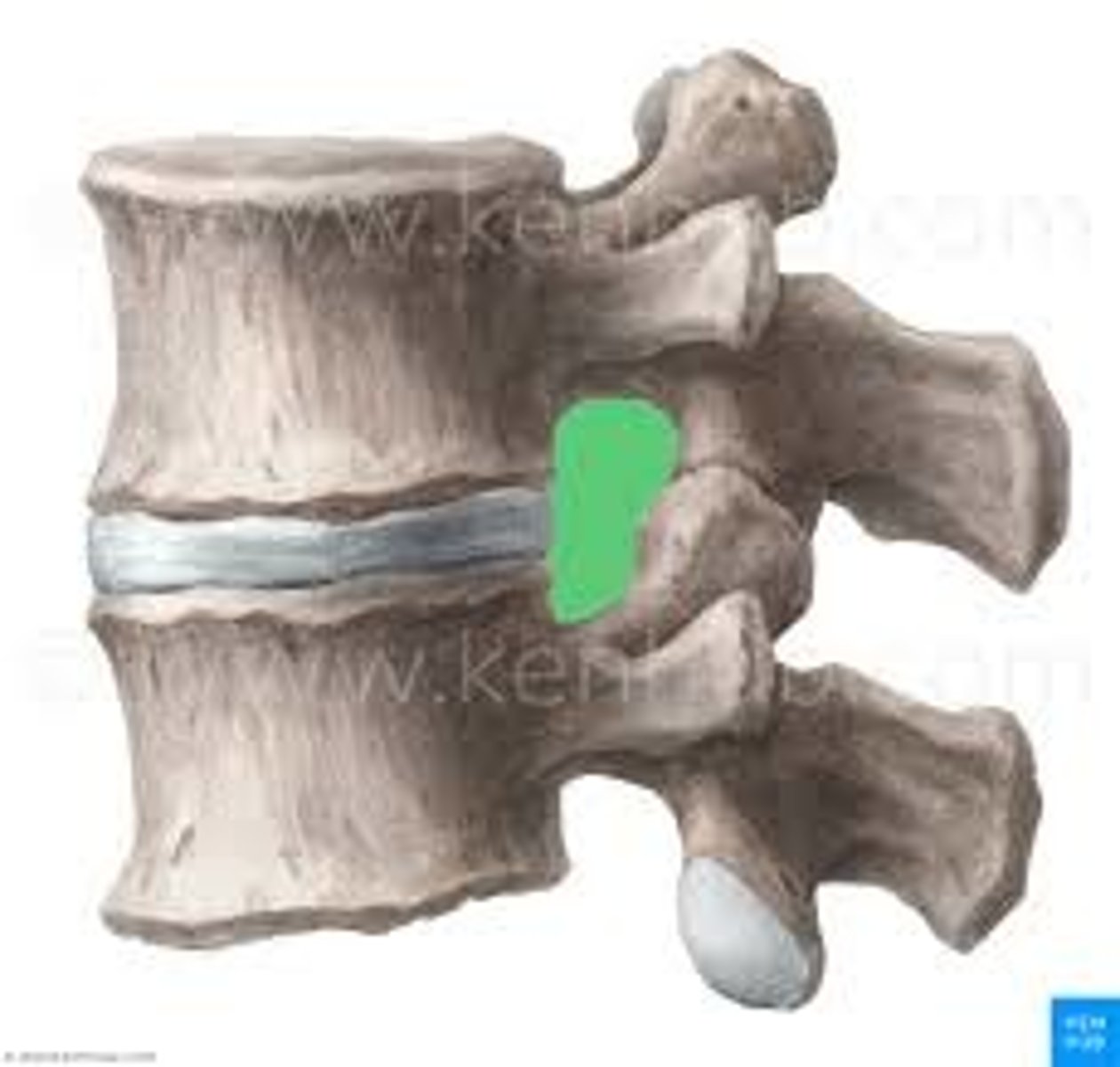
Intervertebral foramen
- holds spinal nerves
superior articular facet
- joints with inferior articular facet of adjacent vertebrae to form facet (Z) joints
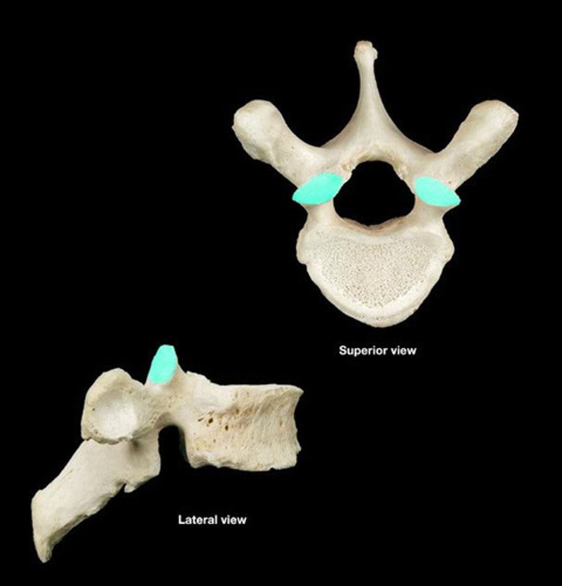
inferior articular facet
uncinate processes (cervical vertebrae)
mammillary process (lumbar vertebrae)
- small tubercles on posterior part of superior articular process
Acromion
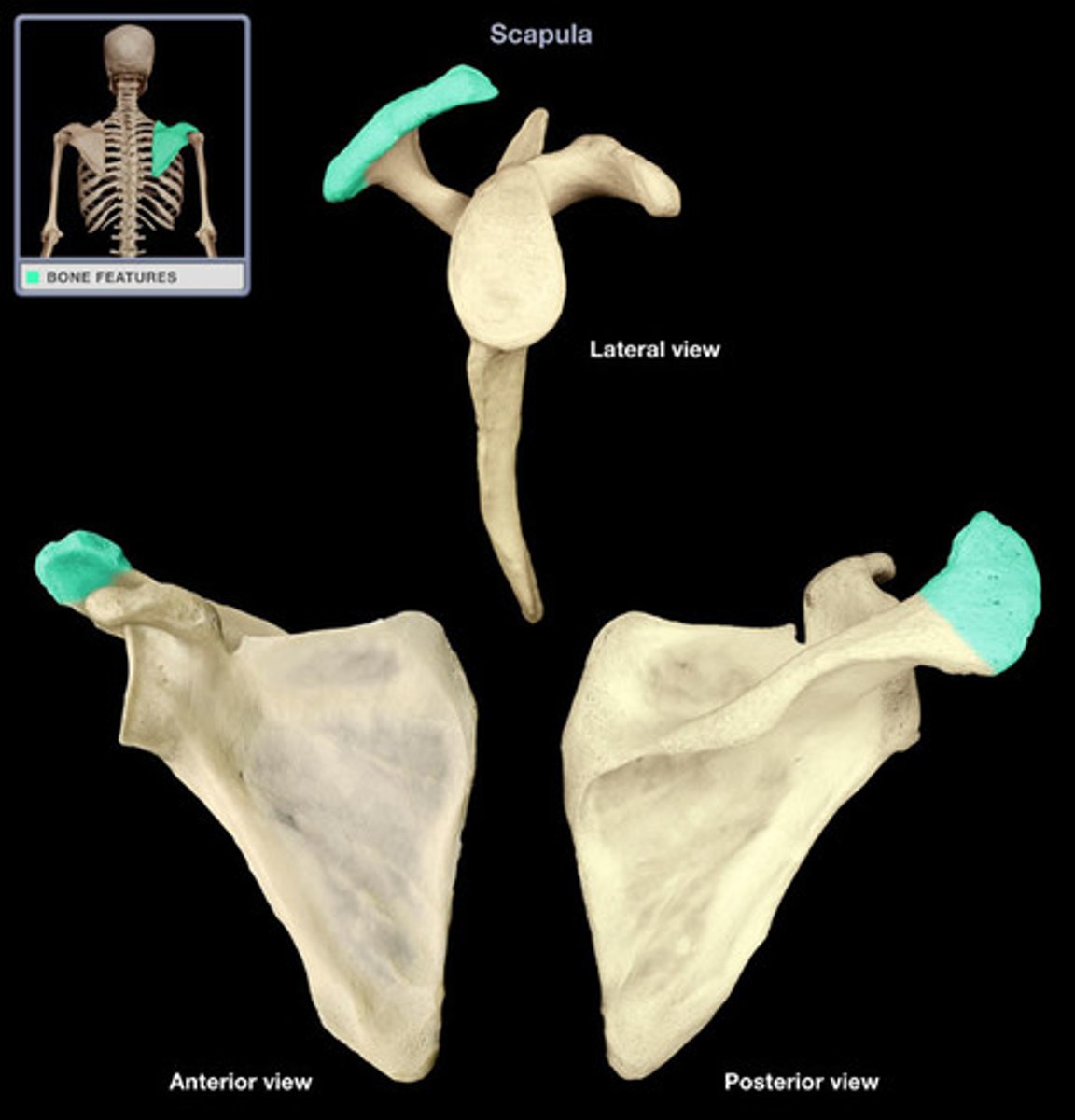
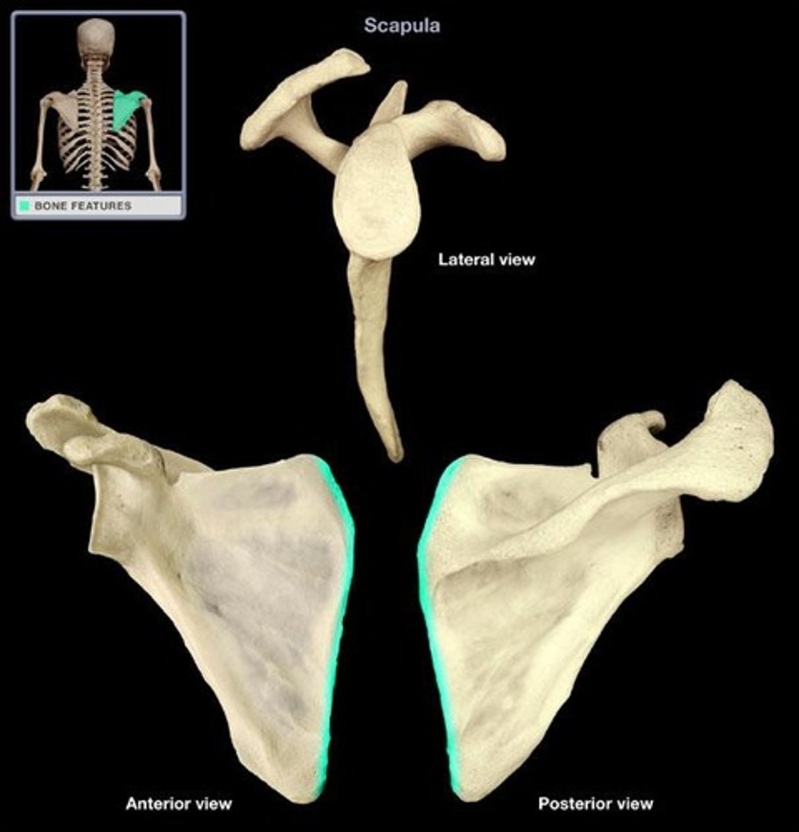
medial (vertebral) border
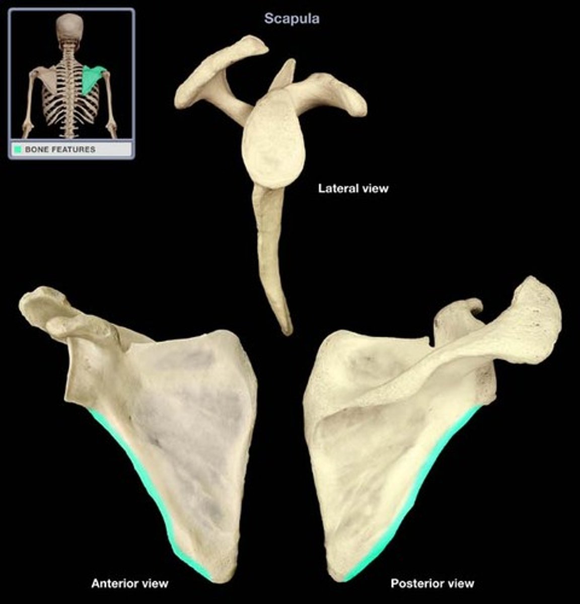
lateral (axillary) border
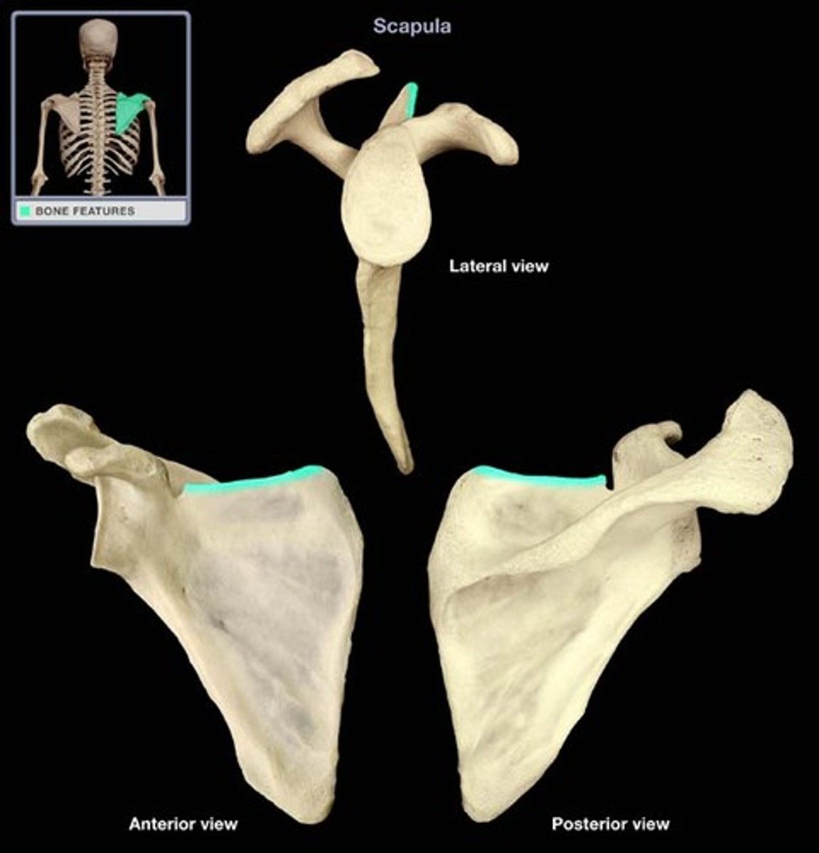
superior border
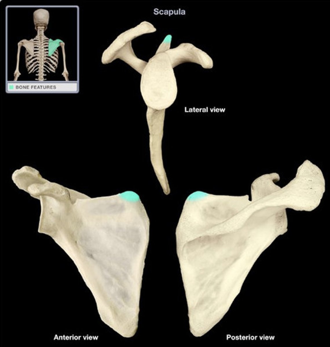
superior angle
inferior angle
- T7 transverse plane
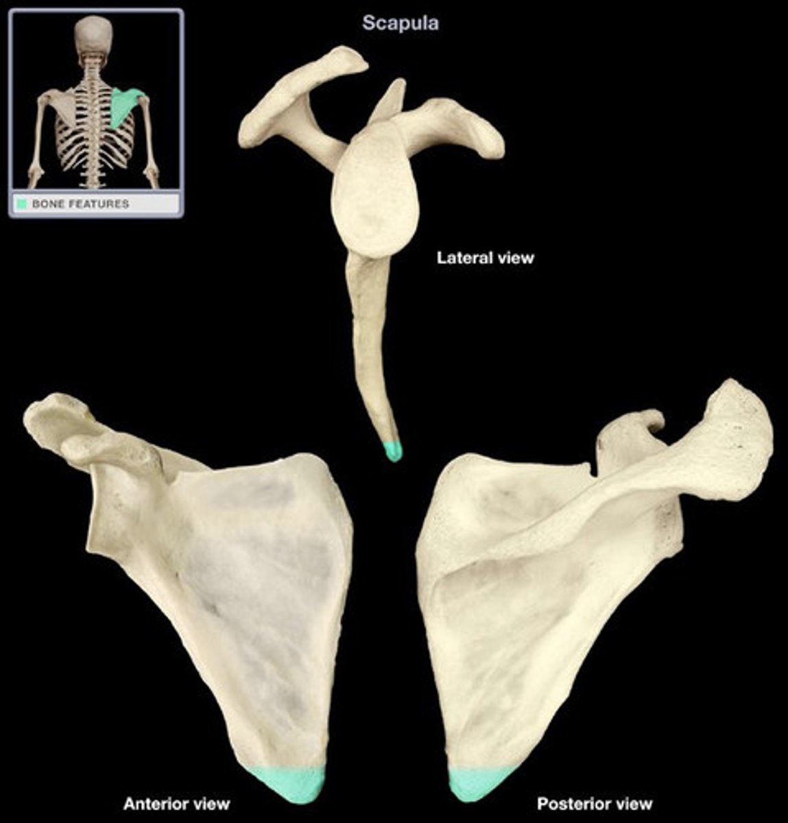
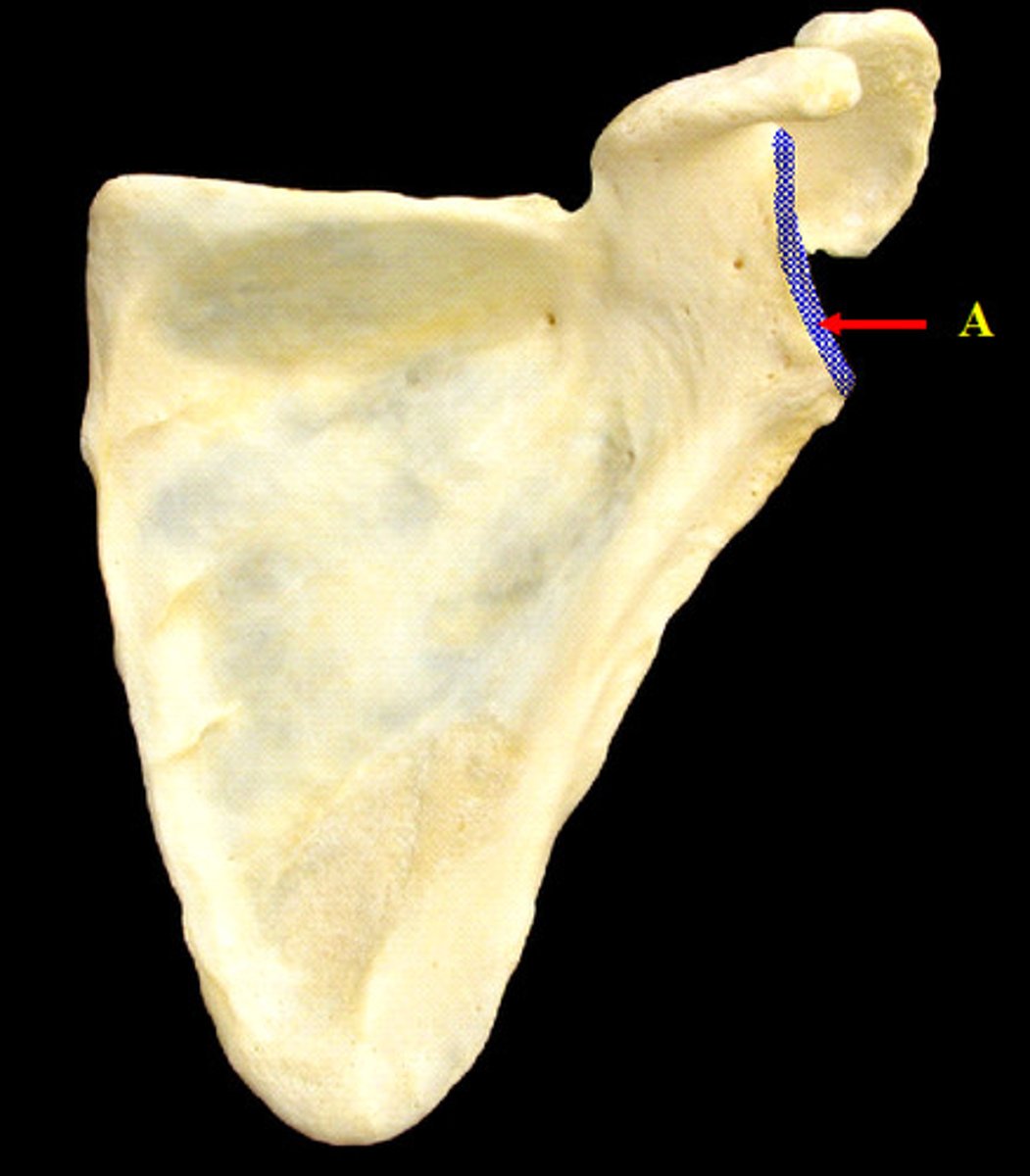
lateral angle (glenoid fossa)
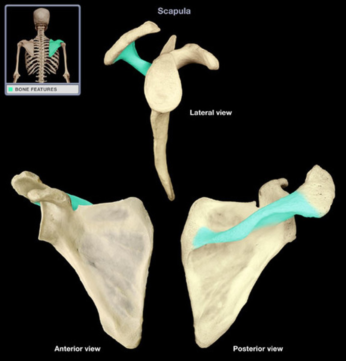
scapular spine
- lies in T3 plane
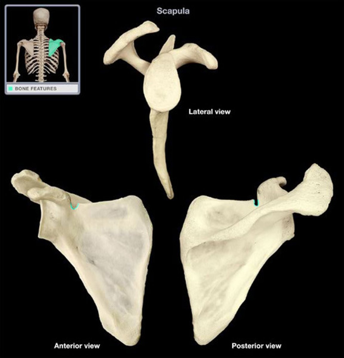
scapular notch
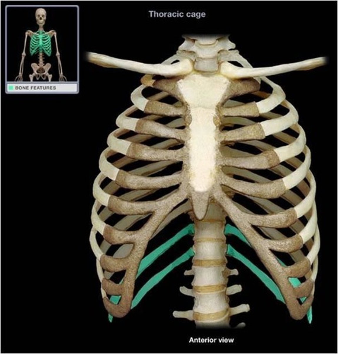
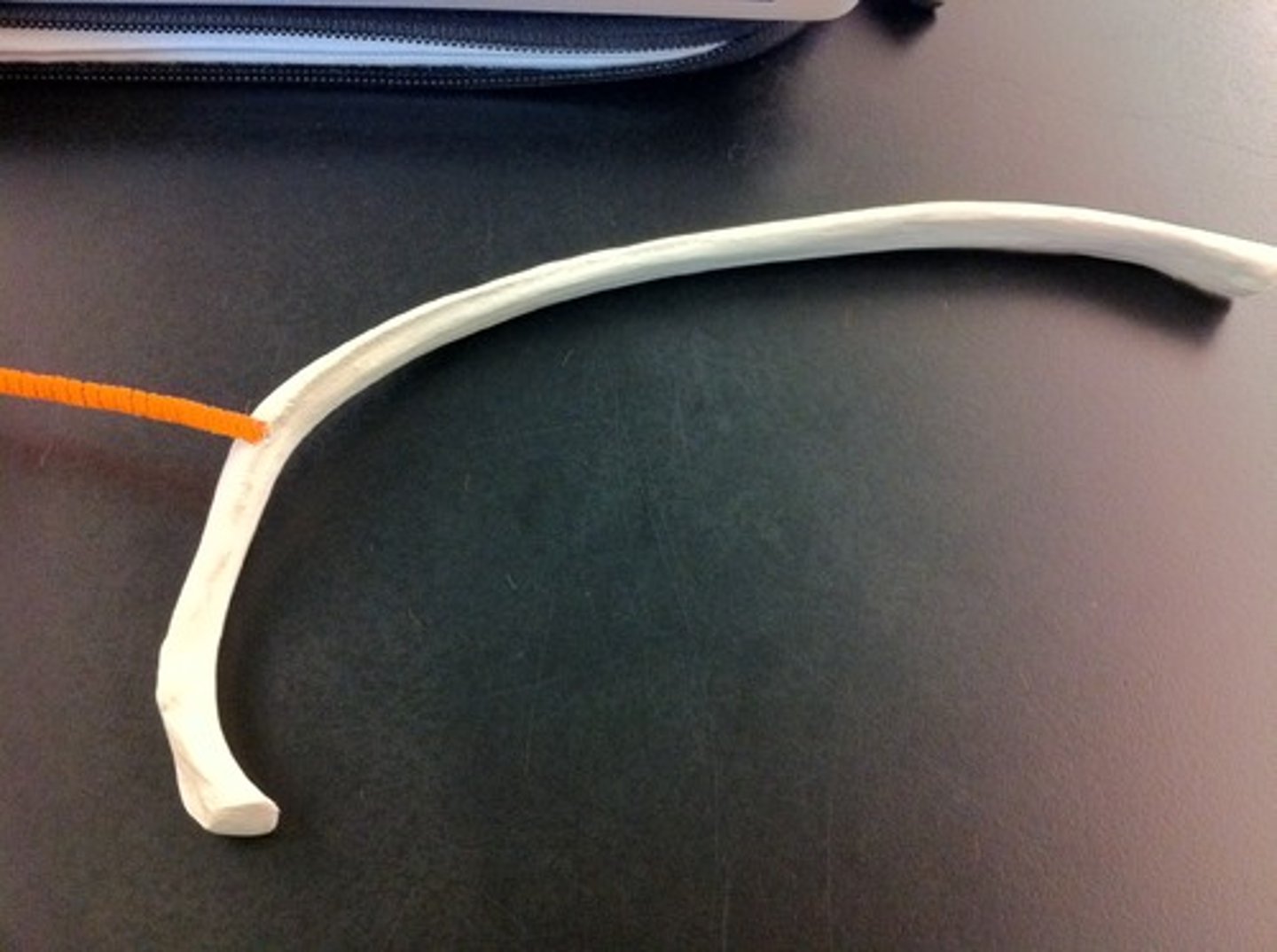
true ribs (1-7)
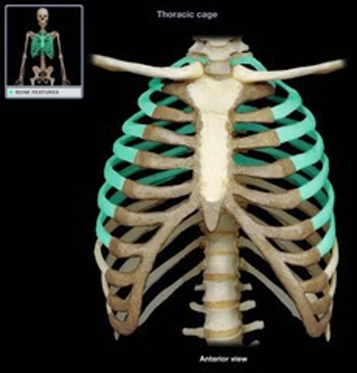
false ribs (8-10)
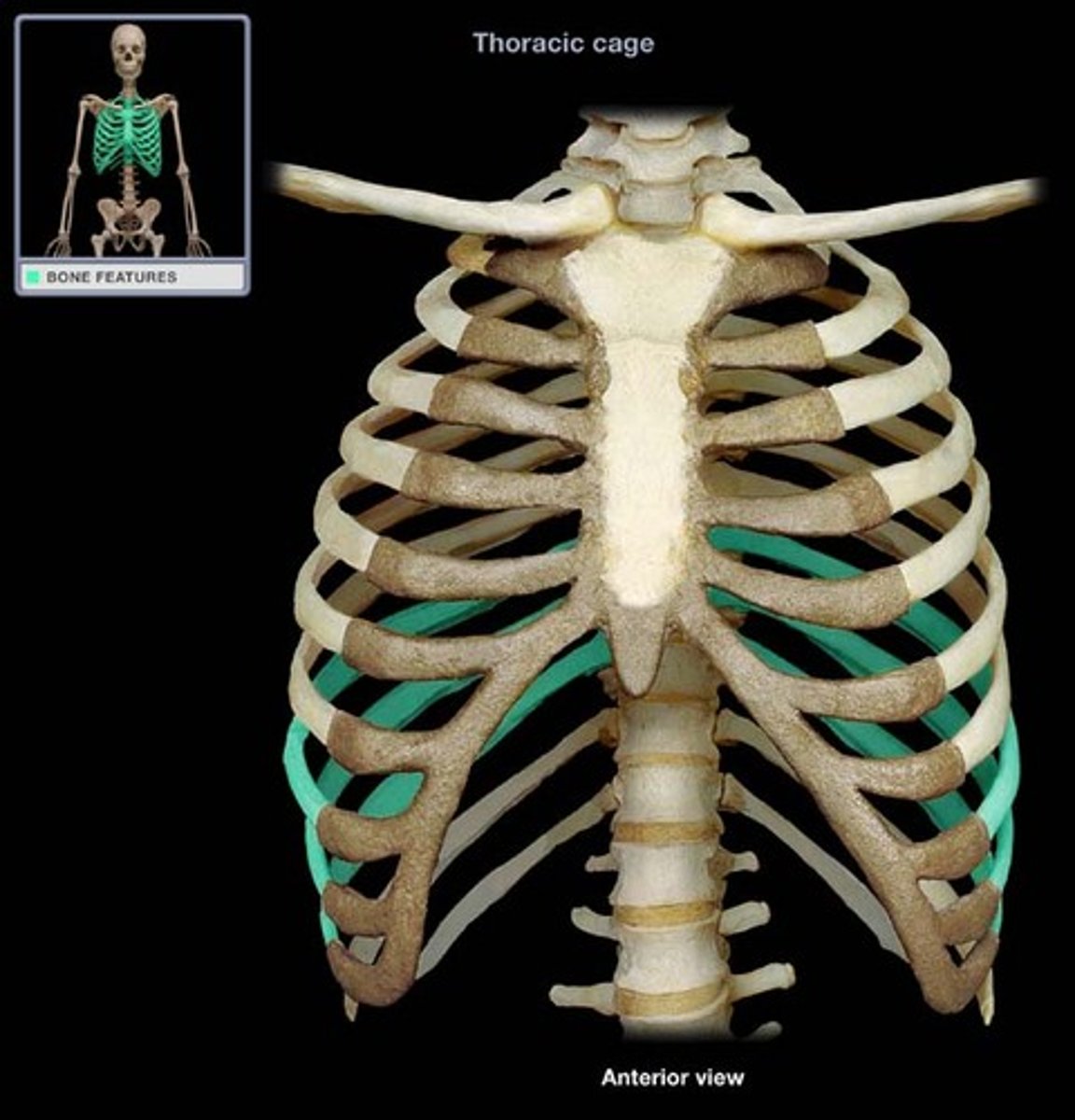
floating ribs (11-12)
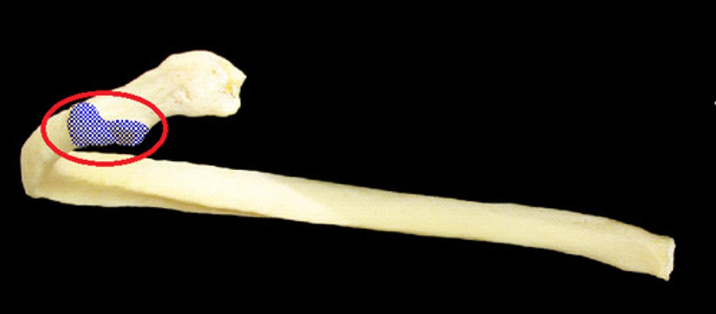
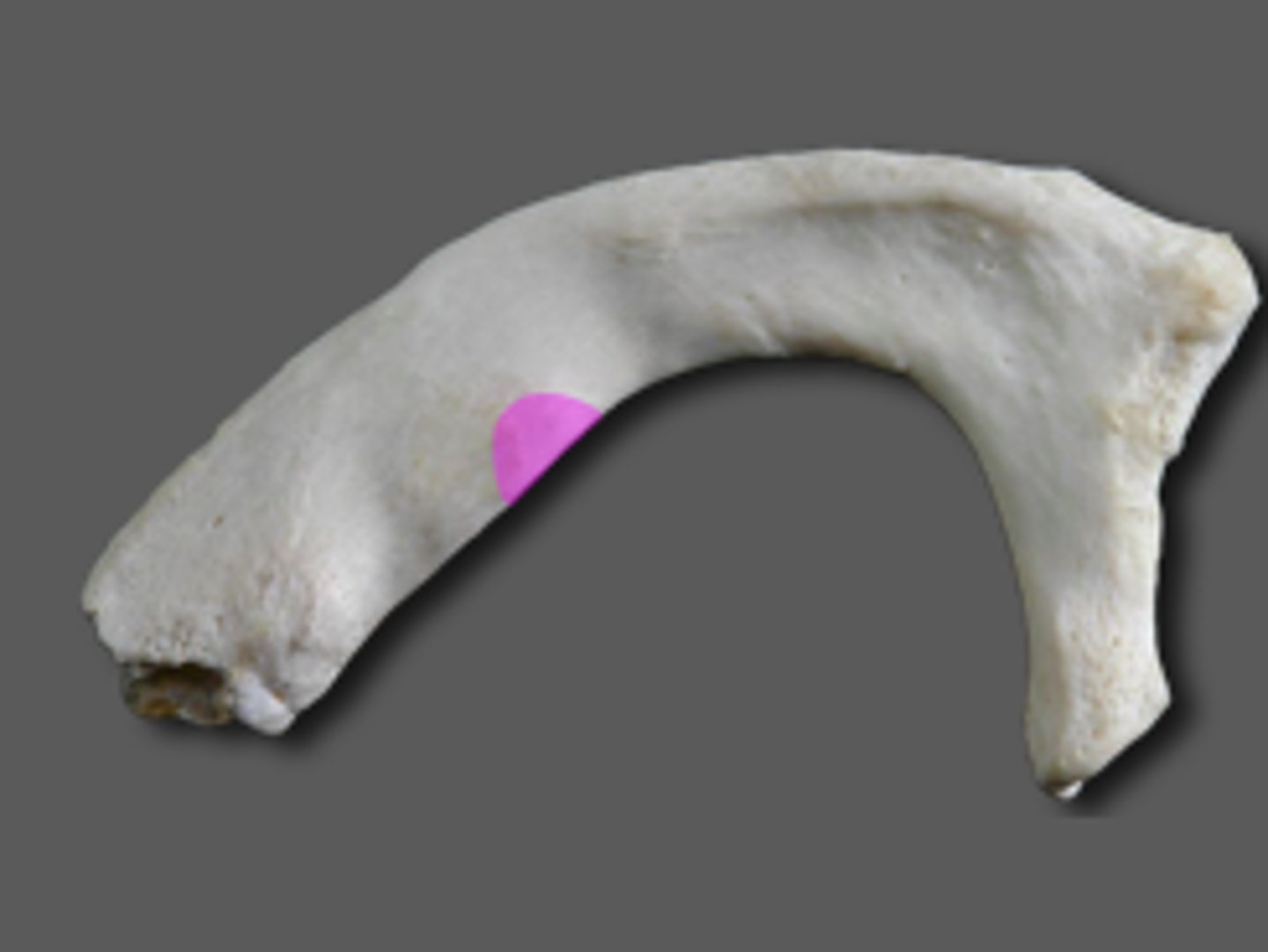
head of rib
- posterior end of a rib that articulates with the bodies of thoracic vertebrae
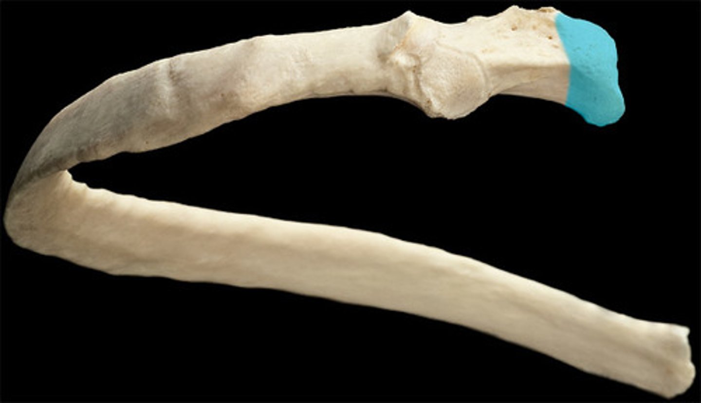
neck of rib
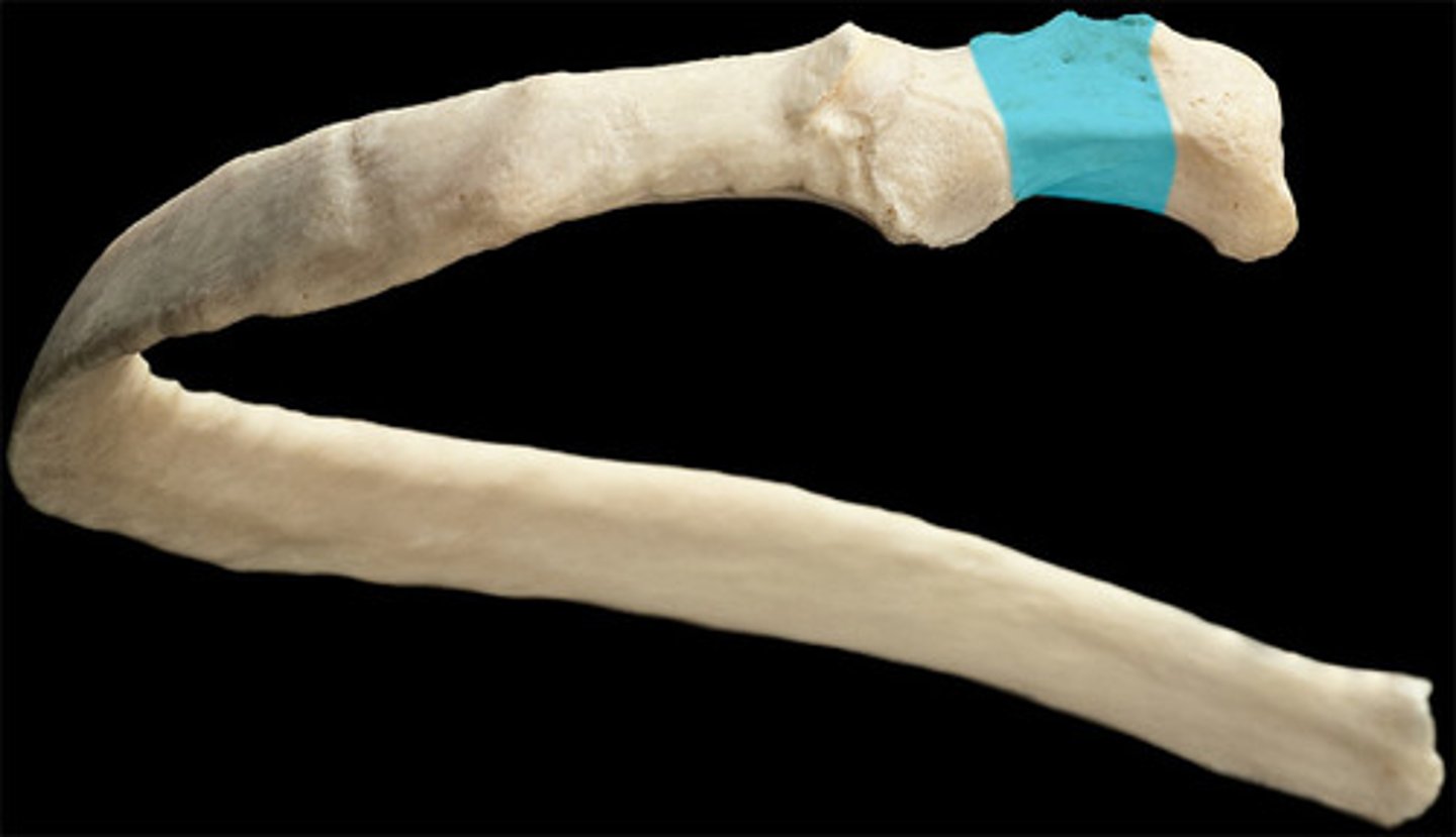
tubercle of rib
- articulates with the costal facet of thoracic vertebra's transverse process.
costal groove
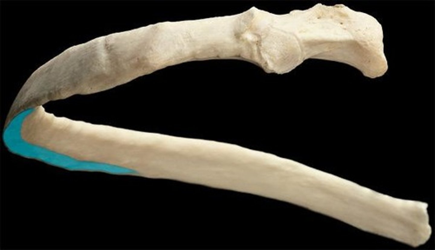
shaft of rib
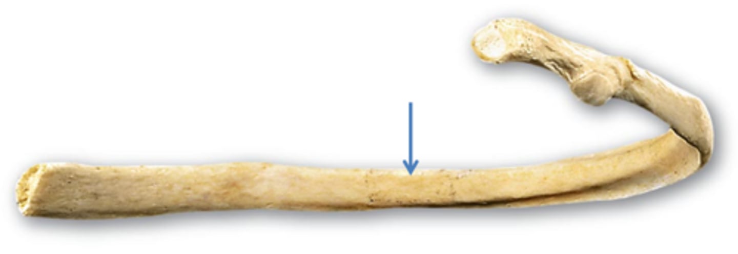
angle of rib (curve of rib)
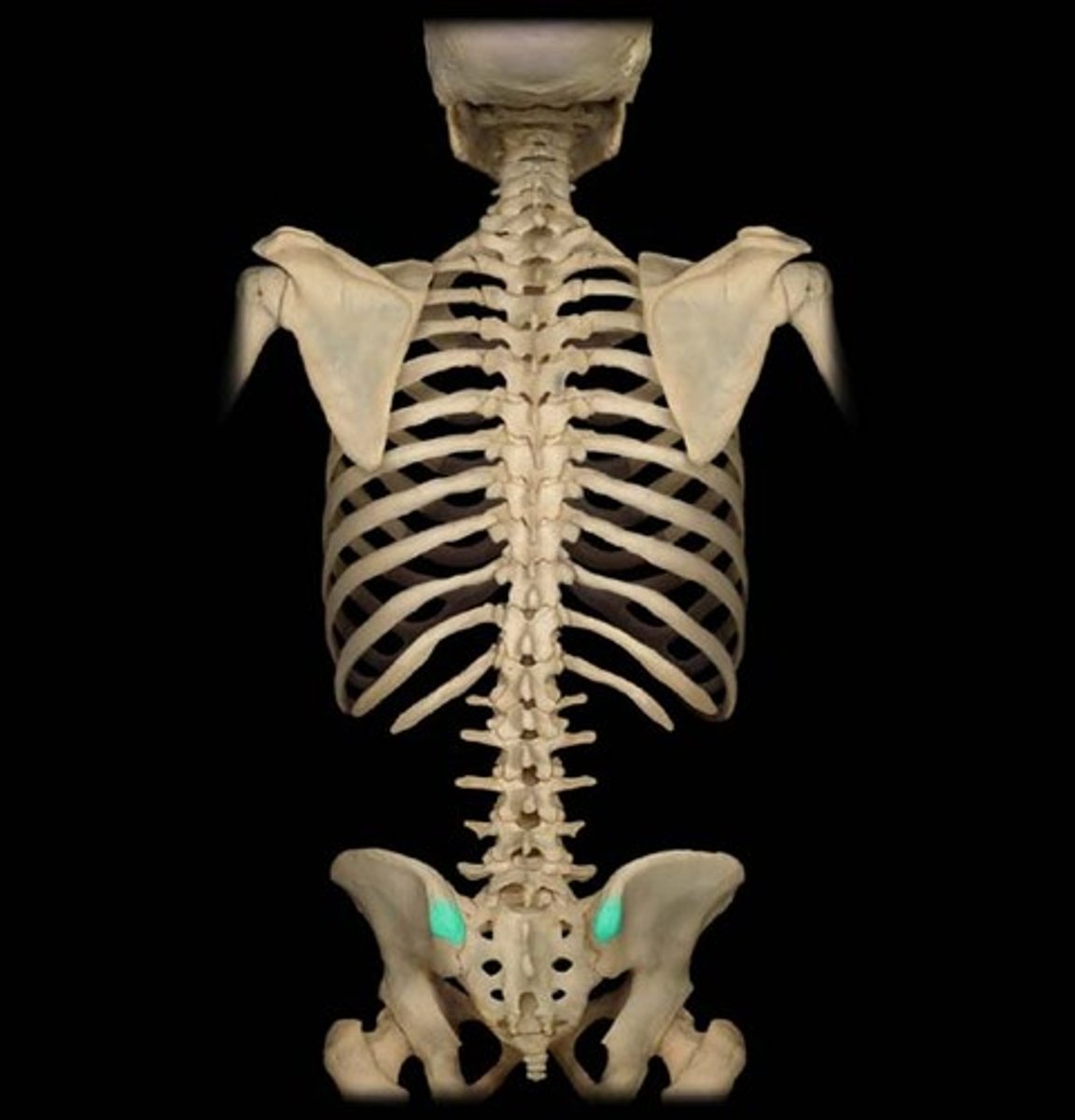
iliac crest
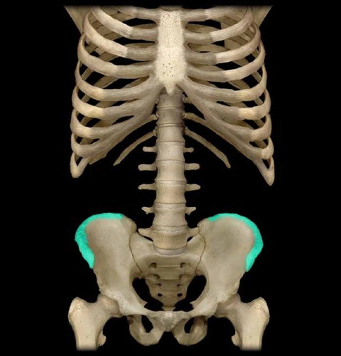
posterior superior iliac spine
median sacral crest
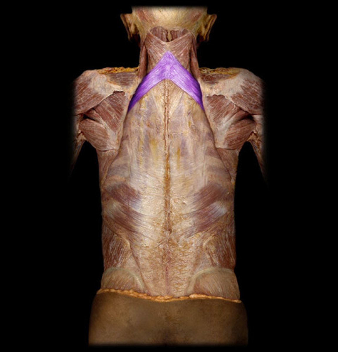
trapezius
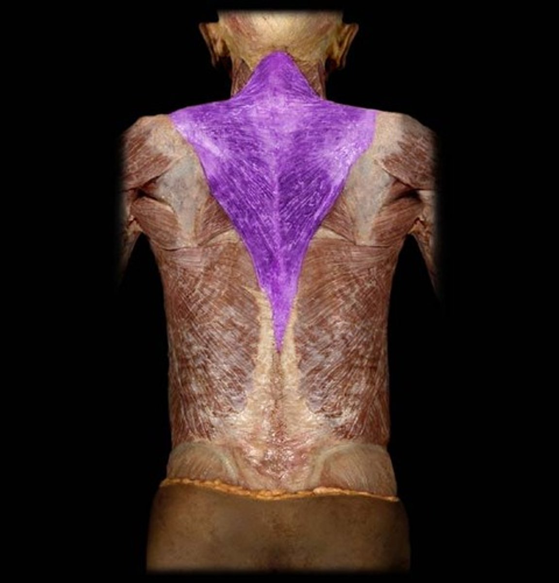
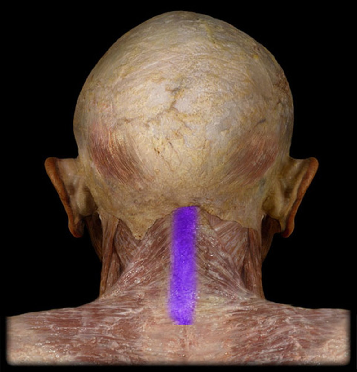
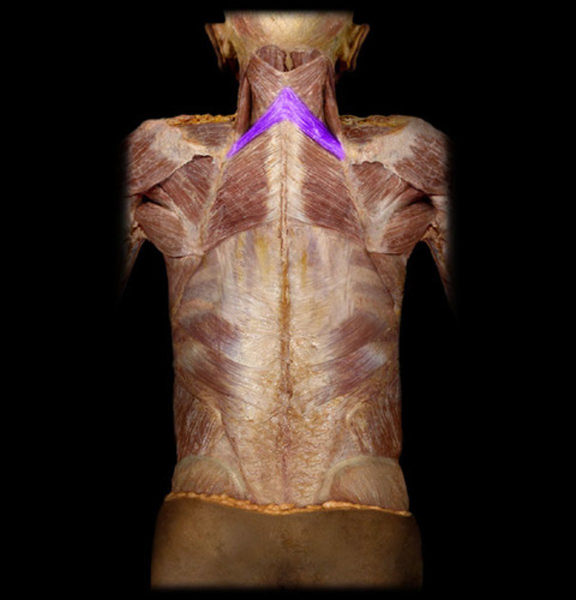
ligamentum nuchae (nuchal ligament)
spinal accessory nerve (CN XI)
transverse cervical artery
latissimus dorsi
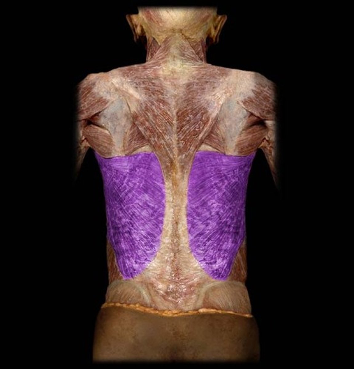
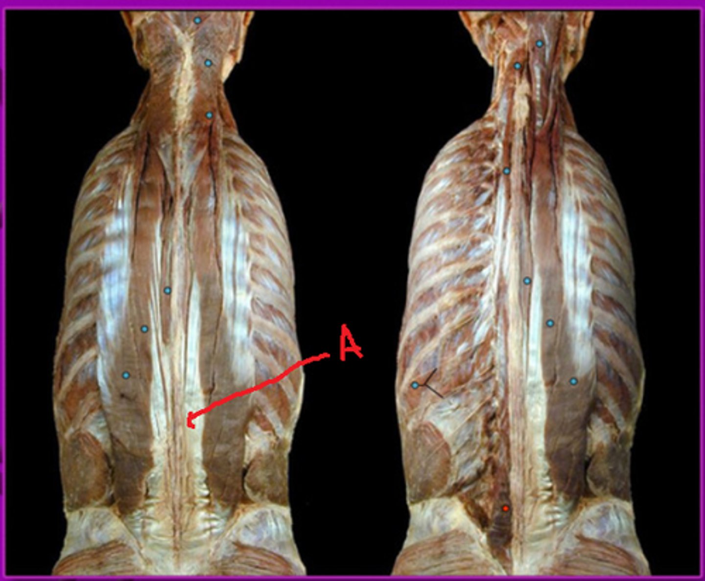
thoracolumbar fascia
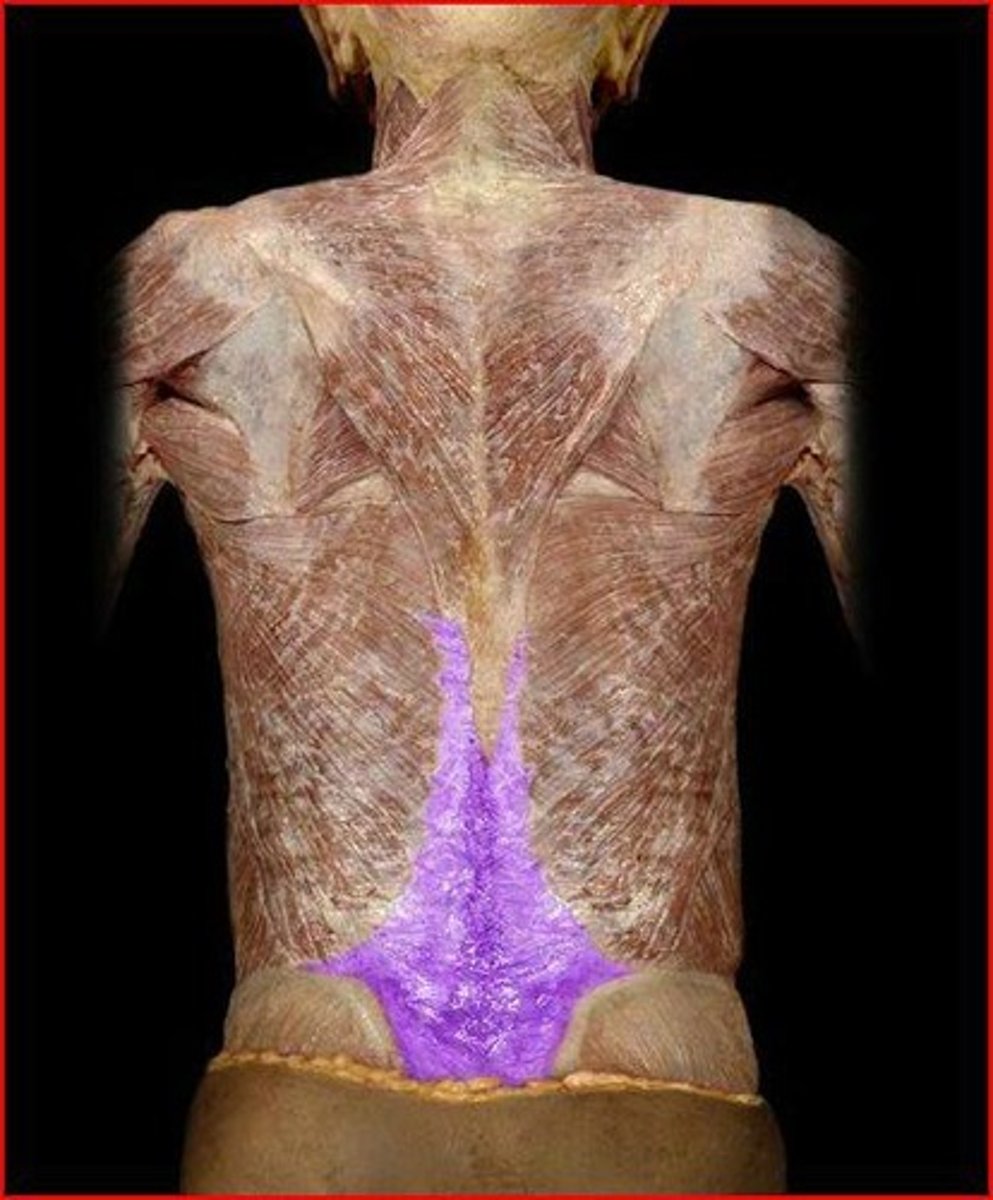
Serratus Posterior Inferior
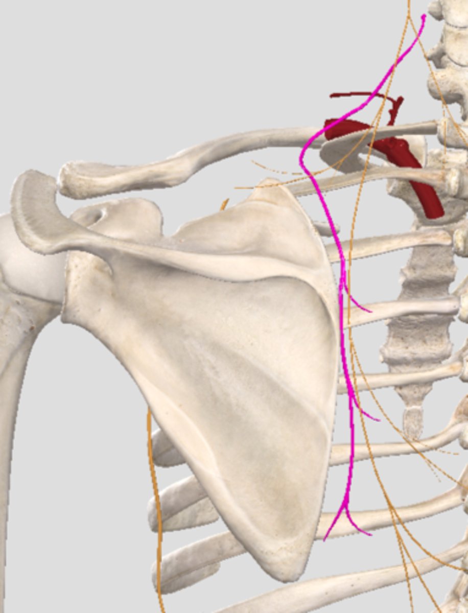
dorsal scapular nerve
rhomboid major
rhomboid minor
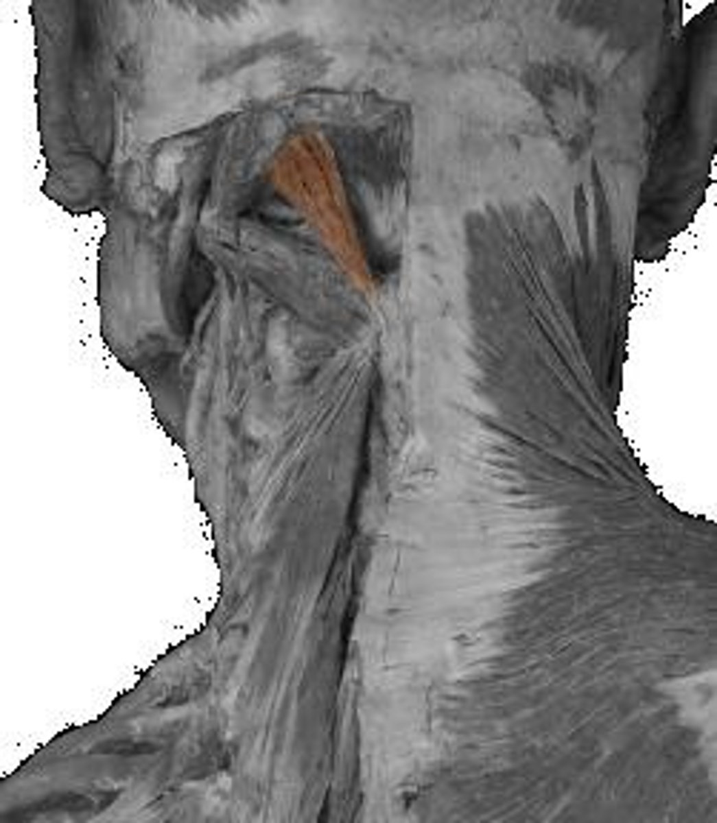
levator scapulae
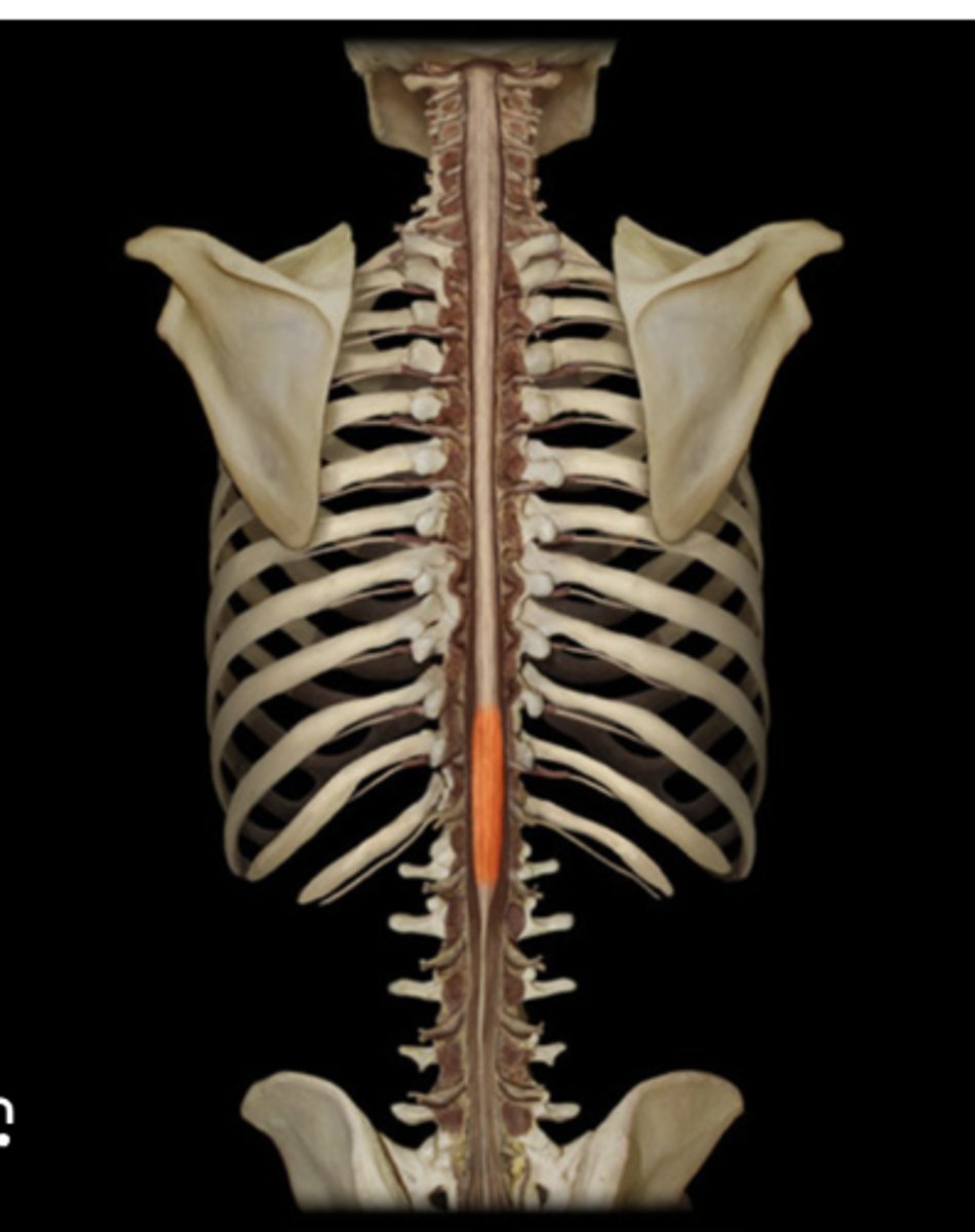
Multifidus lumborum
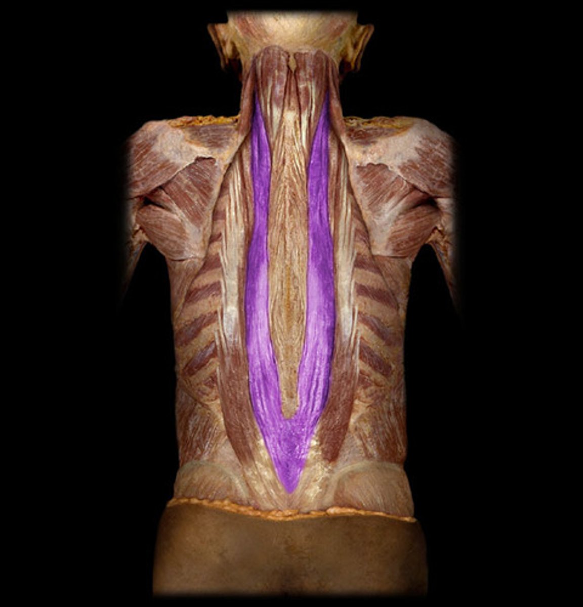
iliocostalis
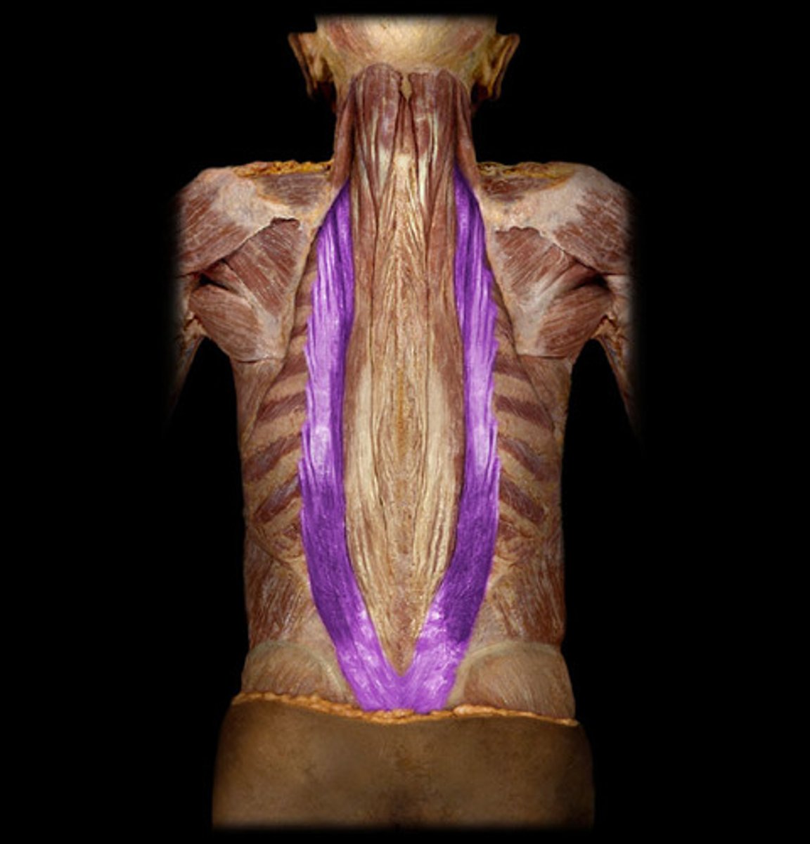
Longissimus
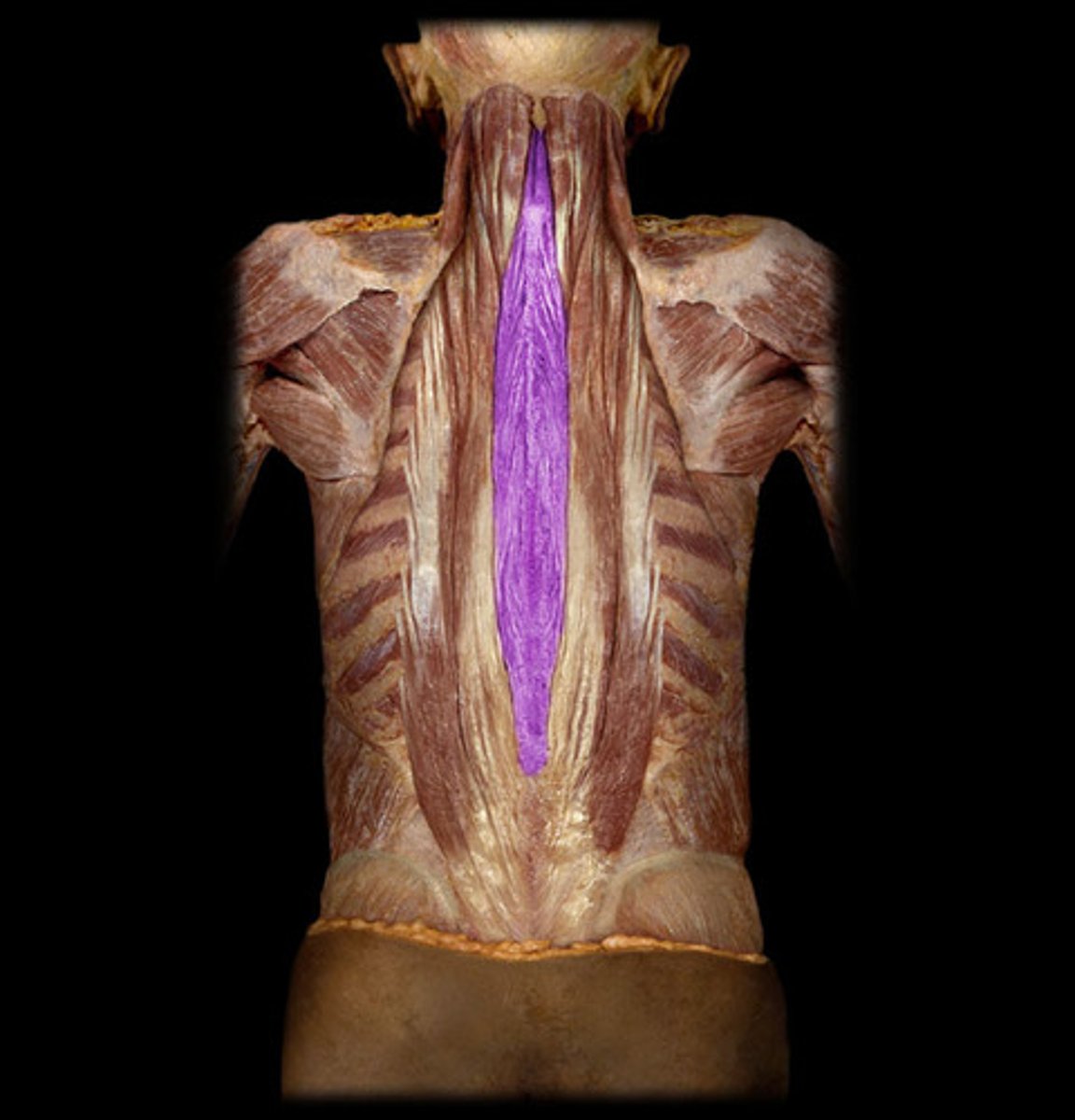
Spinalis
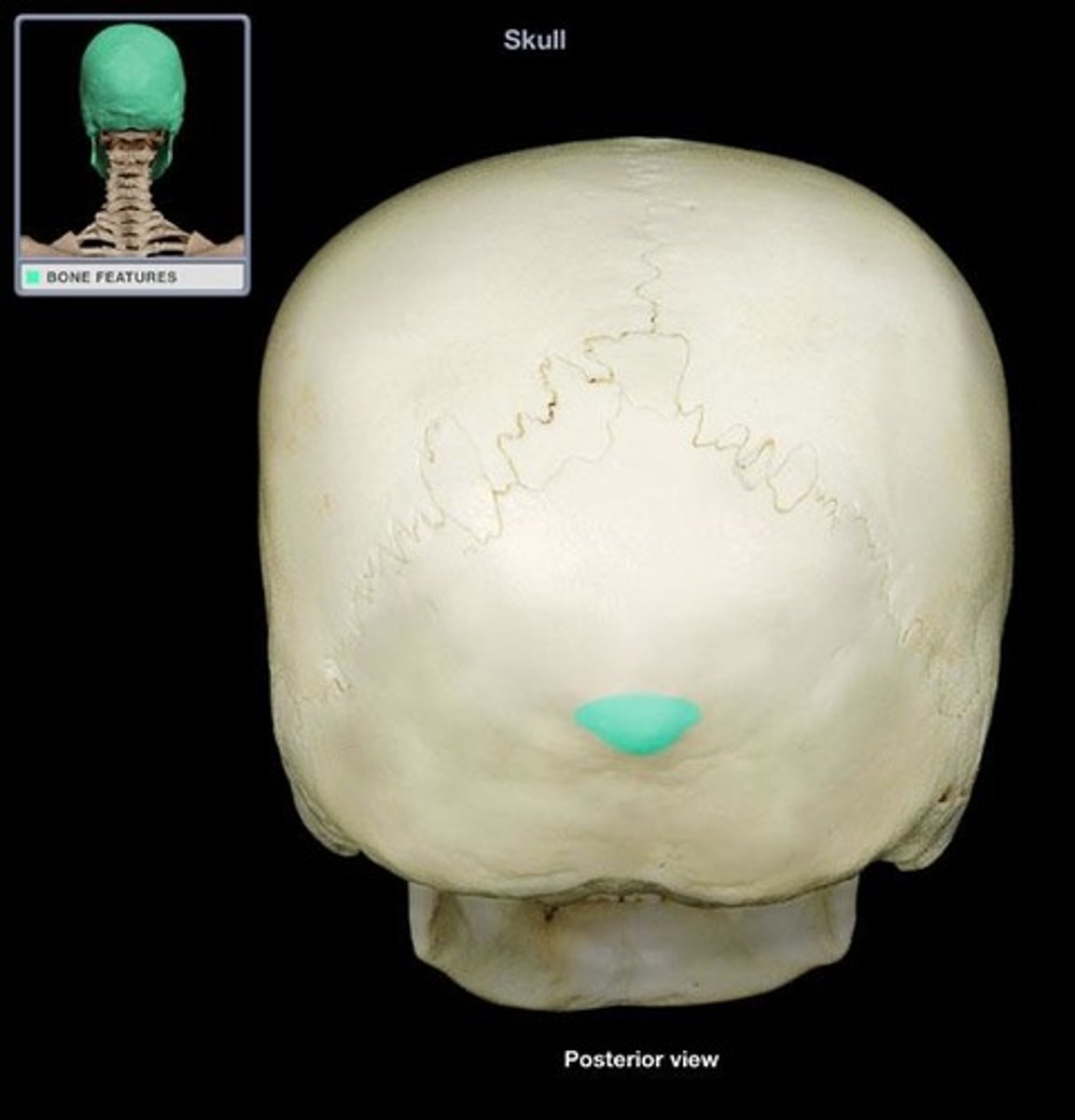
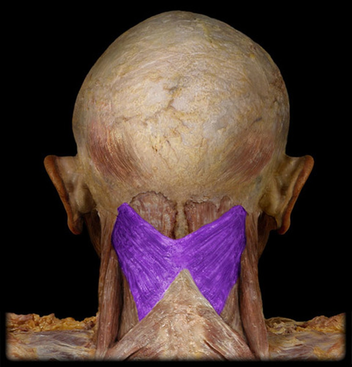
external occipital protuberance
- most posterior projection of cranium
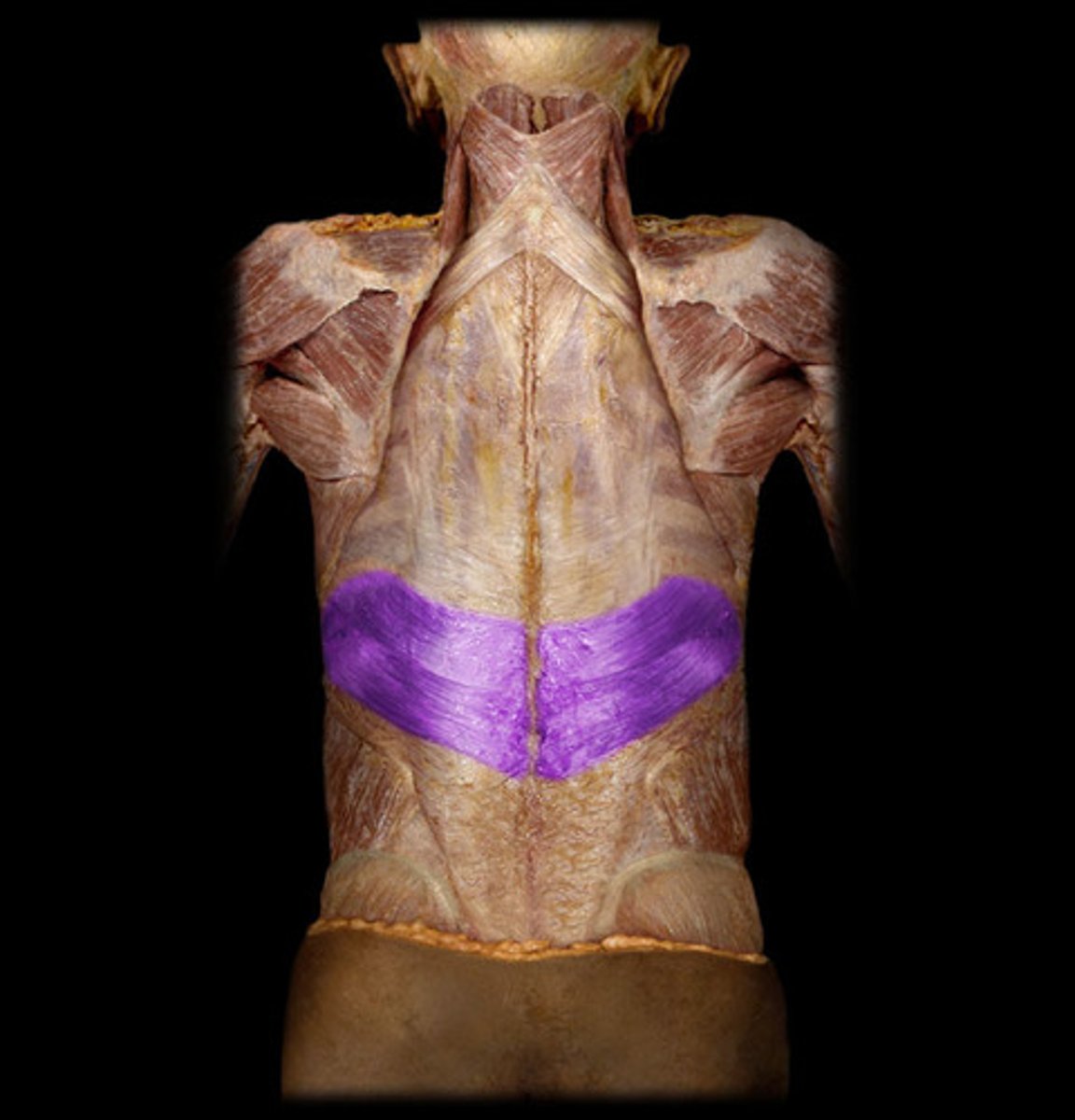
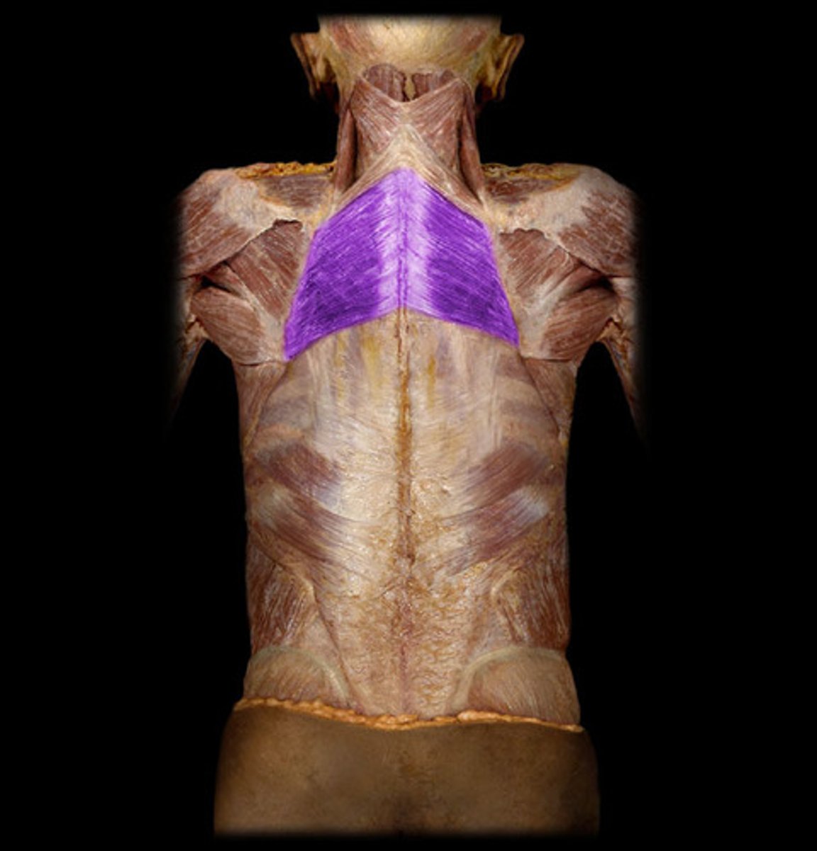
Serratus posterior superior
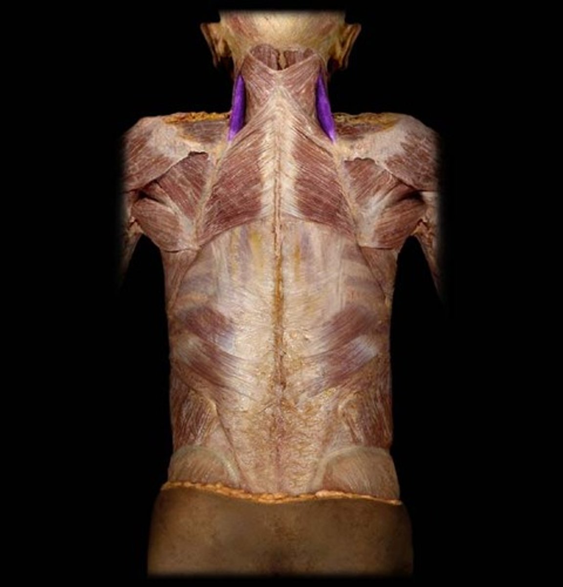
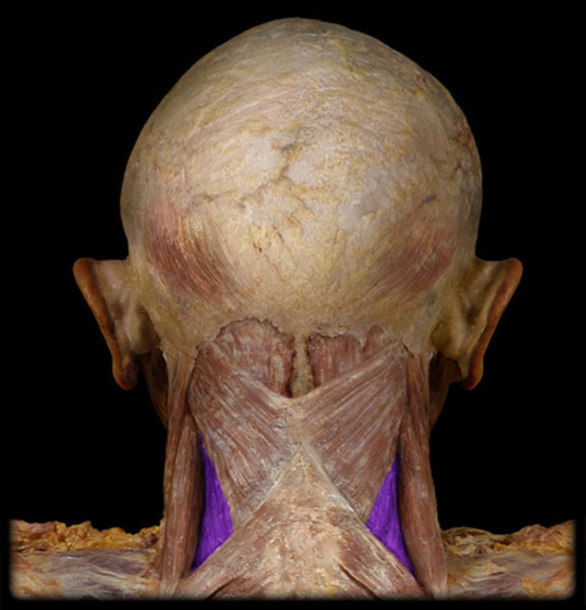
Splenius capitis
splenius cervicis
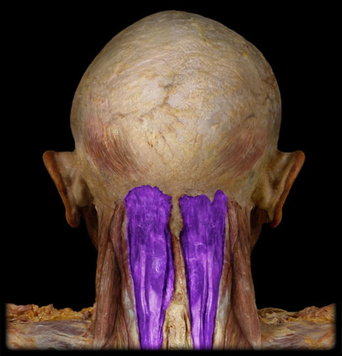
Semispinalis capitis
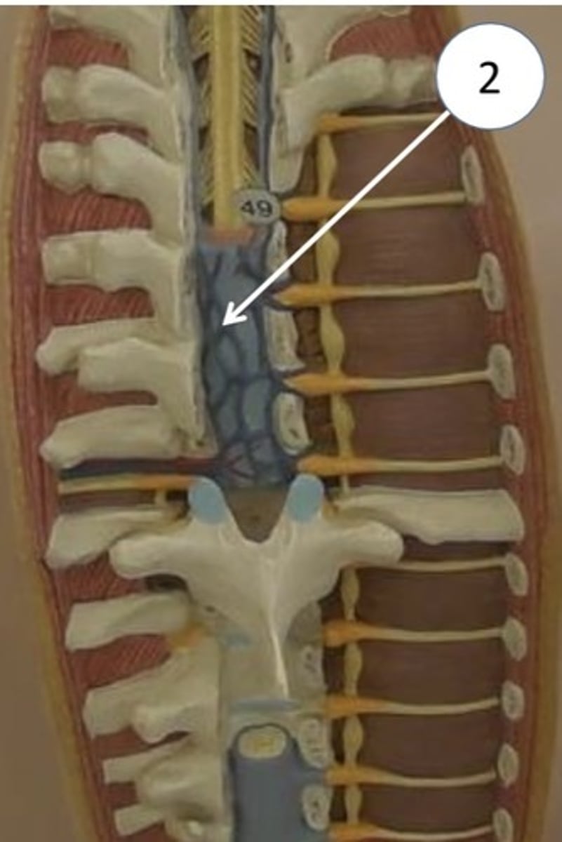
vertebral canal
ligamentum flavum
- thick and yellowish
- connects adjacent laminae
epidural space
- between dura mater and bone
epidural fat
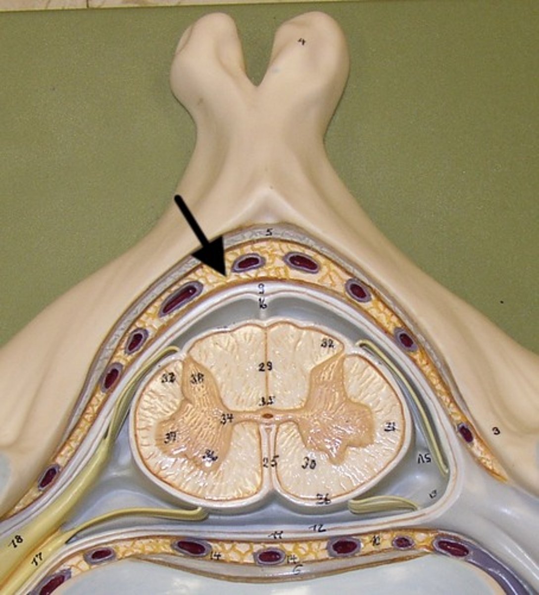
internal vertebral venous plexus
- located in epidural space
meninges
- dura mater (outermost, thickest)
- arachnoid (subarachnoid space has CSF)
- pia mater
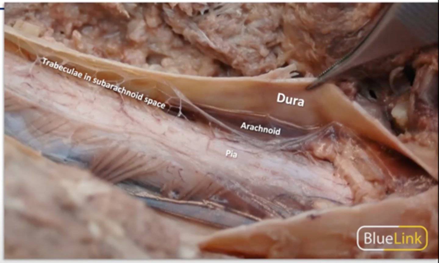
dural (thecal) sac
- ends at S2
?
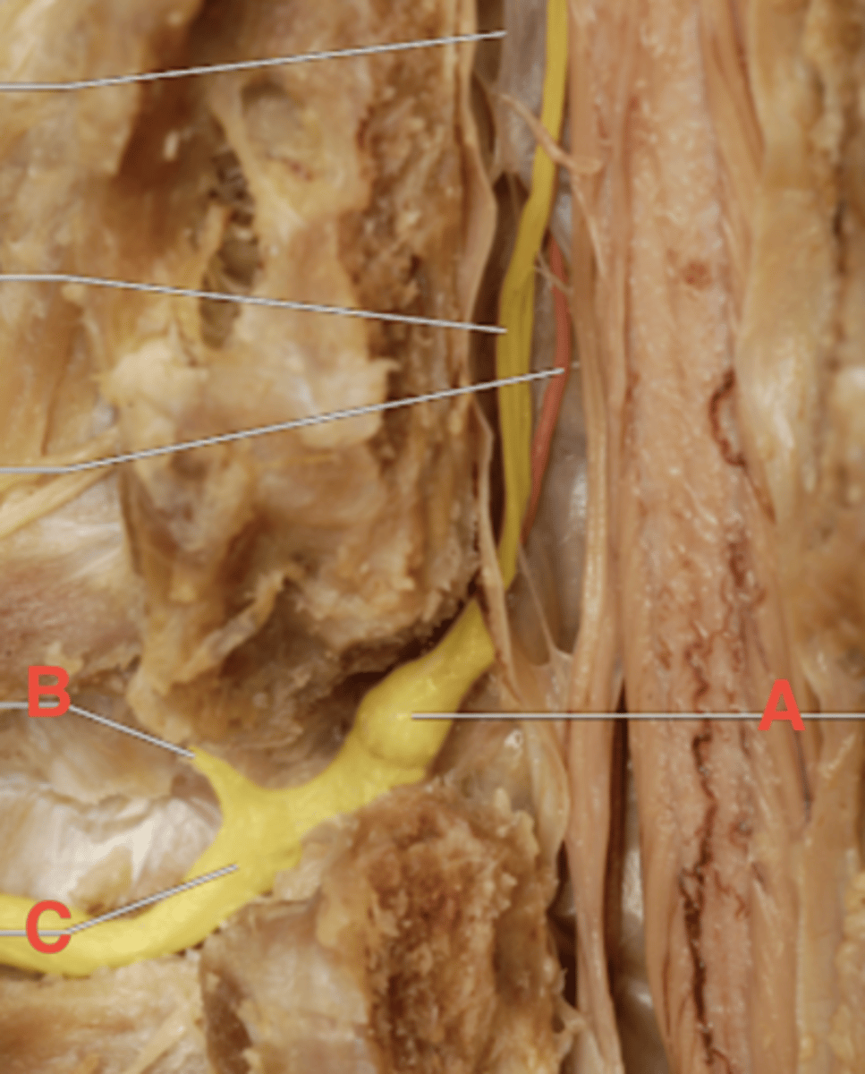
spinal (dorsal root) ganglion
What is 8?
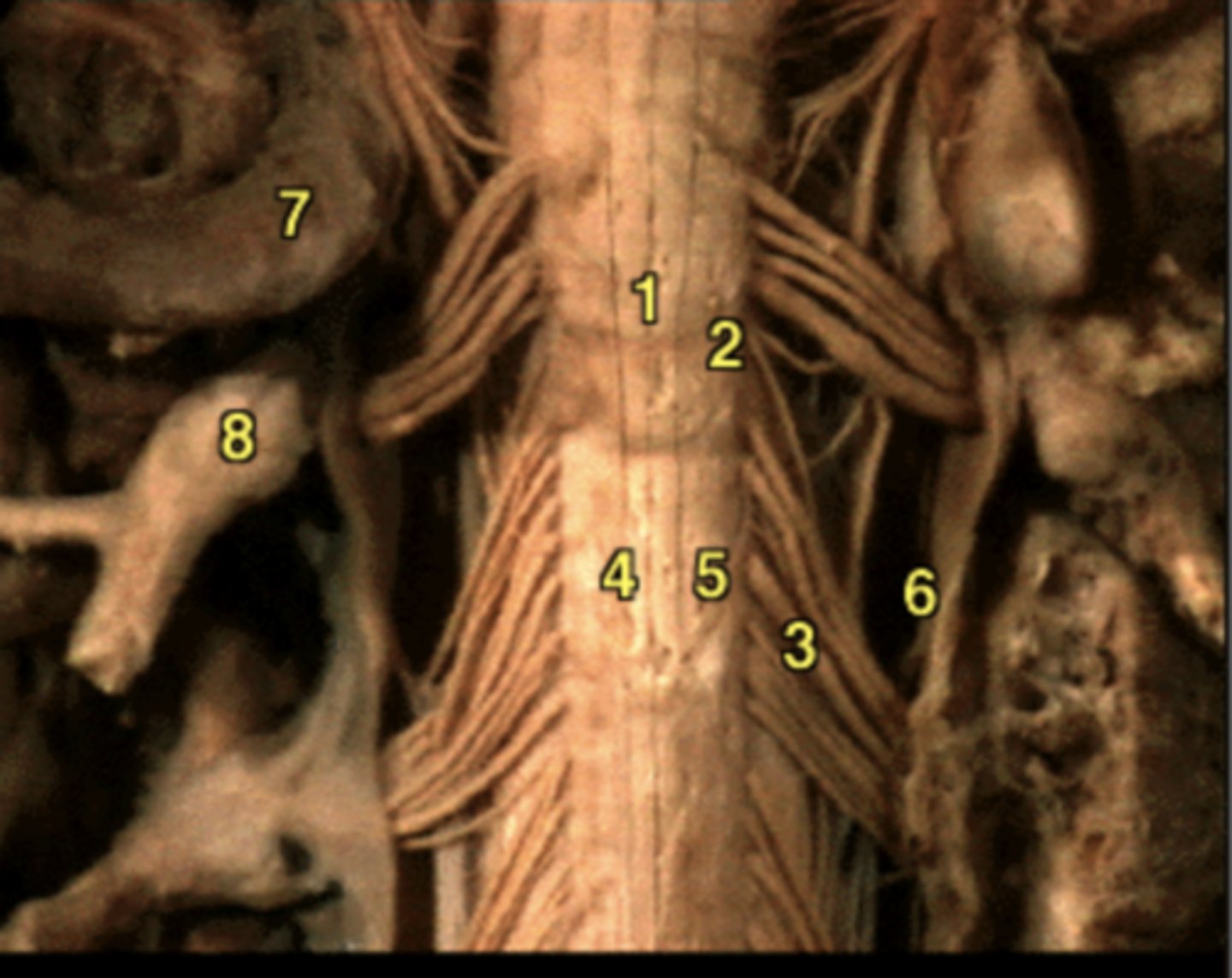
A: dorsal root ganglion
B: dorsal rami
C: ventral rami
cervical enlargement of spinal cord
lumbosacral enlargement of spinal cord
conus medullaris
- tapered end of the spinal cord at L1 and L2
red line
filum terminale
red line
cauda equina
- collection of spinal nerves below the end of the spinal cord
(looks like horse tail)
denticulate ligaments
- extensions of pia mater that secure cord to dura mater
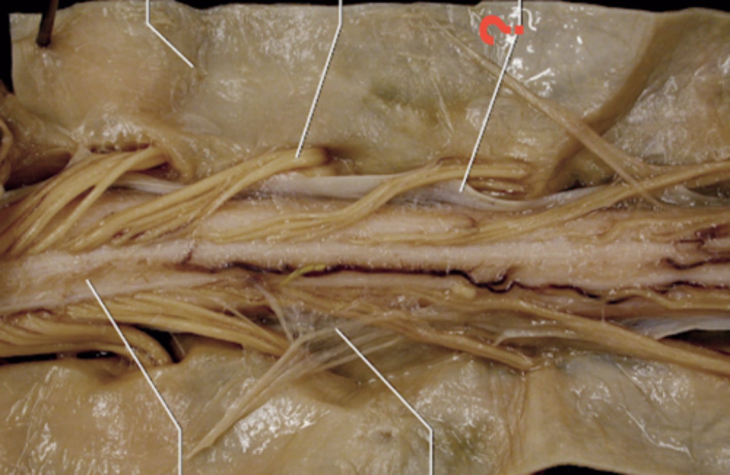
posterior spinal arteries
F
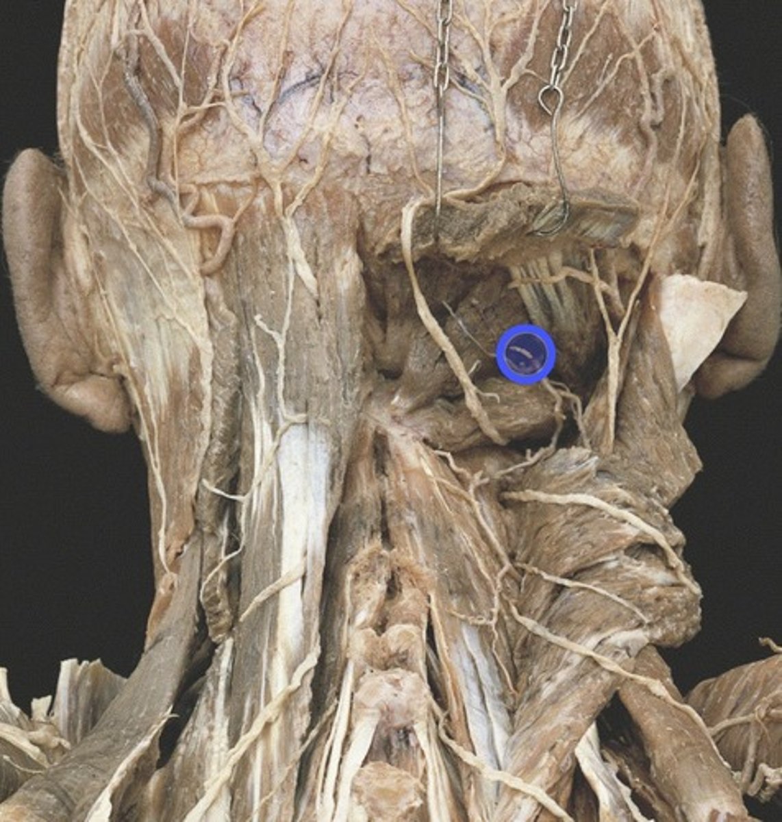
Greater occipital nerve
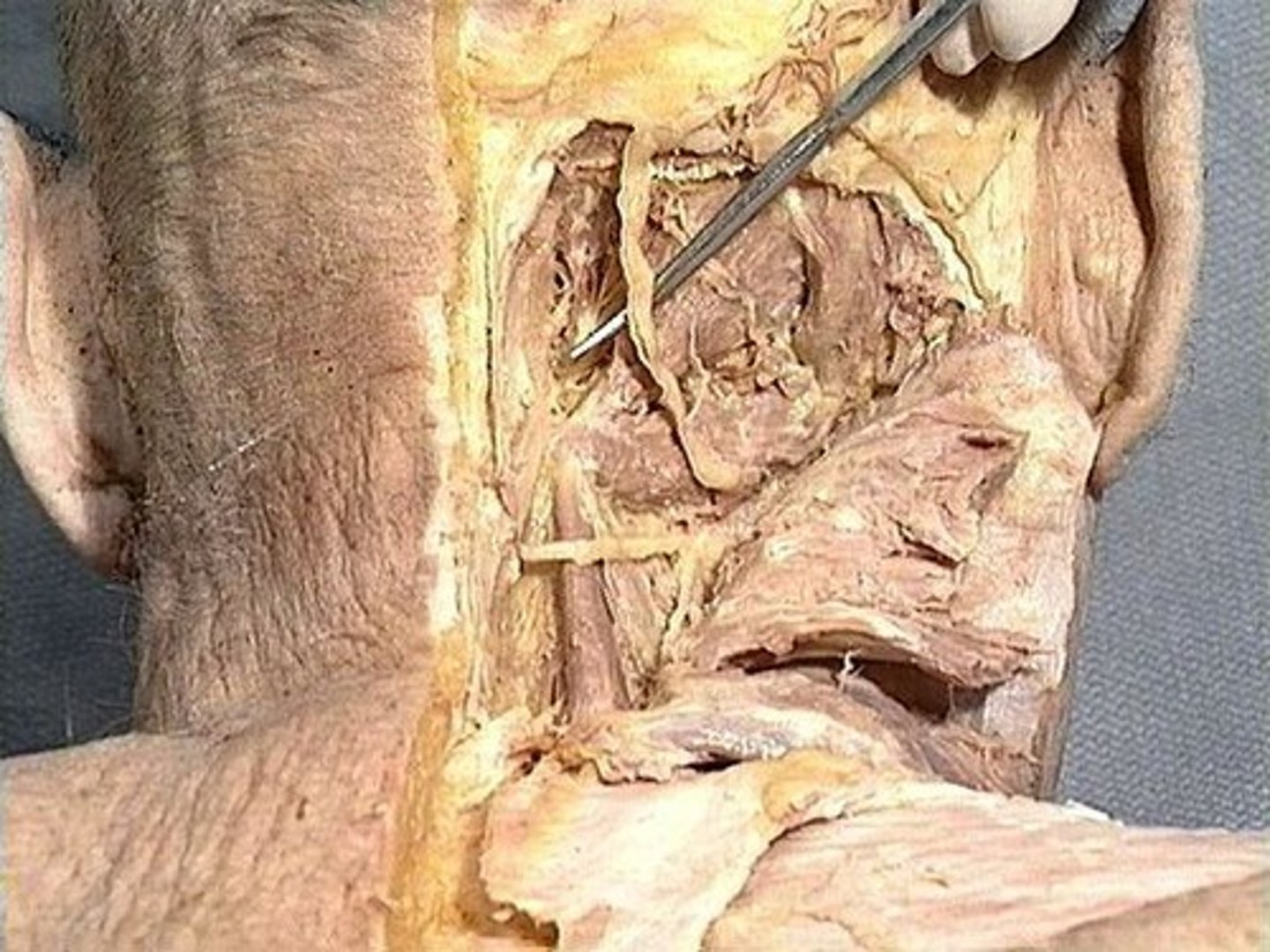
Rectus capitis posterior major
Obliquus capitis inferior
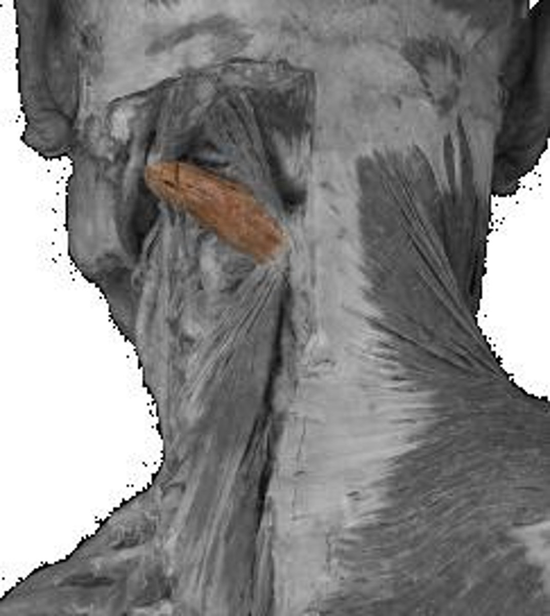
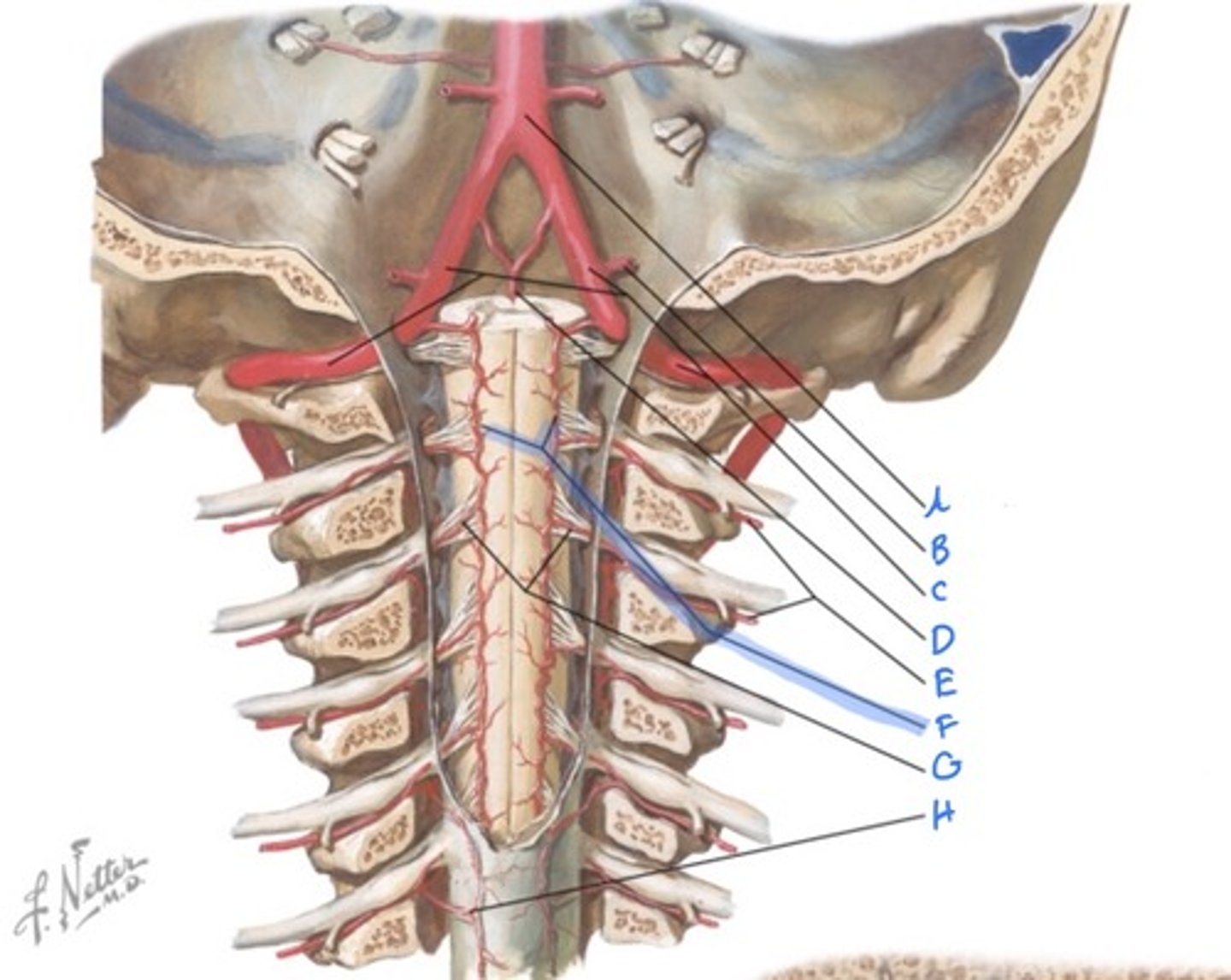
vertebral artery
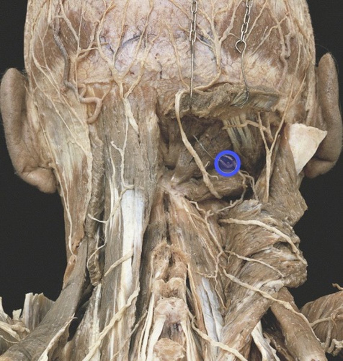
suboccipital nerve
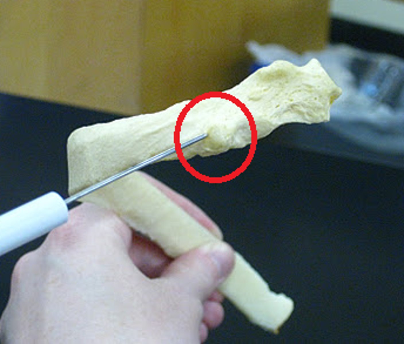
tubercle of a rib
jugular notch

clavicular notch (sternoclavicular joint)
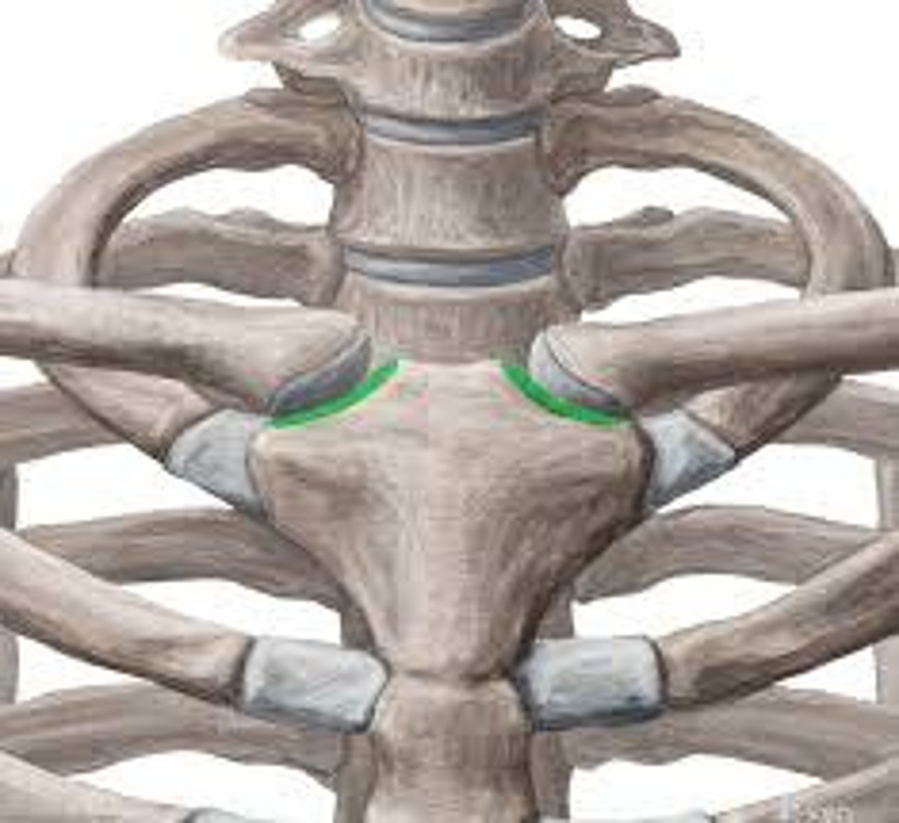
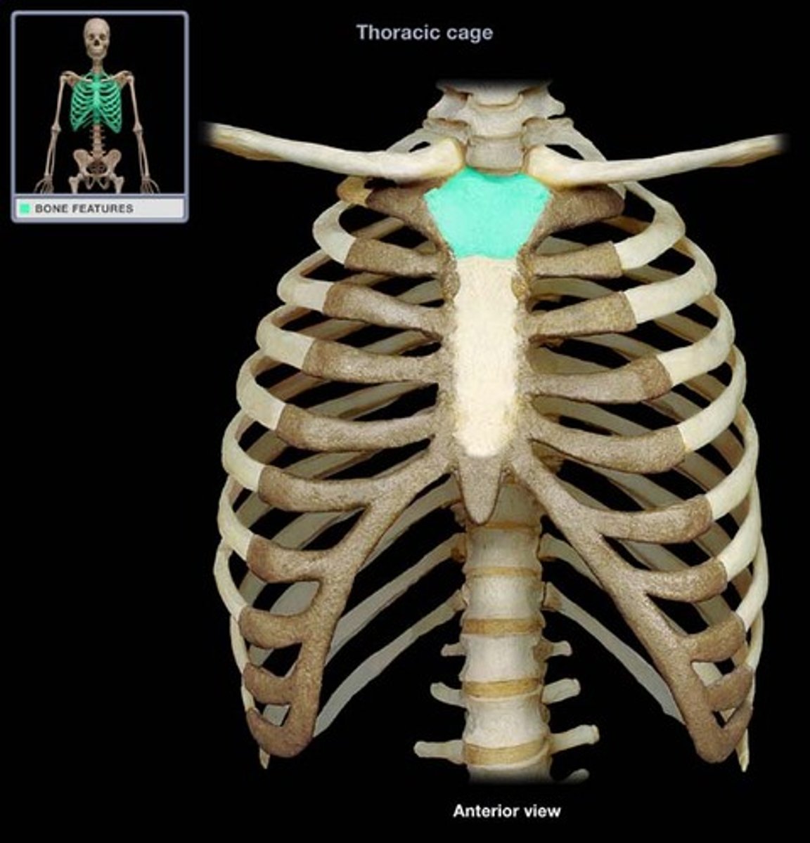
manubrium
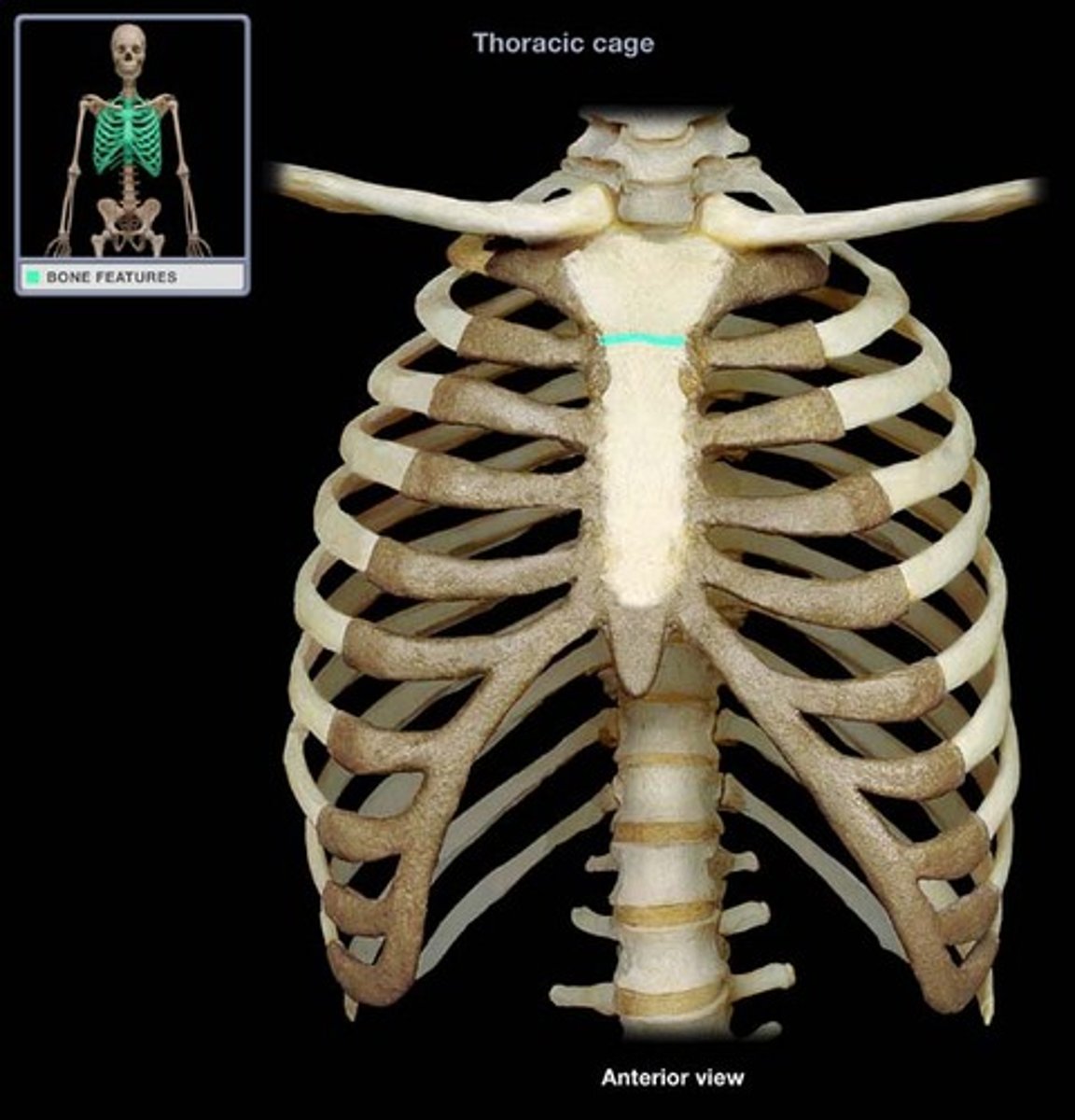
sternal angle
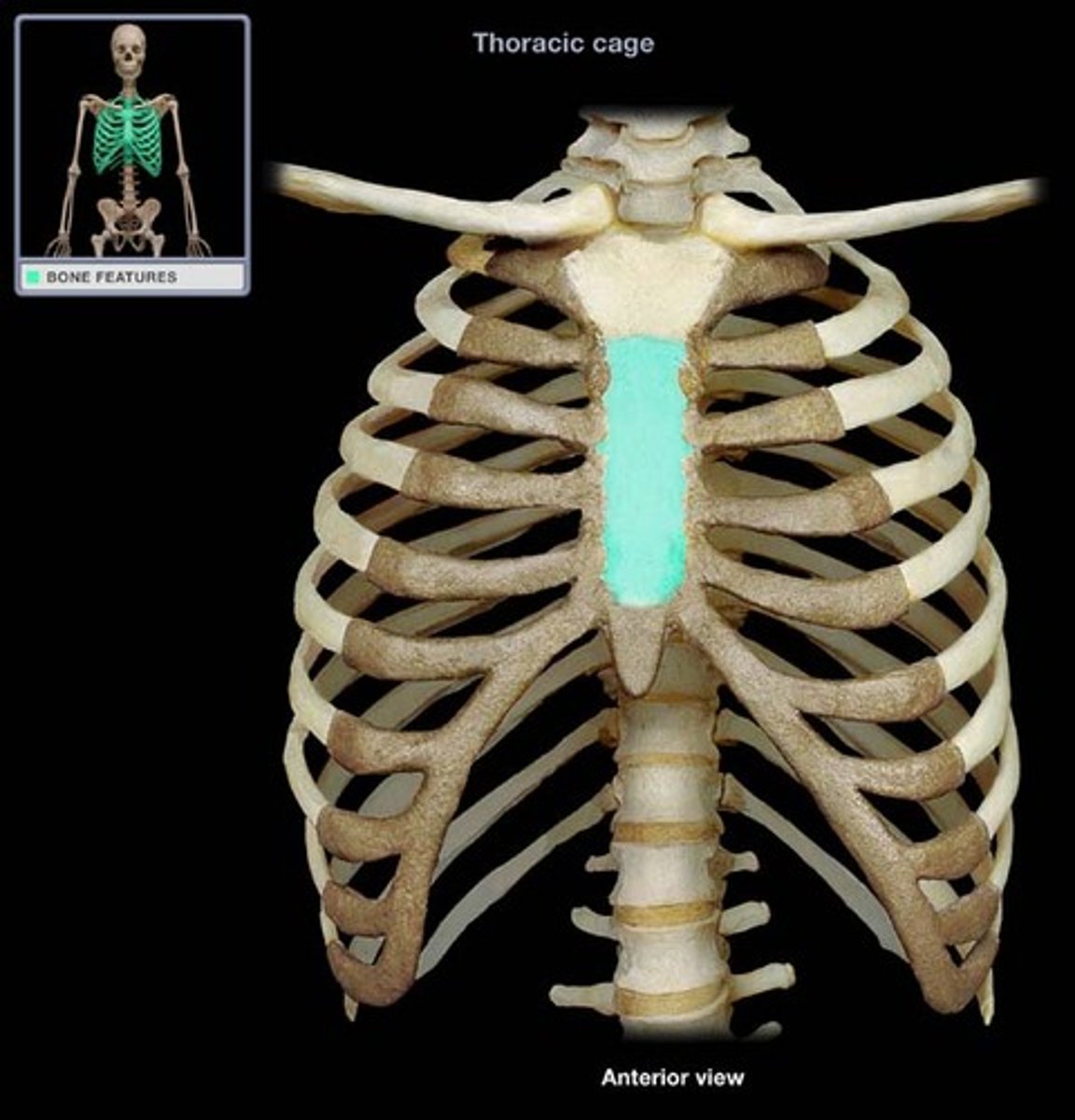
body of sternum (gladiolus)
xiphoid process
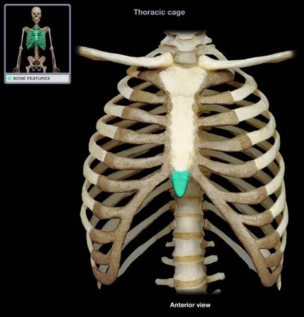
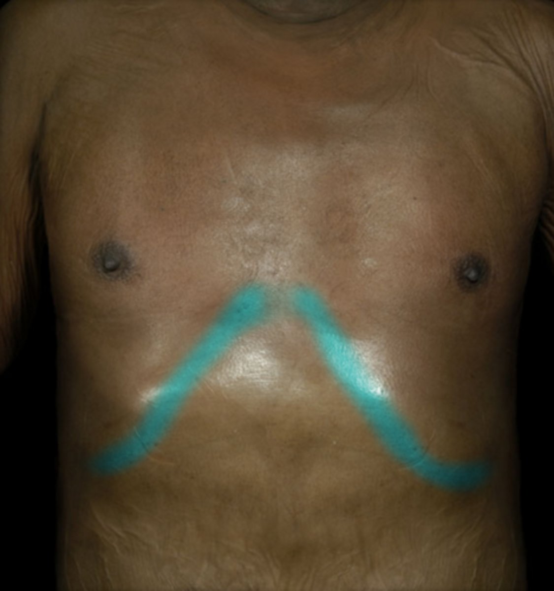
costal margin
- inferior border of thoracic cage formed by downward arc of ribsinferior border of thoracic cage formed by downward arc of ribs
first rib
Manubrium
