FUNCTIONAL ANATOMY
1/48
There's no tags or description
Looks like no tags are added yet.
Name | Mastery | Learn | Test | Matching | Spaced |
|---|
No study sessions yet.
49 Terms
Skeletal muscle
Attached to skeleton by tendons → create movement when they shorten (concentric contraction)
Links 2 bones across its connecting joint and is under voluntary control and creates movement
striated in appearance which means fibres contain alternating light and dark bands perpendicular to fibres
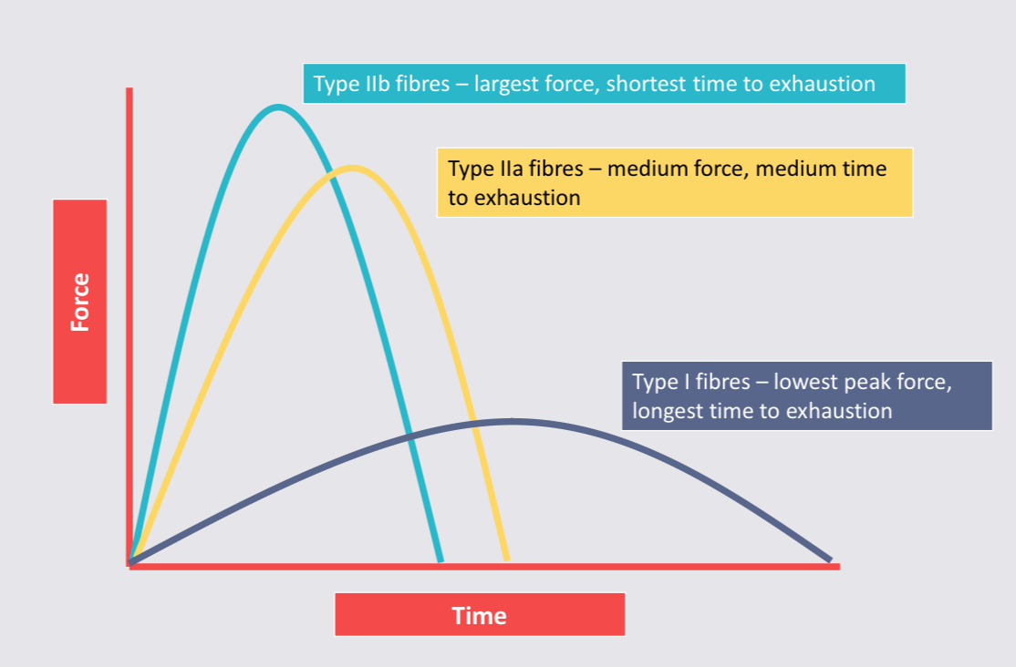
Slow twitch fibres (red) - endurance (type l)
Fast twitch fibres (white) - speed and power (type lla and llb)
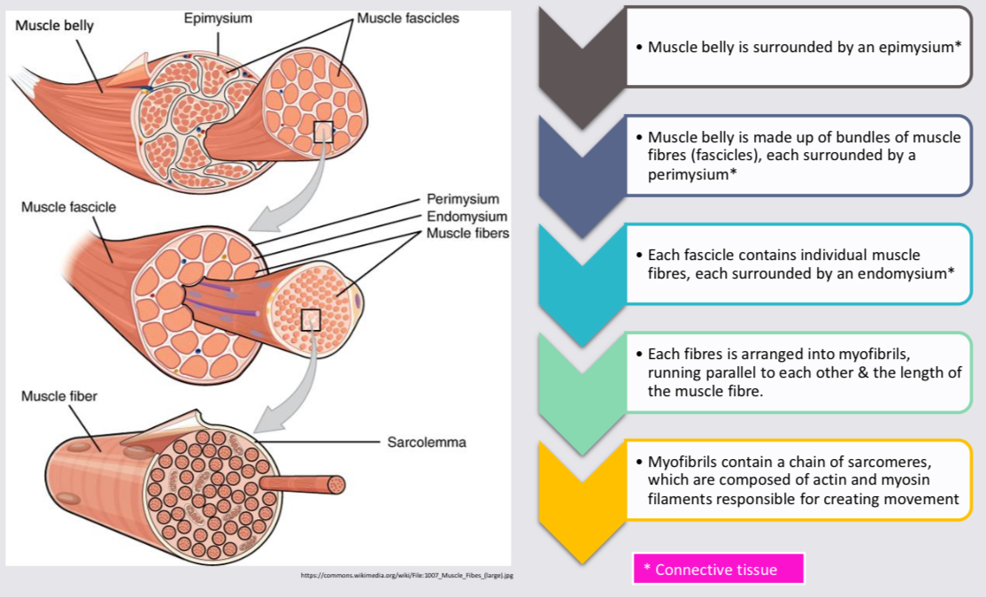
muscular level diagram
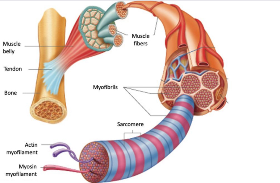
muscle diagram with descriptions
Origin
Attachment to the bone that does not move when the muscle contracts.
attachment point at the proximal end.
Insertion
Attached to the bone which moves more when the muscle contracts.
Attachment point at the distal end
Muscles produce movement
Muscles work in pairs to produce movement
agonist (prime mover) muscle responsible for movement.
Antagonist relaxes to allow the movement to occur
Reciprocal inhibition → coordinated relaxing of muscles on one side of a joint to accomodate contraction on the other.
Types of contraction
Concentric - muscle shortens
Eccentric - muscle lengthens
Isometric - muscle does not change length
greatest potential for force generation → max no. cross bridges attached to actin.
Sliding filament theory - INTRO
movement is created when muscles change in length
Length changes when the myofibril length changes
Myofibril is made up of sarcomeres joined end-to-end
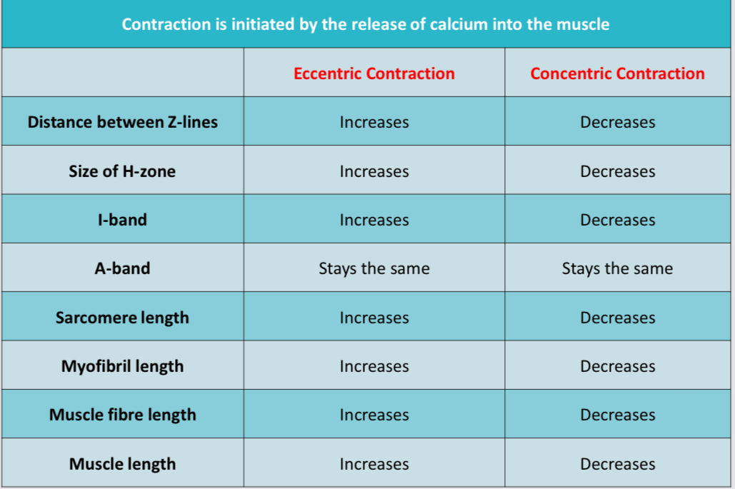
The myofibril changes length when proteins actin and myosin ‘slide over’ each other and increase/decrease the sarcomere length depending on contraction type (eccentric vs concentric)
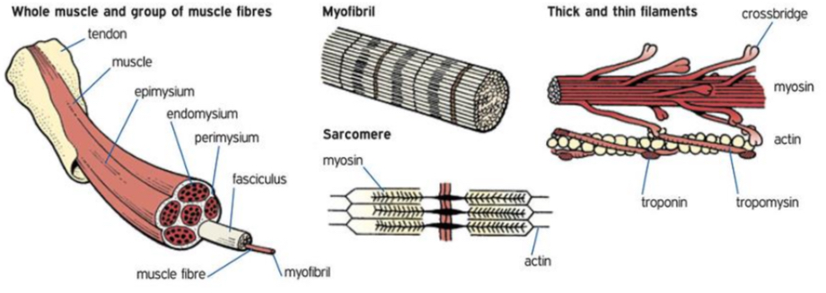
Sarcomere
Comprises the unit between the two Z lines and makes up the functional unit of a muscle fibre
actin and myosin → contractile filaments which change the length of the sarcomere
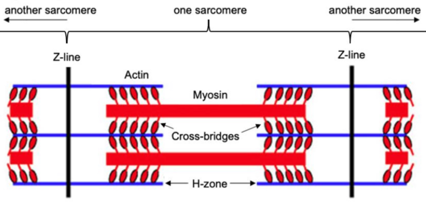
Actin
Thin protein filament attached to the Z line
Cross bridges on the myosin attach to the actin when stimulated by the release of calcium to create movement. Actin filaments are attached to the Z-line and pull the Z-line towards the midline of the sarcomere in a concentric contraction
Myosin
Thick protein filament containing cross bridges.
The myosin cross bridges attach to the actin when stimulated with calcium.
Z-line
Found at either end of the sarcomere.
The Z-lines of a sarcomere come closer together in concentric contractions and spread further apart as the muscle relaxes or in an eccentric contraction.
Cross bridges
Tiny projections from myosin filaments that attach temporarily to actin filaments, pulling the actin filaments towards the midline of the sarcomere making the H-zone shorten or disappear which shortens the sarcomere shortening the myofibril and creating movement (concentric contraction)
H-zone
Space between the actin filaments which gets longer or shorter as the sarcomere changes in length
Diagram
I-band
Light band that contains the thin actin filament
In a relaxed muscle the thin filaments do not completely overlap the myosin thick filaments, and a prominent I-band exists.
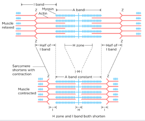
A-band
It contains both thick and thin filaments and is the centre of the sarcomere that spans the H-zone

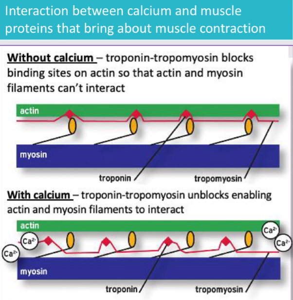
Sliding filament theory
A theory used to explain the mechanism of muscle contraction based on the interaction of actin and myosin filaments to generate movement.
A neurochemical stimulation releases calcium from the sarcoplasmic reticulum into the sarcomere.
This causes the actin filaments to reveal binding site for the myosin head to connect (due to calcium binding to troponin and moving the tropomyosin).
Myosin heads bind to the actin filaments creating a cross-bridge.
Breakdown of ATP releases energy to stimulate the myosin cross bridge to pull the actin filaments towards the midline line of the sarcomere.
This results in the shortening of the sarcomere as the actin and myosin filaments ‘slide over’ each other causing the Z lines to come closer together and the H-zone to shorten.
Shortening each sarcomere shortens the myofibril resulting in the shortening of the muscle fibres and movement occurs.
Cross bridges attach and re-attach at different times to create movement and maintain tension.
The process keeps repeating if the neural impulse is present or the muscle relaxes if the neural impulse ends.
Sliding filament theory diagram
Sliding filament theory summary
Steps of muscle contraction
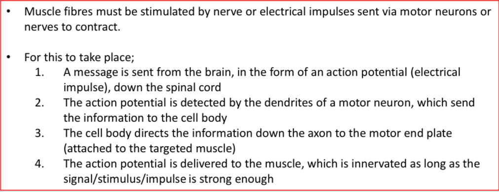
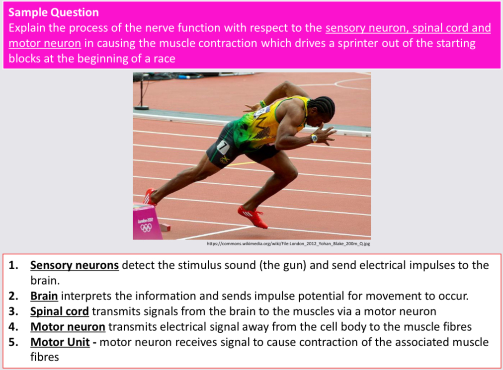
Before a muscle contracts to initiate movement, the muscle fibres must be stimulated by nerve or electrical impulses sent from the brain as an action potential to the spinal chord.
brain → sends message in the form of an action potential to the spinal cord
Spinal cord → responsible for the transmission of the message between the brain and the muscle and from the muscle/body to the brain
Motor neurons → receives the message/action potential from the spinal cord and delivers it to the targeted muscle and movement occurs
Sensory neurons → ends messages back to the brain via the spinal cord
Brain → analyses the information delivered by the spinal chord to determine next action
Process repeats
Nervous system
Three key functions
Through sense organs and sensory nerves, it receives info abt changes in the body and environment and sends this info to the spinal chord and brain.
Brain determines a suitable response
Brain sends commands to muscles to carry out selected response
Parts of the nervous system
Central nervous system
Peripheral nervous system
Central nervous system (CNS)
brain and spinal cord
Spinal cord
Delivering messages from the body to the brain
Delivering messages from the brain to the body
The brain
Analyses info received from the sensory neuron
Determines suitable response
Sends message to targeted muscles via spinal cord and peripheral nerves to contract and movement occurs.
Peripheral nervous system (PNS)
Remainder of the nervous system includes sensory and motor neurons which transmit messages to and from the CNS.
sensory division
Carries messages from the body and environment to the spinal cord and brain → sensory neurons.
motor division
Carries messages from the brain to the muscles to respond as appropriate → motor neurons.
* eyes, ears, taste, smell = straight to brain.
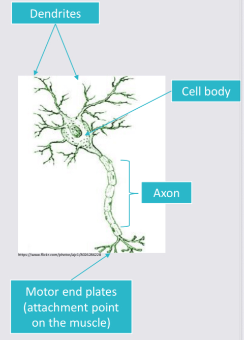
Motor neuron
A cell within the nervous system that transmits impulses/signals to other nerve cells/muscle.
Dendrites → acts as antenna to detect impulse from sensory receptors and then deliver it to the cell body.
Cell body → contains nucleus which detects neuron’s activities and sends message to the axon.
Axon → transmits message away from the cell body to the muscle.
Motor neurons attach to muscles at motor end plates.
Motor unit
Motor neuron and the fibres it activates / innervates are called motor unit.
a single motor neuron joins with muscle fibres which respond (contract) when the motor neurons attach is activated.
Whole muscle contains many seperate motor units which allows the muscle to generate different amounts of force to meet the demands of the situation.
possibly need
order to answer question - check
Size of motor unit
The action potential size required to innervate a motor unit is proportional to the number of fibres in the motor unit.
Precise movement
small number of muscle fibres required
Small motor unit
Small action potential
Large movement
gross motor skill
Many fibres required
Large motor unit
Large action potential
A motor unit requires a signal/electrical impulse/action potential to reach a certain threshold (level/intensity) before it will activate.
Impulse doesn’t reach threshold → doesn’t activate → fibres wont contract
Impulse reaches threshold → all muscle fibres contract @ 100% capacity.
Slow twitch fibres have lower stimulus threshold and will fire before fast twitch fibres.
Effect of weight on number of motor units
increase weight = more units required → more signals to more motor units → increase size of stimulus.
Less weight = size of stimulus decreased
All or none principle
When a motor unit receives stimulation/impulse/action potential that exceeds the threshold, all the muscle fibres associated with it will contract to their maximum potential or not at all.
When a motor unit is stimulated by a signal that exceeds its stimulus threshold, all the muscle fibres in that motor unit contract with maximal force simultaneously.
If the stimulus threshold is not reached, none of the fibres in the motor unit will contract at all.
Motor unit recruitment (muscle fibre recruitment)
Motor units are recruited in order depending on exercise intensity.
Slow twitch motor units (type I)
low activation levels - only small stimulus is required - light to moderate activity
Higher threshold motor units (fast-twitch type IIa)
recruited as exercise intensity increases → slow twitch units are still activated.
Highest stimulus threshold (fast twitch type IIb)
Generating peak force uses motor units that generate the greatest force → slow twitch and IIa motor units still being activated
Increasing force produced by a muscle
Increase number of motor units recruited by increasing stimulus size
Increasing the frequency at which impulses are sent to the motor unit resulting in the motor unit firing repeatedly to increase the force generated.
The increase in signal size/frequency is controlled by the brain - neural process.
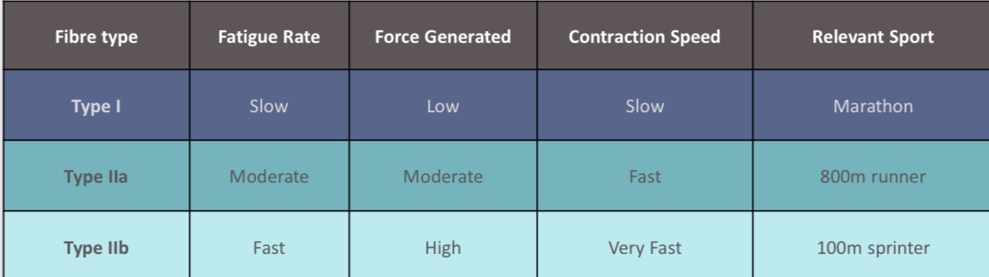
Slow twitch (type I)
Purely aerobic and are suited to events which require continuous activity.
AKA red / slow twitch fibres
Fast twitch (type IIa)
Partially aerobic and are suited to events which require both aerobic and anaerobic elements.
Fast twitch (type IIb)
Purely anaerobic and are suited to events which require explosive movements.
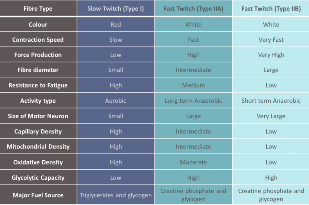
Role of diameter
The greater the diameter of the fibres the larger the neuron needed to activate the muscle fibres
type I - small diameter = small neuron
Type IIa - intermediate diameter = intermediate neuron
Type IIb - large diameter = large neuron
Role of genetics
An athlete can determine their potential to achieve at an elite level by having a muscle
biopsy to determine the relative percentages of Type I and Type II fibres they possess.
• A muscle biopsy is a minor surgical procedure that involves removing a small amount of muscle tissue from the muscle and then examining it to determine relative percentages of fibres.
high % of type II fibres (80% +) = speed/power/explosive
High % of type I fibres (80% +) = endurance
Diagram force - time
Simplified summary table
Full summary table
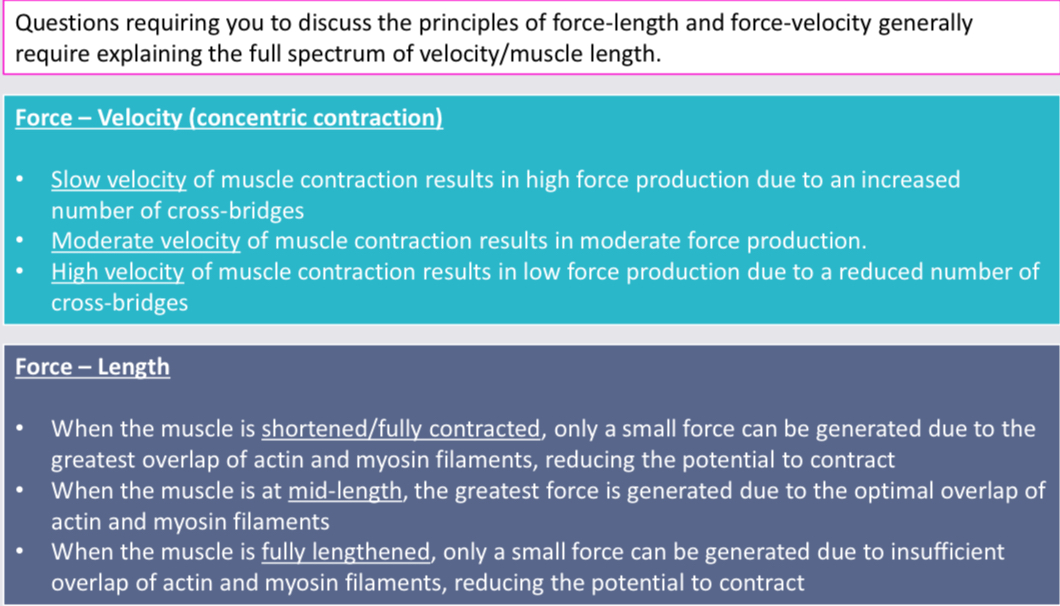
Force-velocity
Describes the relationship between force production and the velocity of movement.
muscle can create larger force with a decrease in velocity of concentric contraction.
easier to lift heavy weight concentrically upwards slowly than it is quickly → more motor units to be recruited
more time for sarcomere to contract fully
Increases number of cross-bridges that can be attached between the myosin and actin
Force-velocity (concentric contraction) graph
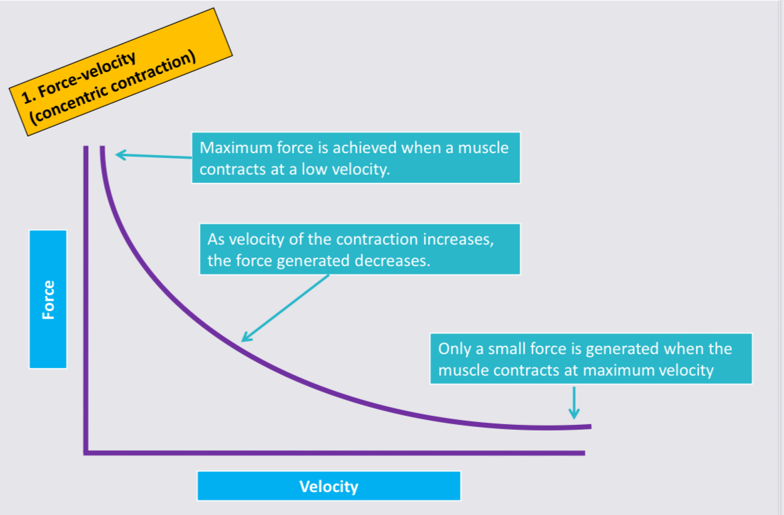
Force-velocity summary
Concentric contraction
greater force required = slower speed of contraction
Due to number of cross-bridges that can be attached between the myosin and actin
Increasing velocity of concentric contraction decreases force that can be produced
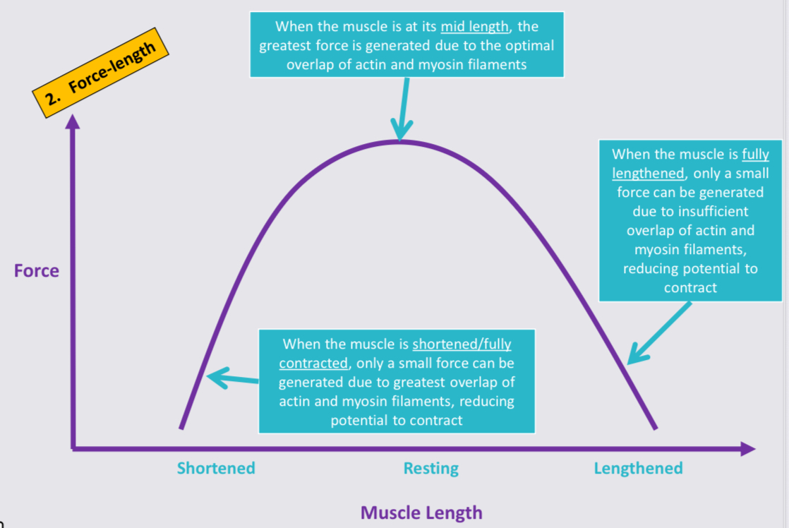
Force-length
Relates to the amount of muscle force that can be produced at varying muscle lengths.
length of muscle affects how much force it can generate
Max tension is best achieved when muscle is @ resting length → increases number of cross-bridges that can be attached between myosin and actin
Less force = contract beyond optimal length
Less force = lengthen beyond optimal length
Greater number of cross-bridges = greater force generated
Force-length graph
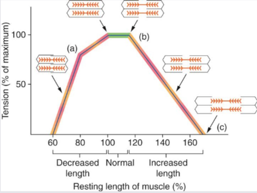
Force-length and joint angle
As a muscle changes length during contraction, the joint angle around which the movement is occurring also changes.
angle of the joint influences how much force can be generated
Joint angle at which the muscle can generate its greatest force varies for different body parts → usually near the middle of the joint’s range of motion → allows greatest number of cross bridges to be attached and greatest overlap of actin and myosin filaments to occur.
Decrease in force when joint angle is large/small → affects number of cross bridges that can attach.
Question help