Atrial Fibrillation
 Call Kai
 Learn
Learn Practice Test
 Spaced Repetition
 Match
Match Flashcards
 Knowt Play
1/73

Earn XP
Description and Tags
AF definition, causes, investigation and management.
Name | Mastery | Learn | Test | Matching | Spaced | Call with Kai | Chat |
|---|
No analytics yet
Send a link to your students to track their progress
74 Terms
What is Atrial Fibrillation (AF)?
A supraventricular tachyarrhythmia with disorganised atrial electrical activity and ineffective atrial contraction. The ventricular response is usually irregular because conduction through the atrioventricular node varies.
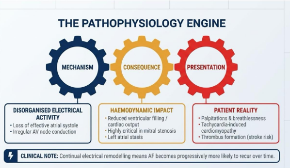
What are the mechanisms and consequent clinical consequences of AF?
Loss of effective atrial systole leads to:
Reduced ventricular filling and cardiac output, particularly with diastolic dysfunction or mitral stenosis.
Irregular and sometimes rapid ventricular response leads to:
Palpitations, impaired exercise capacity and, if persistent, tachycardia-induced cardiomyopathy.
Left atrial and appendage stasis leads to:
Thrombus formation with risk of stroke or systemic embolism.
Atrial structural and electrical remodelling leads to:
AF becomes more likely to recur and progress over time.
What are the four different types of AF, classed according to duration of episodes?
Paroxysmal
Persistent
Long-standing persistent
Permanent
Paroxysmal AF
One or more episodes of AF which terminate spontaneously or with intervention within 7 days.
Persistent AF
One or more episodes that continue for more than 7 days, including episodes terminated by cardioversion after 7 days or longer.
Long standing persistent AF
Continuous AF for 12 months or longer when rhythm control remains a treatment goal.
Permanent AF
When the patient lives with atrial fibrillation long-term, and no further attempt or treatment is panned to restore or maintain sinus rhythm.
What are the different precipitants to AF?
It typically reflects underlying cardiovascular disease, but acute illness may precipitate a first / recurrent episode:
Cardiovascular precipitants
Respiratory precipitants
Metabolic / Endocrine precipitants
Systemic illnesses precipitants
Lifestyle and related factors
What are the main cardiovascular precipitants to AF?
Hypertension
Heart failure
Ischaemic heart disease
Cardiomyopathy
Valvular disease
Myocarditis
Pericarditis
Cardiac surgery
What are the main respiratory precipitants to AF?
Pneumonia
Pulmonary embolism
Hypoxia
Exacerbation of chronic lung disease
What are the main metabolic / endocrine precipitants to AF?
Hyperthyroidism
Hypokalaemia
Hypomagnasaemia
Diabetes
Obesity
What are the main systemic illnesses that can precipitate AF?
Sepsis
Anaemia
Hypovolaemia
Major non-cardiac surgery
What lifestyle factors may precipitate AF?
Alcohol excess
Stimulant drugs
Obstructive sleep apnoea
Physical inactivity
Does AF always present with symptoms?
No, it can be symptomless and detected incidentally. Symptoms can be very minimal or more major.
What are the symptoms AF can present with?
Palpitations / awareness of an irregular heartbeat
Breathlessness, fatigue, or reduced exercise tolerance
Dizziness, presyncope, or syncope
Chest discomfort
Features of heart failure
Stroke, TIA, or systemic embolism
What are some potentially life threatening features AF can present with?
Shock
Syncope with severe / ongoing hypotension
Myocardial ischaemia (chest pain)
Severe heart failure with pulmonary oedema
When first assessing someone presenting with AF, what should you quickly determine?
Whether the AF is stable or unstable - if it is unstable the patient will need immediate treatment.
What does AF stability depend on?
Whether the patient is haemodynamically stable.
What are the signs that a patient has unstable AF?
Shock (BP<90 systolic)
Loss of consciousness/syncope
Myocardial Ischaemia (chest pain or suggestive ECG changes)
Heart Failure
Investigations for AF
Manual pulse palpation
12 lead ECG to confirm
24hr ambulatory ECG monitoring if asymptomatic episodes are suspected / if symptomatic episodes happen less than 24hrs apart.
Longer ambulatory monitoring may be required if symptomatic episodes are happening >24hrs apart.
Blood tests
Transthoracic echocardiography when indicated
Chest X-ray when indicated
Transoesophageal echocardiography if indicated
What is a 12 lead ECG used for when investigating AF?
To confirm the diagnosis
Assess ventricular rate and QRS width
Look for conduction diseases
Look for ischaemic changes and possible pre-excitation
What blood tests might be useful when investigating AF?
FBC
Renal function / electrolytes
Liver tests
Thyroid function if appropriate
Glucose / HbA1c if appropriate
Magnesium if indicated
CRP if indicated
Troponin if indicated
What is a transthoracic echocardiography used for when investigating AF?
Offer only when:
A baseline echo is important for long-term management
Rhythm control/cardioversion is being considered
Structural or functional heart disease is suspected
Refinement of clinical risk assessment is needed
What is a chest X-ray used for when investigating AF?
Offer only when there is suspected:
Heart failure
Infection
Lung disease
What is a transoesophageal echocardiography used for when investigating AF?
Offer only when:
Transthoracic imaging is inadequate for a management decision.
Part of a specialist strategy to exclude left atrial thrombus before early cardioversion.
How is unstable AF managed?
Call for expert help
Monitor ECG, BP, and oxygen sats
Obtain IV access
Record a 12 lead ECG if this does not delay treatment.
Give oxygen if hypoxic (<94%)
Deliver synchronised electrical cardioversion, with sedation and anaesthesia if conscious and it does not cause an unsafe delay.
If cardioversion is unsuccessful, follow the current tachyarrhythmia algorithm and seek expert advice.
Treat reversible causes and assess anticoagulation as soon as possible - do not delay emergency cardioversion though.
How is stable AF managed?
Treat the reversible cause
Rate control is then the first line strategy
Offer rhythm control when symptoms remain after rate control
Assess stroke risk and consider anticoagulation if appropriate
Long term review
There are certain circumstances where this treatment algorithm does not apply.
When is rate control not the first line strategy?
HF is probably primarily caused by AF
AF is new onset
Patient also has an atrial flutter which is suitable for an ablation strategy
Rhythm control is preferable after discussion
In what situations does the usual treatment algorithm for stable AF ot apply?
Pre-excited AF
Acute ischaemic stroke / TIA
Acute coronary syndrome / PCI
Pregnancy
Hyperthyroidism or sepsis associated AF
Post ablation or apparent resolution
What is pre-excited AF?
An irregular broad-complex tachycardia caused by AF conducting through an accessory pathway. E.g. patient with Wolf-Parkinson-White syndrome and AF.
What should you do if a patient has pre-excited AF?
Do not give AV-node-blocking drugs such as beta-blockers, verapamil, diltiazem, digoxin or adenosine.
Seek urgent expert help
RCUK lists procainamide or cardioversion.
What should you do if a patient has AF and an acute ischaemic stroke / TIA?
Follow the stroke pathway.
What should you do if a patient has acute coronary syndrome / PCI and AF?
Combined antiplatelet and anticoagulant treatment increases bleeding risk.
Use the relevant cardiology pathway and minimise combination duration where possible.
What should you do if a patient is pregnant and has AF?
DOACs and warfarin are generally unsuitable in pregnancy.
Obtain urgent specialist obstetric/cardiology/haematology advice.
What should you do if a patient has hyperthyroidism and AF?
Treat the precipitant.
Do not assume stroke risk disappears when the trigger resolves.
Reassess for ongoing AF and anticoagulation.
What should you do if a patient has sepsis associated AF?
Treat the precipitant.
Do not assume stroke risk disappears when the trigger resolves.
Reassess for ongoing AF and anticoagulation.
What should you do if the patient is post-ablation and/or there is apparent resolution of the AF?
Do not stop anticoagulation solely because sinus rhythm is restored or AF is no longer detected.
Reassess CHA₂DS₂-VASc and bleeding risk.
What are the different drugs used for rate control?
Standard beta-blocker (not sotalol) is first line
Rate-limiting calcium channel blocker
Digoxin
Combination treatment
Amiodarone (potassium channel blocker) - not for long term use
When is a standard beta-blocker not recommended for stable AF?
Avoid / cautiously use when the patient has:
Acute decompensated heart failure
Marked bradycardia
Advanced AV block
Bronchospasm
Indication and cautions for rate-limiting calcium channel blockers for patients with AF
Diltiazem or verapamil may be used when appropriate.
Avoid if the patient has left ventricular systolic dysfunction or acute decompensated heart failure.
Do not combine verapamil with a beta-blocker.
Indication and cautions for digoxin for patients with AF
Consider as monotherapy for non-paroxysmal AF when the person does little or no physical exercise, or when other rate-limiting drugs are ruled out.
Less effective during exertion
Dose according to renal function
Check interactions / toxicity
Indication and cautions for combination treatment for patients with AF
Consider two drugs when symptoms persist because of poor ventricular rate control.
Monitor for bradycardia, hypotension and AV block.
Obtain specialist advice where needed.
What are the different rhythm control methods?
Synchronised electrical cardioversion
Pharmacological cardioversion
Rhythm control drugs
Catheter ablation (pacing may also be an option)
What factors affect the preferred method of rhythm control?
Onset and duration of AF
Symptoms
Structural heart disease
Heart failure
Comorbidity
Treatment risks
Patient preference
What is the cardioversion pathway?
Immediate synchronised electrical cardioversion if the patient has unstable AF
Onset clearly < 48 hrs - electrical/pharmacological cardioversion may be considered according to clinical context + specialist expertise.
Onset > 48 hrs or uncertain - If long term rhythm control is planned, delay electric cardioversion (not pharmacological) until anticoagulation therapy has been maintained for >3 weeks.
After cardioversion, how is the patient managed?
Continue anticoagulation for at least 4 weeks when indicated.
Continue anticoagulation long term when indicated by stroke-risk assessment, irrespective of apparent maintenance of sinus rhythm.
When is rhythm control indicated?
When symptoms persist after heart rate has been controlled, or rate-control was not successful.
When is rate control for patients with AF considered successful?
Strict control - resting heart rate of ≤ 80 bpm.
Lenient control - resting heart rate of ≤110 bpm if symptomless and left ventricular systolic function remains normal.
When is cardioversion preferred to long term rhythm control?
Patient has unstable AF.
Patient has new-onset stable AF, and it is clinically appropriate.
What drugs are used for long-term rhythm control?
Standard beta-blocker is typically first line
Flecainide or Propafenone (cardiac sodium channel blocker)
Dronedarone (multi-channel blocker)
Amiodarone (potassium channel blocker)
When is dronedarone indicated for rhythm control?
It is second line (beta-blockers are first)
Not used in unstable of severe heart failure
When is Flecainide or Propafenone indicated for rhythm control?
May be considered when there is no known ischaemic or heart disease.
When is Amioderone indicated for rhythm control?
When left ventricular impairment or heart failure makes other options unsuitable, but long-term toxicity and interactions require monitoring.
When is catheter ablation offered?
May be considered in patients with symptomatic paroxysmal / persistent AF when drug treatment is unsuccessful, unsuitable or not tolerated.
Shared decision making should cover recurrence (20-40%) and procedural risks.
AVN ablation with pacing is a specialist option for refractory symptoms and uncontrolled ventricular rate.
How is stroke risk assessed?
CHA₂DS₂-VASc
When is stroke risk assessed in AF patients?
In all patients diagnosed with AF
It is reassessed when clinical circumstances change, and at review.
Why is anticoagulation important for AF patients?
They are at increased risk of strokes as the uncoordinated contractions within the atria can result in blood clot formation:
20–30% of all strokes occur in people with atrial fibrillation.
Patients with AF have a 142% increased risk of any stroke and a 133% greater risk of ischemic stroke compared to individuals without AF.
Stroke is more severe and debilitating in patients with AF than among those who do not have AF.
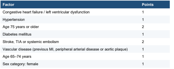
CHA₂DS₂-VASc
A scoring tool to assess stroke risk.
Results:
0 - do not offer stroke-prevention anticoagulation
1:
Men - Consider anticoagulation, taking bleeding risk and preferences into account.
Women (1 is from sex alone) - do not offer stroke-prevention anticoagulation.
2 or more - Offer anticoagulation, taking bleeding risk and preferences into account.
When reviewing anticoagulation, how is bleeding risk assessed?
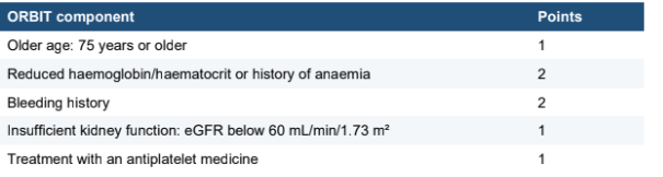
ORBIT
ORBIT score
A scoring tool to assess bleeding risk. Its purpose is to support discussion, monitoring and modification of bleeding risks; a high score is therefore not an automatic reason to withhold anticoagulation.
Alongside assessing bleeding risk with ORBIT, what other actions should be taken when considering prescribing anticoagulation to patients with AF?
Offer monitoring and support to modify risk factors for bleeding, including:
Controlling hypertension.
Review concurrent antiplatelets, NSAIDs and interacting medicines.
Address harmful alcohol use.
Investigate and treat anaemia or thrombocytopenia.
Improve time in therapeutic range for vitamin K antagonist treatment.
Reduce avoidable falls risk and review adherence.
What are the different treatment options for anticoagulation?
DOAC
Vitamin K antagonist (typically warfarin)
Antiplatelet therapy
Left atrial appendage occlusion
When are DOACs indicated for patients with AF?
If indicated after assessing stroke and bleeding risk, these are first line.
Select according to:
renal function
Age
Weight
Interactions
Adherence
Dosing preference
Comorbidity
Contraindications include:
Mechanical heart valves
Moderate to severe rheumatic mitral stenosis
etc.
When are vitamin K antagonists indicated for patients with AF?
When DOACS are contraindicated (second line). Monitor INR and time in therapeutic range.
When are antiplatelets indicated for patients with AF?
These are NOT offered as a monotherapy for anticoagulation in patients with AF.
Patients with AF may have them in addition to other anticoagulants if they are necessary to manage another condition.
When is left atrial appendage occlusion indicated for patients with AF?
Consider only when anticoagulation is contraindicated or not tolerated; specialist assessment is required.
Management of DOACs in patients with AF
Calculate creatinine clearance rather than relying only on eGFR for dosing decisions.
Use the licensed AF dose and apply dose-reduction criteria exactly.
Check renal and liver function, FBC, weight, interactions and adherence at baseline and periodically.
Review more frequently with frailty, renal impairment, intercurrent illness or increased bleeding risk.
Provide counselling about adherence, missed doses, bleeding, procedures and when to seek help.
Long term review in patients with AF
Review at least annually.
Review after treatment changes.
Review more frequently when clinically indicated.
Review:
Symptoms
Ventricular rate
Rhythm strategy
Adherence
Stroke and bleeding risks
Renal function
Adverse effects
Comorbidities
What risk factors should be managed alongside other treatments for AF?
Optimise management of:
BP
Diabetes
HF
IHD
Lipid management
Support weight reduction when overweight / obese.
Reduce alcohol intake and avoid binge drinking.
Encourage regular moderate physical activity while avoiding extreme unaccustomed endurance exercise.
Assess for obstructive sleep apnoea when clinically suspected.
Support smoking cessation and medication adherence.
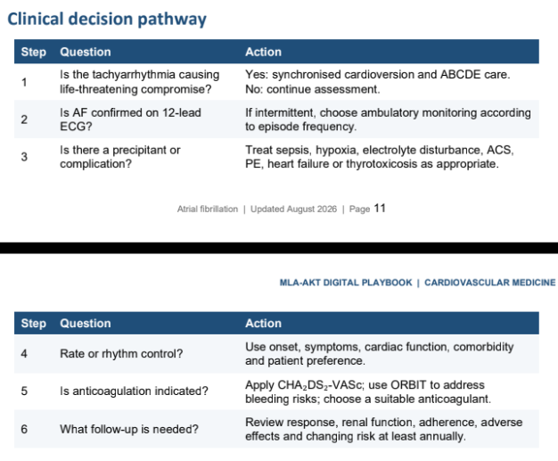
Summary of AF clinical decision pathway
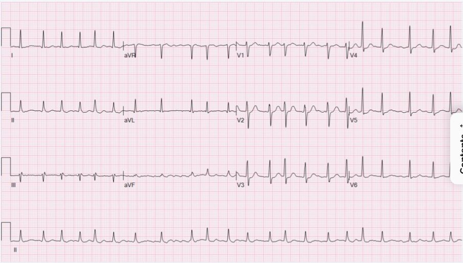
Features of AF on an ECG
Rate = Tachycardic
Rhythm = irregularly irregular
Axis = Typically normal, although there usually is no clear isoelectric baseline
Wave components:
P waves - absent. Instead there are chaotic fibrillatory waves.
QRS complexes - narrow (normal), variable R-R intervals.
Summary: The ECG shows atrial fibrillation with a rapid ventricular response rate of about 132 bpm with a normal axis and QRS duration. R-R intervals are variable, showing an irregularly irregular rhythm. There is normal R progression, but no evidence of discrete P waves either normal or ectopic excluding sinus / atrial tachycardia.
Risk factors for AF
Male sex
Caucasian ethnicity
Increasing age
Alcohol
Cigarette smoking
Obesity
Co-morbidities (e.g. chronic kidney disease and obstructive sleep apnoea)
AF diagnostic criteria
A standard 12-lead ECG recording or a single-lead ECG recording of ≥30 seconds showing a heart rhythm of no discernible repeating P-waves AND
Irregular RR intervals
Possible differential diagnoses in the context of suspected AF include:
Other supraventricular tachycardias: atrial flutter, atrial extrasystoles, multifocal atrial tachycardia, sinus tachycardia
Ventricular ectopics