15. Protozooses of GIT (giardiosis, cryptosporidiosis, isosporosis, sarcocystosis, toxoplasmosis, neosporosis)
1/53
There's no tags or description
Looks like no tags are added yet.
Name | Mastery | Learn | Test | Matching | Spaced | Call with Kai |
|---|
No analytics yet
Send a link to your students to track their progress
54 Terms
What are protozoan parasitic diseases of the GIT?
Giardiosis
Cryptosporidiosis
Cystoisosporosis
Sarcocystosis
Toxoplamosis
Neosporosis
What are the zoonotic protozoan parasites that affect the GIT?
Giardia duodenalis, Giardia enterica
Cryptosporidium parvum
Toxoplasma gondii
What is the species of Giardia affecting mammals?
Giardia duodenalis (Assemblages A-H)
What are examples of Giardia assemblages affecting dogs and cats?
Giardia dudodenalis (Assemblage A)- mammals, dog, cat, humans - Zoonotic
Giardia enterica (Assemblage B) – dog, cat, humans – Zoonotic
Giardia canis (Assemblage C/D)
Giardia cati (Assemblage F)
Where does Giardia spp. locate in the host?
Small intestine (duodenum & jejunum)
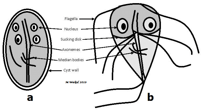
What are the two stages of Giardia spp.?
Trophozoite stage: Replicative stage. Pear-shaped. Adhesion disk in anterior part. 2 nuclei & 4 pairs (8) of flagella.
Cyst stage: Infective stage. consist of 2 trophozoites, with 4 nuclei.
How is Giardia spp. transmitted?
Faecal-oral route, waterborne, foodborne
What is the pathogenesis of Giardia spp.?
Villous atrophy/destruction
Carpet effect (malabsorption; do not destroy villi, just cover them)
What is the life cycle of GIardia?
Ingestion of cysts → go to small intestine → excystation → release of trophozoites which adhere to mucosal surface of intestine (CS) → multiply & form cysts w/ 4 nuclei → cysts are excreted into faeces
What are the clinical signs of Giardiosis?
Asymptomatic in healthy animals.
Acute giardiasis: Diarrhoea (soft to watery), flatulence, anorexia, nausea, vomiting. 1-2 weeks.
Chronic giardiasis: catarrhal diarrhoea, malabsorption, steatorrhea & foul smell (maldigestion).
How is Giardiosis diagnosed?
Flotation method (zinc sulphate/faust solution)
Snap test, ELISA (coproantigen) - faeces must be room temperature
Nested PCR
What is the treatment for Giardiosis?
Metronidazole for 5-10 days
What are species of Cryptosporidium?
C. parvum – dog, human Zoonotic
C. canis – canids
C. meleagridis – dog
C. muris - cat
C. felis - cat, dog
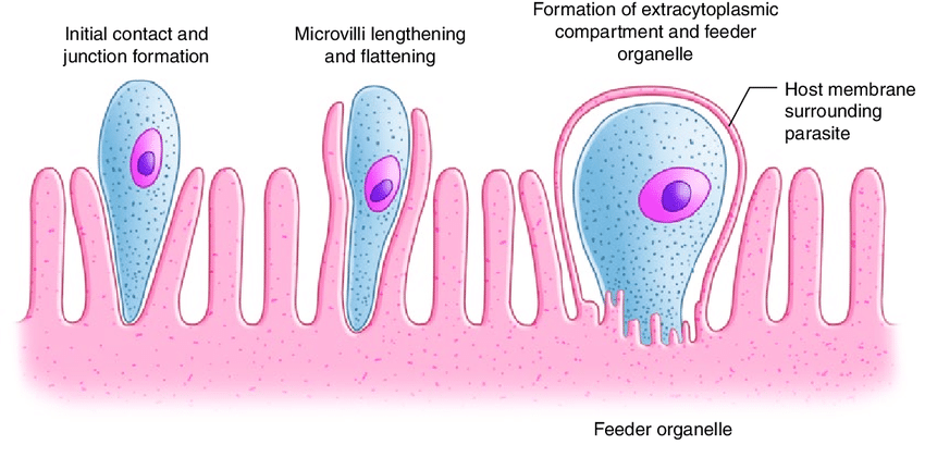
Where does Cryptosporidium spp. locate in the host?
Microvilli cells in SI or epithelial cells of respiratory tract. Intracellular, extracytoplasmatic
What is the infective stage of Cryptosporidium spp.?
Oocyst (5-8 um) with 4 free sporozoites
How is Cryptosporidium spp. transmitted?
Faecal-oral route, ingestion of contaminated food/water
Autoinfection!
What is the life cycle of Cryptosporidium?
Endogenous merogony, gametogony & sporogony
What are the clinical signs of Cryptosporidiosis?
Usually asymptomatic infections. Very young/old or immunosuppressed: necrosis & atrophy of villi → long-lasting, pale, watery-mucoid-diarrhoea.
Puppies: asymptomatic but during parvovirosis → Haemorrhagic diarrhoea
Kittens: asymptomatic period followed by acute diarrhoea with blood
How is Cryptosporidiosis diagnosed?
Direct faecal smear (Kinyoun or Ziehl-Nielsen staining), ELISA, PCR
What is the treatment for Cryptosporidiosis?
No anticrypto meds, antibiotics (nitazoxanide, tylosin, paromomycin, azithromycin)
What are examples of species of Cystoisospora affecting dogs and cats?
Cat: Cystoisospora felis (non-pathogenic - large), C. revolta (pathogenic - small) - small intestine
Dog: Cystoisospora canis (non-pathogenic - large) - small intestine, C. ohioensis complex (pathogenic - small) - SI, caecum, colon
What size of Cystoisospora are more pathogenic?
Small
What is the morphology of Cystoisospora spp. oocysts?
Oocyst with 2 sporocysts with 4 sporozoites each
How is Cystoisospora spp. transmitted?
Faecal-oral route
What is the life cycle of Cystoisospora?
Direct or facultative indirect w/ PH (rodent, pig, sheep, cattle, rabbit). Exogenous sporulation!
Direct development: ingestion of sporulated oocyst intestine villi intracellular multiplication
Facultative indirect development: include PH. Ingestion of sporulated oocyst by PH sporozoites infect extraintestinal tissues where it persists intracellularly as dormozoites (sleeping stage). Remain infectious for 2 years - Can only continue developing after being ingested by FH
What are the clinical signs of Cystoisosporosis?
Asymptomatic infections, bloody diarrhoea, vomiting, anorexia, apathy, impaired growth, abdominal pain, fever, haemorrhagic enteritis
How is Cystoisosporosis diagnosed?
Flotation method with FAUST (sporulated and un-sporulated in fresh sample)
C. canis is very large (36 um)
What is the treatment for Cystoisosporosis?
Toltrazuril (baycox)
What is the morphology of Toxoplasma gondii oocysts?
Oocyst containing 2 sporocysts with 4 sporozoites each
Where does Toxoplasma gondii replicate in the host?
Intracellularly (nucleated cells)
How is Toxoplasma gondii transmitted?
Faecal-oral (oocyst), ingestion of infected IH (tissue cyst), transplacental (tachyzoites)
What type of life cycle does Toxoplasma gondii have?
Facultative heteroxenous
FH: Cats, felids
PH: Mammals, humans
What is the pathogenesis of Toxoplasma gondii infection?
Parasite proliferation causes focal necrosis, inflammatory lesions in many organs.
Cause disease when infected for the first time. Second time: production of antibodies from the first infection will block infection
What are the clinical signs of Toxoplasmosis in cats?
Asymptomatic infections, fever, effusion, reduced appetite, diarrhoea, lethargy, enteritis, lymphadenopathy, weight loss, ocular signs (uveitis), dyspnoea, apathy, pneumonia, hepatitis, encephalitis, nephritis (rare)
What are the clinical signs of toxoplasmosis in dogs?
Dogs: very rare but if then mimic neospora
Acute: ↑ temp, enlarged lymph nodes, nose & eye discharge, diarrhoea, weight loss, pneumonia, dyspnoea, tonsillitis
Chronic: Hepatitis, myocarditis, pneumonia, paresis, paralysis, tremor, apathy, ataxia (CNS disturbances)
How is Toxoplasmosis diagnosed?
Serology (ELISA, IFAT, Latex-agglutination, MAT)
High IgM: acute infection (look for this! increased level in ill cats)
High IgG: Chronic infection (not so helpful, will anyway find antibodies in 50% of healthy cats too)
Coprology: low importance: oocysts difficult to detect in faeces → only shed once
If ascites: puncture ⤍ smears - find free tachyzoites.
What is the treatment for Toxoplasmosis?
Clindamycin, sulfadiazine + trimethoprim, GCCS (for uveitis), symptomatic treatment
Treatment not effective against tissue cysts.
What are examples of species of Sarcocystis?
Sarcocystis bovicanis: IH – Cattle, FH – Canids
S. cruzi: IH – Cattle, FH – carnivores
S. tenella: IH – sheep, FH – carnivores
S. suicanis: IH – swine, FH – canids
What is the transmission route for Sarcocystis spp.?
Predatory-prey (IH ingest oocyst, FH ingest IH containing sarcocyst)
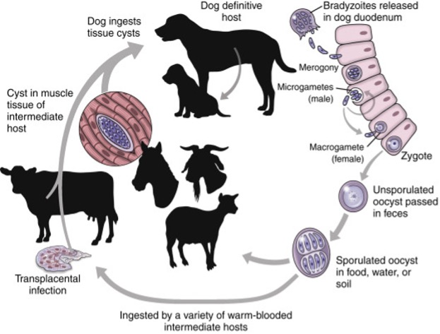
What is the life cycle of Sarcoystis?
In IH:
Merogony: IH ingest sporulated oocyst release sporozoite 1st generation: in arteries, 2nd generation: in endothelial layer of capillaries, 3rd generation: in lymphocytes in bloodstream Sarcocysts containing bradyzoites in muscle & nerve cells
In FH:
Gametogony: in intestine: bradyzoites micro/macrogametocytes fertilised zygote unsporulated oocyst
Sporulation: in intestine: oocyst sporulates: 2 sporocysts w/ 4 sporozoites each shell ruptures, sporocysts shed via faeces
Are Sarcocystis spp. pathogenic for carnivores?
Usually not, they are generally asymptomatic.
How is Sarcocystis diagnosed?
FH: flotation method with faust (only sporocysts seen as oocyst wall ruptures)
IH: serology, digestion method.
What is the treatment for Sarcocystis?
Potentiated sulfonamides for intestinal infection in dogs and cats.
No treatment for tissue cysts
What is the prevention against Sarcocystis infection?
Don’t feed raw meat unless previously frozen to deactivate bradyzoites
What are the hosts of Neospora caninum?
FH: Canids, dog - intestine
IH: Horse, cattle + canids! - skeletal muscle, CNS
Where does Neospora caninum form cysts in the host?
Muscle tissue and neural tissue.
What is the transmission route for Neospora caninum?
Faecal oral, ingestion of meat tissue cyst. Vertical: transplacental & lactogenic.
What type of life cycle does Neospora caninum have?
Indirect. Sporulation in the environment
What are the clinical signs of Neosporosis in puppies?
Neuromuscular signs (hind limb paresis, muscular atrophy, dysphagia, blindness), enteritis, dermatitis, bronchopneumonia
What are the clinical signs of Neosporosis in adults (> 6 months)?
Asymptomatic infection. Old dogs may develop encephalitis or encephalomyelitis. Abortion.
How is Neosporosis diagnosed?
Serology (ELISA, IFAT) GOLD STANDARD)
Coprology: flotation (very short release of oocysts; clinically manifested dogs do not release oocysts)
MRI: tissue cysts
What is the treatment for Neosporosis?
Clindamycin for 8 weeks (will not remove tissue cysts)
Symptomatic treatment: ATB, antiemetics, fluids
How can Sarococystis and Cystoisospora be differentiated in flotation?
Sarcocystis is sporulated, Cystoisospora is not
What is the main difference in clinical signs between giardiasis and cystoisosporosis?
Giardia: extracellular → diarrhoea
Cystoisospora: intracellular → haemorrhagic diarrhoea