Diagnostic Imaging Final Practice
 Call Kai
 Learn
Learn Practice Test
 Spaced Repetition
 Match
Match Flashcards
 Knowt Play
1/137

Earn XP
Description and Tags
Flashcards 1-98 were before the midterm. Flashcards 99- are after the midterm.
Name | Mastery | Learn | Test | Matching | Spaced | Call with Kai | Chat |
|---|
No analytics yet
Send a link to your students to track their progress
138 Terms
A veterinarian orders a diagnostic image for a patient. Which statement best describes what the image can contribute to the clinical picture?
A. It provides a definitive diagnosis that replaces the need for physical examination.
B. It reveals internal structure at the time of the study, which must be interpreted alongside history and physical examination findings.
C. It captures all disease processes occurring in the patient, including biochemical abnormalities.
D. It is most useful when ordered before a physical examination to avoid anchoring bias.
Answer: B
Imaging reveals internal structure at the time of the study and must be interpreted alongside the patient's history and physical examination findings.
Which of the following is the most accurate description of functional imaging?
A. It produces cross-sectional images of soft tissue anatomy with higher resolution than radiography.
B. It uses sound waves to evaluate the movement of internal organs in real time.
C. It produces images based on physiologic activity within tissues rather than their physical appearance.
D. It is used exclusively for evaluating the musculoskeletal system.
Answer: C
Functional imaging produces images based on physiologic or metabolic activity rather than physical anatomy.
Nuclear scintigraphy is classified as a structural imaging modality because it produces detailed images of organ anatomy.
A. True
B. False
Answer: B (False)
Nuclear scintigraphy is a functional imaging modality that maps metabolic activity through radiotracer distribution.
Which statement best describes the principle of stepwise imaging?
A. Always begin with the most advanced modality available to avoid missing findings.
B. Begin with the least invasive, most accessible modality that can answer the immediate clinical question, and escalate only when indicated.
C. Order all available modalities simultaneously to ensure complete information.
D. Select imaging based on owner financial preference before considering clinical need.
Answer: B
Stepwise imaging begins with the least invasive and most accessible modality capable of answering the clinical question and escalates only when needed.
Which of the following clinical questions is one that imaging CAN answer?
A. What is the patient's level of pain?
B. What did the owner observe at home before the visit?
C. Is there a change in the size or position of an internal organ at the time of the study?
D. What is the concentration of a circulating enzyme in the patient's blood?
Answer: C
Imaging can identify changes in organ size, position, and structure but cannot measure pain, owner observations, or biochemical values.
Which statement correctly describes the primary type of information that diagnostic imaging provides - information that cannot be obtained through physical examination or history alone?
a. Imaging provides information about what the owner observed at home before the patient arrived at the clinic, allowing the veterinarian to verify the accuracy of the history.
b. Imaging provides provides information about the patient's response to previous treatments by measuring changes in blood analyte concentrations during the imaging procedure.
c. Imaging provides information about the patient's pain level by measuring nerve conduction velocity in real time during image acquisition.
d. Imaging reveals the internal physical structure of a living patient - the anatomy, size, and condition of organs, bones, and other structures beneath the skin surface - without requiring surgery or autopsy.
d. Correct.
This is the foundational role of diagnostic imaging: to make the interior of a living patient visible without invasive exploration. Before imaging existed, internal pathology could only be confirmed by surgery or post-mortem examination. Imaging allows clinicians to visualize fractures, masses, fluid accumulations, organ enlargement, and other internal findings in the living patient, informing clinical decisions without the risk and cost of exploratory surgery.
Which statement correctly distinguishes structural imaging from functional imaging - and correctly identifies what type of clinical question each is best suited to answer?
a. Structural imaging shows the anatomy of tissues — their size, shape, location, and physical condition. Functional imaging shows the physiologic activity of tissues — what they are doing metabolically. Structural imaging answers "what does it look like?" and functional imaging answers "what is it doing?"
b. Structural imaging is used only for bone and functional imaging is used only for soft tissue — selecting the correct type requires knowing whether the affected tissue is mineralized or not.
c. Functional imaging is more advanced than structural imaging and provides more complete diagnostic information in all clinical situations — structural imaging is retained only for practices that cannot afford functional imaging equipment.
d. The distinction between structural and functional imaging refers to whether the imaging equipment is built into the clinic (structural) or requires referral to a specialist centre (functional).
a. Correct.
This is the fundamental distinction that drives modality selection throughout veterinary imaging. Structural modalities (radiography, CT, MRI, ultrasound) produce images based on physical properties of tissue and reveal anatomy. Functional modalities (nuclear scintigraphy) produce images based on physiologic processes (radiotracer distribution based on metabolic activity) and reveal what tissues are doing. A patient with spinal cord compression has a structural problem — structural imaging answers it. A patient with suspected thyroid overactivity has a functional problem — functional imaging answers it.
Which statement correctly describes how diagnostic imaging fits into the overall diagnostic reasoning process?
a. Imaging findings alone are sufficient to establish a definitive diagnosis in most veterinary patients — additional clinical information (history, examination, laboratory tests) is only needed when imaging is technically inadequate.
b. Imaging should always be performed before physical examination and history imaging findings will guide which examination and history questions are relevant.
c. Imaging is one component of diagnostic reasoning — imaging findings must be interpreted in the context of the clinical history, physical examination findings, and other diagnostic results to contribute meaningfully to diagnosis and clinical decision-making.
d. Imaging replaces physical examination in modern veterinary practice because imaging provides more accurate and complete information than what can be detected through palpation, auscultation, and observation.
c. Correct.
Diagnostic imaging provides specific anatomical or physiological information about the patient. That information only becomes diagnostically useful when interpreted alongside the clinical context. A lytic bone lesion in an 8-year-old Labrador with acute lameness has different implications than the same radiographic finding in a 6-month-old dog. The imaging finding is the same; the clinical context changes the diagnosis, urgency, and management entirely. This is why veterinary radiologists provide reports that integrate findings with the clinical question — and why imaging is ordered with a specific clinical question in mind.
Which statement correctly explains why practical factors — invasiveness, cost, and availability — are legitimate considerations in imaging decisions — even when a more advanced modality would provide more complete information?
a. Practical factors are not legitimate clinical considerations — imaging decisions should be based on which modality provides the most complete information, regardless of cost, invasiveness, or availability.
b. Practical factors are legitimate considerations because imaging decisions affect the patient's safety, the owner's ability to pursue care, and the timeliness of diagnosis — the goal is to obtain the information needed to answer the clinical question with the least burden to the patient and owner, not to obtain the most complete information possible regardless of context.
c. Practical factors are only relevant for large animal patients, where cost and logistics are different from small animal practice. In small animal practice, the most advanced available modality should always be selected.
d. Practical factors are relevant only when the patient is stable — in emergency situations, the most advanced imaging should always be obtained immediately regardless of cost or invasiveness.
b. Correct.
Clinical decision-making in veterinary practice requires balancing diagnostic value against patient safety, owner resources, and clinical urgency. Invasiveness is a patient safety consideration: general anesthesia carries real risk, particularly in compromised patients. Cost is an owner access consideration: a study the owner cannot afford does not benefit the patient. Availability and timeliness affect treatment: a diagnosis delayed by referral may delay critical treatment. These are not excuses for providing inferior care — they are the practical dimensions of providing the best care possible within real-world constraints.
Which statement correctly explains which clinical question imaging can answer directly — and which it cannot — and correctly identifies why?
a. Imaging can answer both questions — radiographs of the radius will show whether a fracture is present and will also reveal behavioral history if the owner has not reported it accurately.
b. Imaging cannot answer either question — a definitive diagnosis requires laboratory testing, and radiographs are only useful after surgery has already been performed.
c. Imaging can directly answer Question 1 — a radiograph reveals the internal structure of the radius and can show whether a fracture or bone lesion is present. Imaging cannot answer Question 2 — behavioral history requires direct questioning of the owner and is not visible on any imaging study.
d. Imaging can answer Question 2 — nuclear scintigraphy can detect behavioral changes because it measures metabolic activity in the brain during the examination. Imaging cannot answer Question 1 because radiographs only show soft tissue, not bone.
c. Correct.
This is the fundamental role of diagnostic imaging: revealing information about the internal physical state of a patient that cannot be obtained by external examination alone. A radiograph passes X-rays through the femur and records differential attenuation, allowing visualization of bone structure, cortical integrity, and any abnormality within the bone. The distal femoral lesion visible on the radiograph cannot be detected by palpation alone in a painful, swollen limb. Question 2 (behavioral history) requires the owner’s account — no imaging modality can reveal what an owner observed at home before the visit.
Which explanation best justifies why the type of imaging information needed determines which modality is selected — and correctly identifies what each type of information reveals that the other cannot?
a. Structural imaging shows the anatomy of tissues — where they are, what they look like, whether they are displaced or compressed. Functional imaging shows the physiologic activity of tissues — what they are doing metabolically. For Barnaby’s problem (suspected physical compression), structural imaging is needed because the clinical question is about anatomy, not metabolic activity.
b. Both types of imaging provide identical information and the choice between them is based on cost and availability, not on what clinical question is being asked.
c. Functional imaging is always preferred over structural imaging because it provides more complete information about what is happening inside the body, making structural imaging obsolete in modern veterinary practice.
d. Structural imaging is only used for bone and functional imaging is only used for soft tissue — selecting the correct type requires knowing which tissue type is affected.
a. Correct.
This is the core distinction that drives modality selection throughout veterinary imaging. Structural imaging (radiography, CT, MRI, ultrasound) produces images based on physical properties of tissue — X-ray attenuation, acoustic impedance, hydrogen proton behavior — and shows anatomy: where structures are, what they look like, whether they are normal in size and position. Functional imaging (nuclear scintigraphy) produces images based on physiologic processes — radiotracer accumulation in metabolically active tissue — and shows what tissues are doing. For suspected spinal cord compression by disc material, the clinical question is anatomical: is the cord being physically compressed and where? This is a structural question requiring structural imaging. If the question were instead "is the affected cord segment metabolically suppressed?" that would be a functional question.
Which recommendation best reflects sound clinical reasoning about which imaging option to use first — and correctly identifies the factors that drive this decision?
a. The principle that "most complete information should always be obtained first" does not reflect sound clinical reasoning. Obtaining the most complete information first regardless of other factors ignores invasiveness (CT requires general anesthesia in cats), time (referral, travel, anesthesia recovery delays diagnosis and treatment), cost (substantially higher than in-clinic ultrasound), and clinical urgency (Mochi needs rapid assessment). For a stable patient where the immediate clinical question is confirming fluid and identifying a likely cause, ultrasound provides the specific information needed rapidly and non-invasively. CT may be appropriate subsequently if ultrasound identifies a complex lesion requiring detailed staging.
b. Perform ultrasound first to answer the immediate clinical question. If ultrasound identifies a finding that requires more detailed characterization, CT can be considered.
c. Neither option is appropriate — Mochi should have laboratory testing completed before any imaging is performed, because laboratory results will determine whether imaging is necessary.
d. Perform CT first because ultrasound requires significant operator skill and the findings are unreliable unless performed by a specialist.
b. Correct.
This recommendation applies the key clinical reasoning principle: match the imaging approach to the immediate clinical question and the patient’s clinical status, weighing information yield against invasiveness, time, and cost. Ultrasound answers the immediate question (confirm fluid, evaluate organs, guide sampling if needed) immediately, non-invasively, at lower cost, and without anesthesia risk. If ultrasound reveals a complex mass or finding that requires cross-sectional staging, CT is then specifically justified. This stepwise approach is more efficient, safer for the patient in the immediate term, and more cost-effective than proceeding directly to the most advanced available modality.
X-rays are produced when high-speed electrons strike the anode of an X-ray tube. Which statement correctly describes bremsstrahlung radiation?
A. It is produced when an electron ejects an inner-shell electron from a tungsten atom, releasing a photon at a specific energy.
B. It is produced when an electron is decelerated by the electric field of a tungsten nucleus, releasing its lost kinetic energy as an X-ray photon.
C. It produces photons at fixed, discrete energy levels specific to the target material.
D. It accounts for only a small proportion of total X-ray output from a diagnostic tube.
Answer: B
Bremsstrahlung ("braking radiation") occurs when electrons decelerate near tungsten nuclei and release their lost kinetic energy as X-ray photons. It produces a continuous spectrum of energies and accounts for most X-ray production.
A radiograph shows the lumbar spine of a dog. Dense cortical bone appears bright white, while the gas-filled colon appears nearly black. Which physical property is the primary reason for this difference in appearance?
A. Bone reflects X-rays back toward the tube while gas transmits them to the detector.
B. Bone has higher atomic number and density than gas, so it attenuates more X-rays and allows fewer photons to reach the detector.
C. Gas absorbs more X-rays than bone because it is less dense, producing fewer photons at the detector.
D. The kVp setting determines which tissues appear white and which appear black, independent of tissue composition.
Answer: B
Bone has a higher atomic number and density than gas, causing greater attenuation of X-rays. More attenuation results in a brighter (whiter) appearance on the radiograph.
On an abdominal radiograph of a cat, the liver and a large hepatic mass are both visible, but their shared border cannot be identified. Which of the following best explains this finding?
A. The radiograph was taken at too low a kVp, reducing contrast between the structures.
B. The liver and the mass are both soft tissue/fluid in composition and attenuate X-rays to a similar degree, so no contrast exists at their shared border.
C. The mass is located behind the liver on the lateral view and is hidden by superimposition.
D. Hepatic masses always appear as gas opacity and should be dark on the radiograph.
Answer: B
Soft tissue and fluid share the same opacity category and attenuate X-rays similarly. When structures of identical opacity are in contact, their border disappears.
A single lateral thoracic radiograph is sufficient to confirm the location of a pulmonary nodule because projection radiography captures structures at all depths simultaneously.
A. True
B. False
Answer: B (False)
A single projection cannot determine depth because all structures in the beam path are superimposed. A second orthogonal view is needed to accurately localize findings.
Which of the following most accurately describes the primary advantage of CT over radiography for evaluating a dog with a suspected nasal tumor?
A. CT uses a lower radiation dose than radiography, making it safer for repeated studies.
B. CT eliminates superimposition by acquiring cross-sectional data from multiple angles, allowing structures to be separated by their actual three-dimensional location.
C. CT provides higher spatial resolution than radiography for fine cortical bone detail.
D. CT does not require anesthesia in most veterinary patients, making it more practical than radiography.
Answer: B
CT reconstructs cross-sectional images from multiple projections, eliminating superimposition and allowing structures to be evaluated based on their true three-dimensional location.
On a CT image, which would have the lowest Hounsfield unit (HU) value?
A. Cortical bone
B. Soft tissue organ (e.g., liver)
C. Fat
D. Air
Answer: D
Air has the lowest Hounsfield unit value at approximately −1000 HU. Fat is around −80 to −100 HU, water is 0 HU, soft tissues are positive values, and cortical bone is approximately +1000 HU.
On a radiograph, which statement correctly describes the relationship between X-ray attenuation and image brightness
a. Structures that attenuate more X-rays appear darker on the image, because the detector records more radiation passing through them.
b. All structures appear the same brightness on a radiograph — differences in brightness only arise in differences in image processing after acquisition.
c. Structures that attenuate more X-rays appear brighter on the image, because fewer X-rays reach the detector — the detector records a lower radiation intensity at that location.
d. Structures that attenuate more X-rays appear brighter because the absorbed radiation is re-emitted toward the detector, increasing the signal at that location.
c. Correct.
Attenuation means absorption and scattering of X-rays before they reach the detector. A structure that attenuates more X-rays allows fewer photons through — the detector receives less radiation at that location and records a lower intensity value, which displays as a brighter (whiter) region on the image. This is why dense, high-atomic-number materials like bone and metal appear white: they absorb most of the beam, and very little reaches the detector beneath them.
Which list correctly names the five standard radiographic opacity categories in order from highest attenuation (brightest on the image) to lowest attenuation (darkest on the image)
a. Metal → bone/mineral → soft tissue/fluid → fat → gas/air.
b. Bone/mineral → metal → soft tissue/fluid → gas/air → fat.
c. Soft tissue/fluid → fat → bone/mineral → metal → gas/air..
d. Gas/air → fat → soft tissue/fluid → bone/mineral → metal.
a. Correct.
This is the standard five-category radiographic opacity hierarchy ordered from highest to lowest attenuation. Metal has the highest atomic number and density of any material routinely encountered — it attenuates almost all X-rays and appears uniformly bright white. Bone contains calcium mineral with high atomic number and density. Soft tissue and fluid are water-equivalent in composition and attenuate moderately. Fat has lower density than soft tissue and attenuates slightly less — it appears marginally darker. Gas and air have negligible density and attenuate essentially no X-rays — they appear dark to black.
A radiograph of the canine skull shows the naval cavity, teeth, mandible, and skull bones all appearing in the same image simultaneously. Which statement best explains why structures at many different depths all appear in the same 2D image?
a. The radiographic detector electronically separates structures by depth during image acquisition, then displays all depth layers simultaneously in a single composite image.
b. Each structure emits a characteristic signal based on its tissue type, which the detector records independently — multiple signals from different depths are then combined into a single image.
c. Multiple X-ray exposures are taken from slightly different angles and superimposed digitally, producing an image that shows all structures from all depths in a single view.
d. The X-ray beam travels in a straight line from the tube through the entire patient to the detector — every structure in the beam path attenuates the beam and contributes to the same 2D image, regardless of how it is deep it is within the patient.
d. Correct.
This is the fundamental principle of projection radiography. The beam is generated at the X-ray tube, travels in a straight line through the patient, and is attenuated by every structure it encounters along that path. Each structure contributes its attenuation to the cumulative signal recorded at the corresponding point on the detector. A structure at the surface of the patient and a structure deep within the patient both lie along the same beam path and both contribute to the same detector point — there is no mechanism to separate their contributions by depth. This is why all structures at all depths appear in the same 2D image.
On a feline abdominal radiograph, the liver, spleen, and kidneys are often difficult to distinguish from one another where their margins contact adjacent organs. Which explanation best accounts for this limitation?
a. The feline abdomen is too small for the X-ray beam to resolve individual organs — larger patients have better abdominal organ differentiation on radiographs because there is more space between structures.
b. Adjacent abdominal organs are composed of soft tissue with similar atomic composition and physical density — they attenuate X-rays nearly identically, so the radiographic contrast between them is inherently low regardless of technical quality.
c. Abdominal organs are always superimposed on each other in the lateral projection — obtaining a VD projection would eliminate the organ overlap and allow clear margin identification.
d. This limitation only occurs when the radiograph is underexposed — with optimal exposure technique, all abdominal organs can be clearly distinguished from each other on a radiograph.
b. Correct.
Radiographic contrast between two adjacent structures depends on the difference in their X-ray attenuation. The liver, spleen, kidneys, and intestinal walls are all soft tissue organs with similar water-equivalent atomic composition and physical density. Because their composition is similar, they attenuate X-rays by nearly identical amounts — and similar attenuation means similar brightness on the radiograph, which means low contrast at their shared margins. This is a physical limitation of the modality, not a technical failure. Organ margins become visible on abdominal radiographs only where an adjacent contrasting opacity (fat, gas) provides a natural interface.
Which statement correctly describes how CT produces a cross-sectional image — and identifies the key difference from standard projection radiography?
a. CT uses a stationary X-ray tube and a single exposure, but the detector is curved to capture depth information — this curvature allows structures at different depths to be recorded separately.
b. CT uses a higher-energy X-ray beam than standard radiography — the higher energy allows the beam to penetrate structures selectively by depth, recording each depth layer separately on the detector.
c. CT rotates the X-ray tube around the patient, acquiring hundreds of projections from different angles — these are mathematically reconstructed into cross-sectional slices in which each point represents the attenuation at a specific location in 3D space.
d. CT uses a stationary X-ray source and moves the patient rapidly through the beam — the movement of the patient creates the cross-sectional effect by ensuring the beam intersects the patient at different angles.
c. Correct.
This is the fundamental mechanism of CT image formation. The X-ray tube (and detector array) rotates around the patient, typically acquiring projections through 360 degrees. Each projection captures a 2D attenuation map from one angle. Mathematical reconstruction algorithms use the angular diversity of these projections to calculate the attenuation value at each specific point in the cross-sectional plane — separating structures by their actual 3D location rather than combining them into a single superimposed projection. The result is a cross-sectional image in which each pixel represents a defined spatial location in the patient.
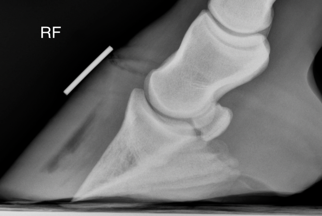
High Voltage undergoes radiography of the right front foot following a 3-day history of lameness.
The radiograph shows multiple structures with clearly different brightness levels.
The clinician reviewing the image identifies: the hoof wall and coronary band region (soft tissue), the coffin bone (dense cortical bone), a small gas pocket within the foot (laminar gas), periarticular soft tissue swelling, and the surrounding air outside the limb.
The student is asked to correctly rank these structures from most attenuating (brightest) to least attenuating (darkest).
Lateromedial projection of the right front foot.
Which ranking correctly orders these structures from highest X-ray attenuation (brightest on the image) to lowest X-ray attenuation (darkest on the image)?
a. Soft tissue → coffin bone → surrounding air → laminar gas — ordered by structural visibility, since the most clearly visible structures must be attenuating the most X-rays.
b. Coffin bone → laminar gas → soft tissue → surrounding air — bone is brightest, gas is next because it is a distinct structure, then soft tissue, then air.
c. Coffin bone → soft tissue (hoof wall/coronary band) → laminar gas → surrounding air — ordered from highest to lowest attenuation based on density and atomic composition.
d. Surrounding air → laminar gas → soft tissue → coffin bone — ordered from lowest to highest attenuation, since the image is read from dark to bright.
c. Correct.
Cortical bone has the highest density and calcium content, giving it the greatest attenuation — it appears brightest. Soft tissue structures (hoof wall, coronary band, digital cushion) have intermediate density and water-equivalent atomic composition — they attenuate moderately and appear gray. Gas (the laminar pocket) has negligible density and attenuates almost no X-rays — it appears very dark. Surrounding air behaves identically to gas — both have essentially zero attenuation and appear black. This ordering reflects the five-category opacity hierarchy: bone > soft tissue > fat > gas/air.
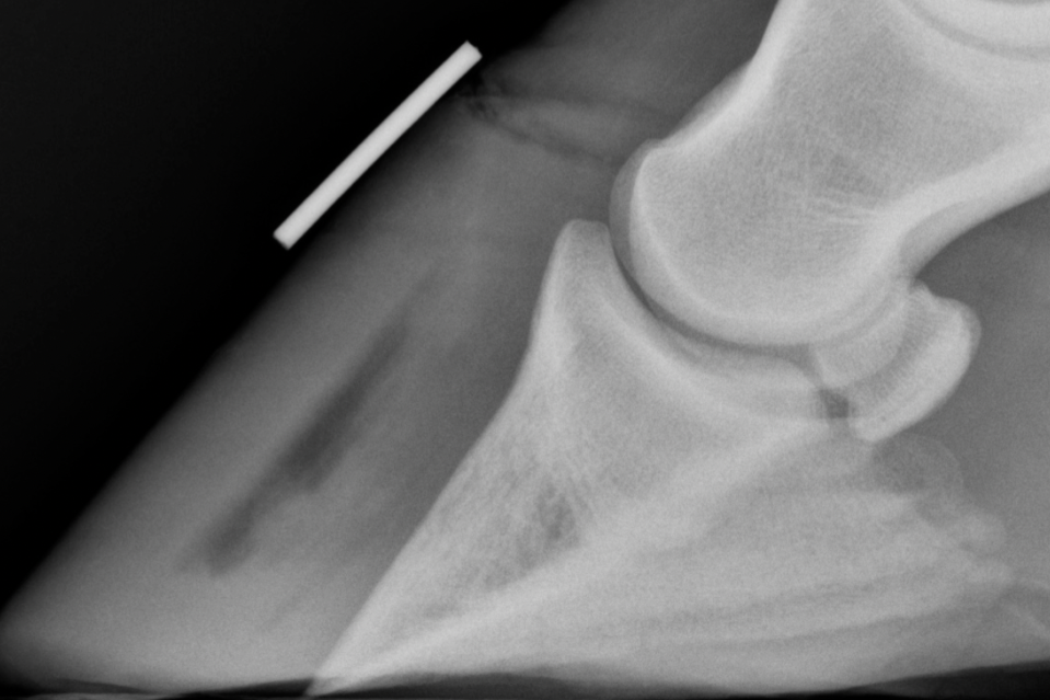
Same lateral equine distal foot radiograph. Close-up emphasis on the dorsal hoof wall region where the lamellar interface is visible. A broad, linear gas opacity is present at the dorsal lamellar zone between the hoof wall and coffin bone. Adjacent hoof wall soft tissue appears intermediate gray. Coffin bone appears bright cortical white adjacent to the gas crescent.
Which explanation correctly accounts for why the lamellar gas is visible on this radiograph AND how its opacity category confirms it is gas rather than fluid?
a. The gas pocket is visible because gas has negligible X-ray attenuation relative to the adjacent cortical bone and soft tissue — this large attenuation difference creates high local contrast, making the gas crescent clearly visible. Its very dark appearance (darker than all adjacent soft tissue) confirms it is gas rather than fluid, since fluid would attenuate similarly to the surrounding hoof wall soft tissue.
b. The gas pocket is visible because it is located at the lamellar interface, a clinically important region — its visibility reflects the clinical significance of the finding rather than a physical property of gas. Fluid would appear identically because both gas and fluid are non-mineralized.
c. The gas pocket is visible because it is denser than the surrounding soft tissue, causing it to attenuate more X-rays and appear distinctly bright against the adjacent structures. Its bright appearance distinguishes it from fluid, which would appear darker.
d. The gas pocket is visible because it has accumulated sufficient volume to be detectable by the radiographic detector — small volumes of gas would be invisible regardless of their attenuation properties. Fluid would appear similarly dark if the same volume were present.
a. Correct.
Radiographic visibility requires contrast — a sufficient difference in attenuation between a structure and its surroundings. Gas has essentially zero attenuation; adjacent cortical bone has very high attenuation; adjacent soft tissue has intermediate attenuation. This creates a large contrast differential that makes the gas crescent clearly visible against both neighbors. Crucially, the very dark appearance — darker than adjacent soft tissue — is what confirms gas rather than fluid. Fluid would attenuate similarly to soft tissue and would not create this distinctive dark crescent. This reasoning directly applies the opacity hierarchy to a clinical finding.
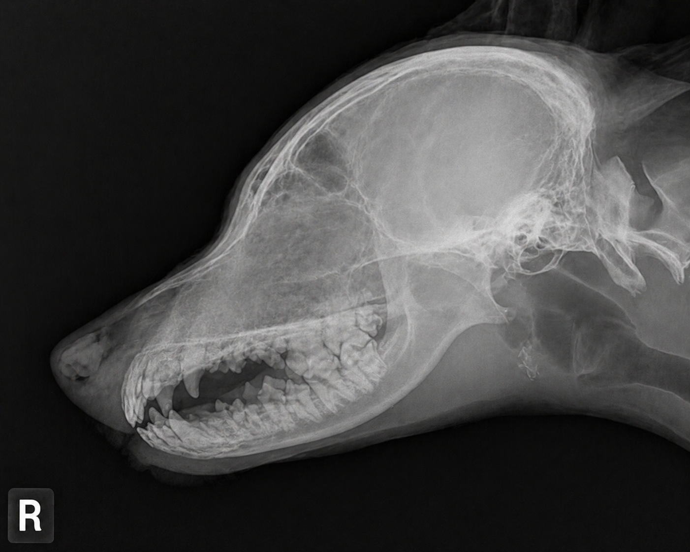
Barnaby presents for evaluation of chronic nasal discharge. A lateral skull radiograph is obtained.
Projection radiography works by passing the X-ray beam through the entire patient in a straight line — every structure in the beam path attenuates the beam and contributes to the same 2D image, regardless of depth. Structures at the skin surface and structures deep within the skull both appear in the same image plane.
On reviewing the radiograph, the clinician identifies a region of mixed opacity in the nasal cavity. A student says: "There's an opacity change in the nasal region — I think I can see the mass."
The supervising clinician asks: "Based on how projection imaging works, what exactly can you determine from this single lateral view — and what would you need to confirm your suspicion?"
Right lateral canine skull radiograph - Barnaby
Which statement best explains what this single lateral projection can and cannot determine about the nasal cavity finding — and what additional imaging would most directly address the limitation?
a. The lateral projection can confirm the presence, precise lateral location, and depth of any nasal mass — additional imaging is only needed to characterize tissue type, which requires MRI.
b. The lateral projection is non-diagnostic for nasal evaluation because superimposition makes all nasal findings invisible — the nasal cavity can only be imaged with CT, and additional radiographic projections would not add useful information.
c. The lateral projection determines both the presence and the side of the lesion, since left-sided lesions appear to the left of the midline and right-sided lesions appear to the right of the midline on the radiograph.
d. The lateral projection can detect that an opacity change is present in the nasal region but cannot determine its precise location within the nasal cavity, laterality, or depth — a VD or open-mouth projection, or CT, would be needed to separate superimposed structures and localize the finding.
d. Correct.
This is the fundamental clinical consequence of projection imaging: because the beam passes through every structure simultaneously, presence of an abnormality can often be detected but precise localization, laterality, and extent cannot be determined from a single projection — superimposed structures from the full depth of the nasal cavity are combined into one image. Additional orthogonal projections (VD, open-mouth) help by changing beam orientation, partially separating what was superimposed in the lateral view. CT eliminates the superimposition problem entirely through cross-sectional acquisition, making it the definitive modality for nasal cavity evaluation.
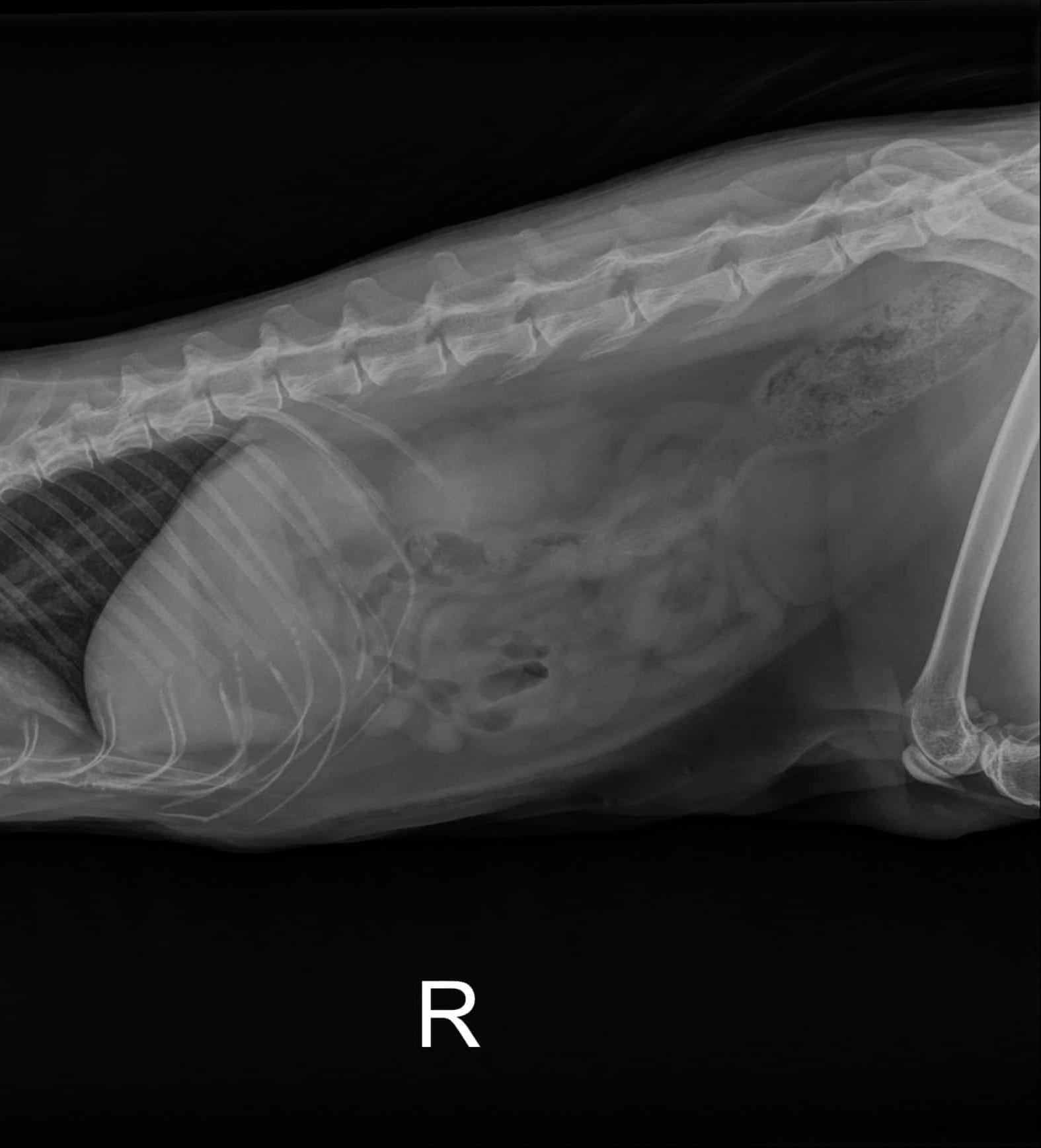
Mochi presents with 2 days of vomiting and reduced appetite. Abdominal palpation is uncomfortable.
A lateral abdominal radiograph is obtained.
On reviewing the image, the clinician observes: the liver, stomach, spleen, kidneys, and intestines are all visible as gray structures, but their margins blend into one another in many regions — it is difficult to identify where one organ ends and another begins.
A student says: "On the thoracic radiograph from Case 2 we could see bone and soft tissue and air separately — why can't we see the abdominal organs separately here?"
Lateral feline abdominal radiograph - adequate exposure, well defined abdominal serosal margin detail
Which explanation best accounts for why the abdominal organ margins are difficult to distinguish from each other — and what does this tell us about the appropriate use of radiography for abdominal soft tissue evaluation?
a. The abdominal organs are difficult to distinguish because the radiograph is underexposed — adequate exposure would restore contrast between organs. Radiography is fully capable of soft tissue organ differentiation when technical quality is optimized.
b. Adjacent abdominal soft tissue organs attenuate X-rays nearly identically because they have similar atomic composition and density — radiography cannot reliably distinguish between them where they are in direct contact, which is an inherent physical limitation of the modality for soft tissue evaluation.
c. The abdominal organs are indistinguishable because the feline abdomen is too small for the X-ray beam to resolve individual structures — larger patients like horses show better abdominal organ differentiation on radiographs.
d. The abdominal organs are indistinguishable because they are overlapping in a projection image — a VD projection would resolve the superimposition and allow each organ to be seen separately.
b. Correct.
This is the fundamental limitation of radiography for abdominal soft tissue evaluation. The five-opacity hierarchy from Case 2 shows that all soft tissue structures occupy the same category — they attenuate X-rays similarly because they have similar composition. Organ margins are visible on abdominal radiographs only where fat or gas provides an adjacent contrasting opacity (falciform fat adjacent to the liver, gas within intestinal loops). Where soft tissue organs contact each other directly, there is insufficient contrast to define the margin. This limitation is why ultrasound — which is highly sensitive to soft tissue interfaces — is the preferred initial modality for abdominal organ evaluation.
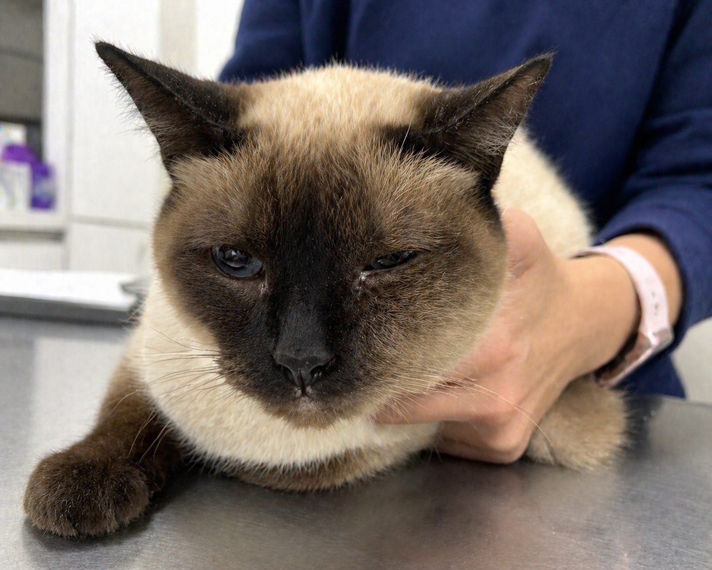
Mochi presents with a 3-week history of progressive left-sided facial swelling and reduced appetite.
Physical examination reveals firm, non-painful swelling of the left maxillary region. The left eye is mildly displaced dorsally.
The primary differential is a mass lesion — nasal or maxillary — potentially involving bone, soft tissue, or both.
Relevant context: CT eliminates superimposition by rotating the X-ray tube around the patient and reconstructing cross-sectional slices — separating structures by their actual 3D location. CT provides superior contrast resolution for soft tissue structures of similar density. Radiography provides higher spatial resolution for fine cortical bone detail, is faster, less expensive, and more widely available. CT delivers a higher radiation dose than a single radiographic projection.
The clinician must select the initial imaging modality.
Both radiography and CT are available at this practice.
Budget is a consideration — the owner has indicated cost is a factor in decision-making.
Mochi left facial eye swelling
Which recommendation best integrates the physical capabilities and practical limitations of both modalities — and provides the strongest reasoning for the recommended approach in this specific clinical scenario?
a. Begin with CT — the soft tissue mass and bone involvement require cross-sectional imaging and superior contrast resolution to characterize the lesion fully. CT provides definitive information in one study, justifying the additional cost over radiography.
b. Obtain radiography of the thorax rather than the skull — thoracic radiography should be performed first to evaluate for pulmonary metastases before local imaging is obtained, since treatment planning depends on metastatic status.
c. Begin with radiography — skull radiographs can assess gross bone involvement and confirm the presence of a mass lesion. If radiographs confirm the clinical suspicion and detailed characterization or surgical planning is needed, CT should be obtained. This stepwise approach respects the cost constraint while providing clinically useful initial information.
d. Obtain ultrasound of the maxillary swelling — ultrasound provides superior soft tissue contrast compared to radiography and would characterize the mass without ionizing radiation, making it the safest and most informative initial modality.
c. Correct.
Radiography is an appropriate initial step for a maxillary mass with suspected bone involvement. While superimposition limits fine detail, gross cortical bone destruction, periosteal reaction, and significant mass effect on adjacent structures are often detectable on skull radiographs. Confirming the presence of a lesion radiographically provides objective evidence to support the clinical decision and client communication about CT. If the clinical question requires precise extent, soft tissue characterization, or 3D reconstruction for surgical planning — all of which CT provides — that step is justified after radiographic confirmation. This stepwise approach is clinically sound and cost-respectful. It is important to note: if the clinical index of suspicion is very high and the owner is prepared for CT, proceeding directly to CT is also justifiable — the reasoning must match the clinical situation.
Which physical property of tissue determines the strength of an ultrasound echo at a tissue boundary?
A. The atomic number and density of each tissue — the same properties that determine X-ray attenuation.
B. The difference in acoustic impedance between the two tissues on either side of the boundary.
C. The hydrogen proton content of each tissue and the rate at which protons relax after stimulation.
D. The speed at which the ultrasound beam travels through each tissue in the direction of the transducer.
Answer: B
Ultrasound contrast is produced by differences in acoustic impedance at tissue interfaces. Larger impedance differences create stronger echoes and brighter interfaces.
The urinary bladder of a dog appears uniformly dark on ultrasound with no internal echoes. Which echogenicity term correctly describes this appearance, and what does it indicate about the fluid inside?
A. Hyperechoic — indicating strong reflection at the fluid-wall interface.
B. Hypoechoic — indicating that the fluid contains some suspended particles producing weak echoes.
C. Anechoic — indicating homogeneous fluid with minimal internal acoustic impedance variation.
D. Isoechoic — indicating that the fluid has the same echogenicity as surrounding soft tissue.
Answer: C
Anechoic structures appear completely black because they produce virtually no internal echoes. Simple urine is a classic example.
Ultrasound is the preferred modality for evaluating the brain and spinal cord in dogs because it does not use ionizing radiation and does not require general anesthesia.
A. True
B. False
Answer: B (False)
Ultrasound cannot effectively penetrate bone. The skull and vertebral column block the ultrasound beam, making MRI the preferred modality for neurologic imaging.
Which statement correctly explains why real-time imaging is a diagnostically significant feature of ultrasonography?
A. It allows the machine to average multiple frames, reducing image noise and improving resolution.
B. It enables continuous observation of dynamic processes — such as cardiac motion, peristalsis, and needle placement — that cannot be captured by a single static image.
C. It eliminates the need for patient preparation because motion artifacts are automatically corrected.
D. It means the entire examination can be completed faster than radiography, reducing patient stress.
Answer: B
Real-time imaging allows clinicians to observe movement and function as they occur, including cardiac activity, gastrointestinal motility, fetal movement, and procedural guidance.
MRI uses a magnetic field and radio waves to generate images based on the behavior of hydrogen protons in different tissues. Which statement best describes why different tissues appear differently on MRI?
A. Different tissues absorb X-rays at different rates, which is detected by the MRI scanner and converted into an image.
B. Different tissues contain different numbers and arrangements of hydrogen protons, which respond to the magnetic field differently and produce different signal intensities on the image.
C. The MRI scanner emits sound waves that bounce off tissue boundaries, and the returning echoes are converted into signal — denser tissues produce brighter images.
D. All soft tissues appear identical on MRI — contrast between structures is only possible when contrast agents are administered.
Answer: B
MRI contrast is generated by differences in hydrogen proton behavior within tissues. Variations in proton content and molecular environment create different signal intensities.
Cortical bone appears bright white on radiography, produces an acoustic shadow on ultrasound, and appears dark on MRI. Which statement best explains why the same tissue looks so different across these three modalities?
A. Each modality uses different software processing algorithms that apply different color maps to the same underlying data.
B. Each modality measures a different physical property of tissue — X-ray attenuation, acoustic impedance, and hydrogen proton relaxation — so the same tissue produces a different signal in each.
C. The appearance of bone varies because kVp, transducer frequency, and magnetic field strength are set differently for each modality.
D. Bone appears differently because its water content changes depending on which form of energy is used to image it.
Answer: B
Radiography measures X-ray attenuation, ultrasound measures acoustic impedance, and MRI measures hydrogen proton behavior. Because each modality evaluates a different physical property, the same tissue can appear very different.
Which set of definitions correctly describes the three standard ultrasound echogenicity terms?
a. Anechoic: produces many echoes, appearing bright white. Hyperechoic: produces moderate echoes, appearing gray. Hyperechoic: produces no echoes, appearing black.
b. Anechoic: appears uniformly gray with a speckled texture. Hyperechoic: appears brighter than surrounding tissue. Hypoechoic: appears darker than surrounding tissue.
c. Anechoic: without echoes, appearing uniformly dark — characteristic of simple fluid. Hyperechoic: producing strong echoes, appearing brighter than surrounding tissue. Hypoechoic: producing fewer echoes than surrounding tissue, appearing darker than the reference tissue.
d. Anechoic: appears dark due to high acoustic impedance preventing echo return. Hyperechoic: appears bright due to low acoustic impedance allowing maximum sound transmission. Hypoechoic: intermediate acoustic impedance producing moderate brightness.
c. Correct.
Anechoic (an- = without; -echoic = echoes) describes structures that produce minimal echo return and appear uniformly dark — simple fluid such as urine, bile, and effusions are the classic anechoic structures. Hyperechoic (hyper- = above) describes structures producing stronger echoes than the surrounding reference tissue, appearing brighter. Hypoechoic (hypo- = below) describes structures producing fewer echoes than surrounding tissue, appearing darker. These are relative terms — hyperechoic and hypoechoic describe brightness relative to adjacent tissue, not on an absolute scale.
When an ultrasound beam encounters a boundary between two tissues, what determines how much of the beam is reflected back toward the transducer?
a. The difference in acoustic impedance between the two tissues — a larger impedance difference produces stronger reflection and a brighter interface; similar acoustic impedance between adjacent tissues produces weak reflection and a poorly defined interface.
b. The thickness of the boundary layer between tissues — thicker boundaries reflect more sound energy and appear brighter, while thin boundaries allow more sound to pass through and appear darker.
c. The temperature difference between the two tissues — warmer tissues have higher molecular motion, producing more acoustic emissions and brighter interfaces on the ultrasound image.
d. The depth of the interface below the transducer — deeper interfaces reflect more sound energy because the beam has been focused and amplified by the time it reaches them, producing brighter appearances at greater depths.
a. Correct.
Acoustic impedance is a measure of resistance to sound wave propagation, determined by a material's density and compressibility. At a boundary between two materials, the proportion of the incoming sound beam that is reflected (rather than transmitted) depends directly on the difference in acoustic impedance between them. A large impedance difference (such as soft tissue meeting air) causes most of the beam to be reflected back — producing a strong echo and bright (hyperechoic) interface. A small impedance difference (such as fluid meeting adjacent soft tissue) causes little reflection — producing a weak echo and poorly defined interface on the image.
Why does bone create an acoustic shadow on ultrasound — and what does this mean for evaluating structures located behind bone?
a. Bone absorbs all incoming ultrasound energy completely, covering it to heat — no sound energy remains to reach deeper structures, so structures behind bone produce no signal.
b. Bone refracts ultrasound waves at a steep angle, scattering them in multiple directions and preventing them from reaching deeper structures in a coherent beam.
c. Bone generates its own acoustic signal that interferes with the reflected echoes from deeper structures, producing noise that obscures the underlying anatomy on the image
d. The very large acoustic impedance difference between soft tissue and cortical bone causes near-total reflection of the ultrasound beam at the bone surface — very little sound is transmitted through to deeper structures, producing a dark region (acoustic shadow) posterior to bone. Structures located behind bone cannot be evaluated by ultrasound.
d. Correct.
Cortical bone has an acoustic impedance far greater than that of soft tissue — among the largest impedance mismatches encountered in clinical ultrasound. At this interface, the reflection coefficient approaches 1.0, meaning nearly all incoming sound energy is reflected back toward the transducer rather than transmitted through the bone. The region deep to the bone surface receives essentially no sound energy and produces no echoes — appearing as a dark region (acoustic shadow) on the image. This is why ultrasound cannot evaluate the brain (behind the skull), the spinal cord (behind the vertebral column), or the interior of joints when overlying cortical bone blocks the beam path.
Which statement correctly describes the fundamental physical mechanism by which MRI produces images — and correctly identifies what determines signal intensity differences between tissues?
a. MRI uses a rotating X-ray beam to acquire multiple projections from different angles around the patient — mathematical reconstruction of these projections produces cross-sectional images in which signal intensity reflects X-ray attenuation differences between tissues.
b. MRI places the patient in a strong magnetic field, causing hydrogen protons in body tissues to align with the field. Radiofrequency pulses disturb this alignment; as protons relax back to equilibrium they emit signals that the scanner detects. Different tissues contain hydrogen in different molecular environments — fat, water, and fibrous tissue protons relax at different rates — producing different signal intensities and creating soft tissue contrast.
c. MRI uses high-frequency sound waves directed through the patient from multiple angles simultaneously — the pattern of sound wave reflections from tissue interfaces is mathematically reconstructed into cross-sectional images in which signal intensity reflects acoustic impedance differences.
d. MRI detects gamma radiation emitted by radioactive tracers that accumulate preferentially in metabolically active tissues — areas of higher metabolic activity produce stronger signals and appear brighter on the image.
b. Correct.
MRI signal is generated by the behavior of hydrogen protons (present in all water- and fat-containing tissues) in a magnetic field. The strong external field aligns protons; a radiofrequency pulse at the resonant (Larmor) frequency disturbs the alignment. As protons return to equilibrium (relax), they emit radiofrequency signals. The rate and character of relaxation differs between tissue types based on the molecular environment of their hydrogen protons: fat, free water (fluid), and bound protons (fibrous tissue, cortical bone) all relax differently. These differences produce the varied signal intensities that distinguish soft tissue structures from each other — the basis of MRI's superior soft tissue contrast resolution compared with radiography and CT.
Which statement best explains what ''real-time imaging'' means in the context of ultrasound — and identifies a clinical scenario where this capability provides information that a static imaging modality cannot?
a. Real-time imaging means the ultrasound machine processes images faster than other modalities — it produces the same static image as radiography but displays it immediately rather than after processing delay, making it more efficient in urgent situations.
b. Real-time imaging means the ultrasound can be performed while the patient is awake and moving — it captures the patient's natural movement rather than requiring the immobility needed for radiography and CT.
c. Real-time imaging means ultrasound continuously acquires and displays images during the examination — allowing the clinician to observe dynamic processes as they occur. A clinical example: repositioning a patient during bladder evaluation to distinguish mobile sediment (which shifts with gravity in real time) from fixed calculi (which remain positionally stable), providing information that a single static radiograph cannot.
d. Real-time imaging means ultrasound images are automatically interpreted by the machine as they are acquired — the system identifies abnormal findings in real time, reducing reliance on operator skill and making ultrasound more reproducible than other modalities.
c. Correct.
Real-time ultrasound produces a continuously updated image throughout the examination — the transducer continuously emits sound pulses and displays returning echoes as a live image rather than a single captured frame. This enables observation of motion, fluid flow, organ movement, and material redistribution as they occur. The clinical example illustrates this precisely: a static radiograph captures one moment and cannot show whether intraluminal material moves with patient repositioning. Real-time ultrasound allows the clinician to deliberately reposition the patient and watch whether echogenic material shifts (sediment, matrix) or remains fixed (calculi with acoustic shadowing) — a distinction with direct clinical implications for diagnosis and management.
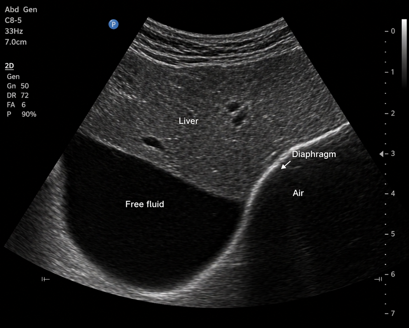
Barnaby presents with progressive abdominal distension over 5 days.
Background: ultrasound produces images by emitting sound waves from the transducer and recording the echoes that return. Structures that reflect many echoes appear bright (hyperechoic); structures that reflect few echoes appear dark (hypoechoic); structures that transmit sound with almost no reflection appear uniformly dark (anechoic).
An abdominal ultrasound is performed. The image shows three distinct regions:
• Region 1: a uniformly dark region between the liver and diaphragm with no internal architecture
• Region 2: the hepatic parenchyma showing a granular speckled gray texture
• Region 3: a bright linear boundary at the lung diaphragm interface
The clinician asks a student to correctly identify each region using standard echogenicity terminology.
Canine abdominal ultrasound:
Which statement correctly identifies all three regions using standard echogenicity terminology and explains the echo-return mechanism responsible for each?
a. Region 1 is hyperechoic (many echoes returning from dense fluid); Region 2 is anechoic (liver parenchyma produces no echoes); Region 3 is hypoechoic (the diaphragm partially reflects sound).
b. Region 1 is anechoic (free fluid transmits sound with minimal reflection, producing few returning echoes); Region 2 is hypoechoic to intermediate (liver parenchyma produces moderate echoes from tissue interfaces); Region 3 is hyperechoic (the diaphragm-lung interface causes near-total reflection of the sound beam).
c. Region 1 is hypoechoic (fluid partially absorbs sound waves, reducing but not eliminating echo return); Region 2 is hyperechoic (the dense liver tissue strongly reflects all incoming sound); Region 3 is anechoic (the diaphragm transmits sound into the air-filled lung without reflection).
d. Region 1 is anechoic (correct); Region 2 is hyperechoic (liver parenchyma is the brightest structure on abdominal ultrasound because it contains the most tissue); Region 3 is hypoechoic (diaphragm tissue is less reflective than liver).
b. Correct.
Region 1 is free fluid — homogeneous fluid has minimal acoustic impedance variation internally, so few echoes return to the transducer and the region appears uniformly dark (anechoic). Region 2 is liver parenchyma — the many microscopic tissue interfaces within the liver each produce small echoes, creating a granular speckled appearance at intermediate (hypoechoic to isoechoic) brightness. Region 3 is the diaphragm-air interface — the very large acoustic impedance difference between soft tissue and air causes near-total reflection of the sound beam back toward the transducer, producing a strongly hyperechoic (bright) line.
Mochi presents for evaluation of vomiting. An abdominal ultrasound is performed.
During the scan, the clinician notes a bright hyperechoic linear structure at the far boundary of the liver, separating the liver from the lung.
A student asks: "Why is that boundary so bright? The liver looks gray — why does the edge look so different?"
A second student adds: "And I noticed that when I moved the probe earlier, the fluid pocket between the liver and spleen had no visible boundary between the fluid and the liver — I could barely tell where one ended and the other began."
The clinician asks both students to explain both observations using acoustic impedance.
Which explanation best accounts for both observations — why the lung-diaphragm boundary is strongly hyperechoic AND why the boundary between fluid and liver parenchyma is poorly defined?
a. The lung-diaphragm boundary is bright because the diaphragm muscle is denser than the liver, causing more X-ray attenuation at that interface. The fluid-liver boundary is invisible because fluid is less dense than soft tissue and produces fewer X-ray photons.
b. The lung-diaphragm boundary is bright because the diaphragm actively emits sound waves back toward the transducer. The fluid-liver boundary is invisible because fluid absorbs all incoming sound energy before it reaches the liver surface.
c. Both observations result from differences in tissue temperature — the diaphragm is warmer than the liver, causing stronger sound wave emission at the interface, while fluid is cooler than liver tissue, reducing reflection.
d. The lung-diaphragm boundary is strongly hyperechoic because soft tissue and air have a very large acoustic impedance difference — nearly all incident sound is reflected back to the transducer at this interface. The fluid-liver boundary is poorly defined because fluid and liver parenchyma have similar acoustic impedance — little sound is reflected at their interface, producing a weak echo and indistinct boundary.
d. Correct.
Acoustic impedance is a measure of how much resistance a material offers to sound wave propagation, determined by its density and compressibility. When a sound wave encounters a boundary between two materials with different acoustic impedance, a portion of the wave is reflected back toward the transducer. The greater the impedance difference, the larger the proportion reflected. Soft tissue and air have a very large impedance mismatch — nearly all sound is reflected at this interface, producing the strongly hyperechoic diaphragm-air line. Fluid and liver parenchyma have similar acoustic impedance — little sound is reflected at their shared boundary, producing a weak echo and a poorly defined interface on the image.
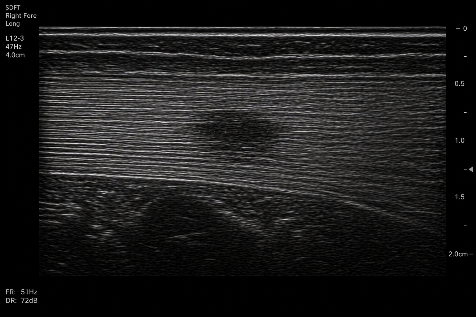
High Voltage presents with acute right forelimb lameness and marked swelling of the palmar metacarpal region following exercise. Palpation reveals heat and pain over the superficial digital flexor tendon (SDFT).
Context: bone creates acoustic shadowing on ultrasound — the large impedance mismatch at a bone surface causes near-total reflection of the sound beam, preventing visualization of structures deep to bone. This means ultrasound cannot evaluate structures behind cortical bone. Ultrasound can, however, evaluate soft tissue structures accessible from the skin surface with no intervening bone.
Ultrasound of the SDFT is performed using a linear high-frequency transducer in longitudinal orientation.
The image shows the SDFT as a parallel fibrillar echotexture (multiple bright parallel lines representing the organized collagen fiber bundles). A focal region of decreased echogenicity (hypoechoic) is visible within the tendon substance, disrupting the normal fibrillar pattern.
A colleague reviewing the images comments: "I see the lesion — but I notice the fiber alignment looks different in the right quadrant of the image. Is that part of the injury or something else?"
Ultrasound equine: Right forelimb, SDFT Longitudinal
Which explanation best accounts for the difference in echogenicity between the bright diaphragm–lung boundary and the comparatively weaker fluid–liver boundary on ultrasound?
a. Ultrasound is appropriate because the SDFT is a superficial soft tissue structure with organized fibrillar architecture — each collagen interface produces echoes detectable by the transducer. The altered fibrillar appearance in the right quadrant most likely represents anisotropy: a probe angle artifact that occurs when the beam strikes the tendon at a non-perpendicular angle, reducing echo return from the fibers and making them appear hypoechoic.
b. Ultrasound is appropriate because tendons contain a high proportion of water, making them anechoic on ultrasound and easy to identify. The altered appearance in the right quadrant represents hemorrhage within the tendon sheath, which appears hypoechoic because blood products reduce sound transmission.
c. Ultrasound is appropriate because MRI and radiography cannot image soft tissue structures at all. The altered appearance in the right quadrant represents a second separate tendon lesion that the referring veterinarian missed on physical examination.
d. The diaphragm–lung boundary is strongly hyperechoic because aerated lung and soft tissue have a large acoustic impedance mismatch, causing strong reflection of ultrasound waves. In contrast, the fluid–liver boundary produces weaker reflections because fluid and liver tissue have more similar acoustic impedances, resulting in a lower reflection coefficient and less echogenic interface.
CONFIRM IF A CORRECT
a. Correct.
The SDFT is an ideal ultrasound target: it is superficial (no intervening bone), consists of organized parallel collagen fibers (each fiber-to-fiber interface produces a small echo), and its fibrillar architecture produces a characteristic echo pattern that changes predictably with injury. Anisotropy is a well-recognized operator-dependent artifact in tendon ultrasound: when the transducer beam strikes the tendon at an angle rather than perpendicular, fewer echoes return to the transducer and the fibers appear artificially hypoechoic. Distinguishing anisotropy from true pathology requires the operator to adjust probe angulation — a skill that makes tendon ultrasound highly operator-dependent.
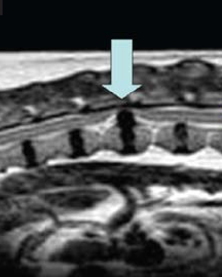
Barnaby develops progressive hindlimb weakness over 3 weeks. Neurological examination localizes the lesion to the thoracolumbar spinal cord.
Context: ultrasound cannot evaluate the spinal cord because the vertebral column creates acoustic shadowing — the large impedance mismatch at bone surfaces reflects sound before it reaches the cord.
MRI of the thoracolumbar spine is performed.
The MRI image shows clear differentiation between the spinal cord, intervertebral disc material, epidural fat, vertebral bone marrow, and surrounding paraspinal muscle — structures that would appear as nearly uniform gray on a radiograph because they all attenuate X-rays similarly.
A student asks: "How does MRI produce such clear contrast between these soft tissues when radiography cannot?"
Canine thoracolumbar spine MRI- Sagittal T2
Which explanation best accounts for why MRI produces clear contrast between these soft tissues — and correctly identifies the physical property that determines the signal intensity of each tissue?
a. MRI produces contrast by measuring differences in X-ray attenuation between tissues — soft tissues with higher water content absorb more X-rays and appear brighter, while tissues with lower water content appear darker.
b. MRI produces contrast by detecting sound wave reflections from tissue interfaces — tissues with more organized structure (like the spinal cord) reflect more sound energy and appear brighter, while disorganized tissues appear darker.
c. MRI places the patient in a strong magnetic field, causing hydrogen protons in body tissues to align with the field. A radiofrequency pulse disturbs this alignment; as protons relax back to equilibrium they emit signals. Different tissues have different hydrogen proton environments — fat, water, and bound protons all relax at different rates — producing different signal intensities and creating soft tissue contrast.
d. MRI produces contrast by detecting radioactive tracer uptake in different tissues — tissues with higher metabolic activity accumulate more tracer and appear brighter on MRI, while less active tissues appear darker.
c. Correct.
MRI signal arises from the behavior of hydrogen protons (present in all water- and fat-containing tissues) in a magnetic field. The strong external magnetic field causes protons to align. A radiofrequency pulse at the resonant frequency disturbs the alignment. As protons return to equilibrium (relax), they emit radiofrequency signals that the scanner detects. The rate and character of relaxation — described by T1 and T2 relaxation times — differs between tissue types based on their molecular environment. Fat protons relax differently from water protons; tightly bound protons in fibrous tissue relax differently from free water in fluid. These differences in relaxation produce the varied signal intensities that create soft tissue contrast on MRI.
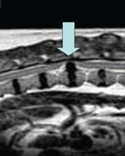
The MRI has confirmed intervertebral disc herniation at T12-T13 with spinal cord compression. The disc material, cord, and epidural fat are all clearly visualized and distinguished from each other.
The surgical team asks: "Could we have answered the same clinical question with radiography or ultrasound — and if not, why not?"
The student summarizes the modality limitations established in this session:
• Radiography: limited soft tissue contrast between cord, disc, and adjacent tissues (all attenuate X-rays similarly)
• Ultrasound: acoustic shadowing from the vertebral column prevents sound waves from reaching the spinal cord
The team asks the student to explain exactly what MRI provides that these modalities cannot — and in which specific clinical contexts this advantage is most important.
Canine thoracolumbar spinal MRI
Which statement best explains what MRI provides that radiography and ultrasound cannot for this case — and correctly identifies the clinical contexts where this advantage is most important?
a. MRI provides faster image acquisition than radiography and ultrasound, making it the preferred modality for any emergency evaluation where speed is critical.
b. MRI provides superior soft tissue contrast resolution — it distinguishes soft tissue structures with similar physical composition (spinal cord, disc material, epidural fat) that appear identical on radiography and are inaccessible to ultrasound due to acoustic shadowing. This advantage is most important for neurologic disease (brain and spinal cord) and musculoskeletal joint disease (ligaments, cartilage, menisci) where the clinical question requires distinguishing between adjacent soft tissue structures.
c. MRI provides better bone detail than radiography because the magnetic field interacts more strongly with the calcium mineral in bone, producing higher signal intensity and clearer cortical margins.
d. MRI eliminates the need for anesthesia or sedation compared with radiography and ultrasound, making it safer for compromised patients who cannot tolerate the positioning required for other modalities.
b. Correct.
MRI's primary advantage is contrast resolution for soft tissues with similar X-ray attenuation — structures that appear as undifferentiated gray on radiography are clearly distinguished on MRI based on their different hydrogen proton relaxation properties. This advantage is most clinically important for two categories: (1) neurologic disease, where the spinal cord, brain, and surrounding structures must be differentiated from each other and from compressive lesions, and (2) musculoskeletal joint disease, where ligaments, cartilage, and menisci must be evaluated for integrity — all structures inaccessible to radiography due to similar attenuation and inaccessible to ultrasound due to overlying bone.
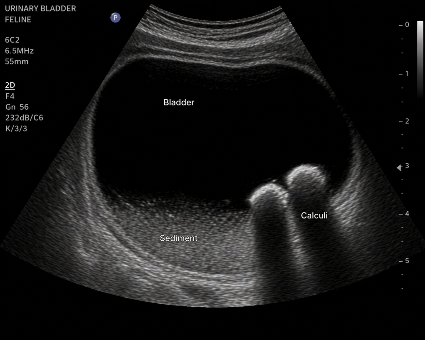
Mochi presents for evaluation of vomiting. An abdominal ultrasound is performed.
During the scan, the clinician notes a bright hyperechoic linear structure at the far margin of the liver, corresponding to the diaphragm and adjacent lung.
A student asks, “Why is that boundary so bright? The liver parenchyma looks relatively gray—why does that interface stand out so much?”
As the examination continues, a small pocket of free fluid is identified adjacent to the liver. Another student observes, “I can see the liver margin outlined by the fluid, but the fluid–liver boundary is much less bright than the diaphragm–lung boundary. Why is that?”
The clinician asks both students to explain the difference using principles of acoustic impedance and ultrasound reflection.
Feline urinary bladder ultrasound - Mochi
Which explanation best justifies why ultrasound is the most appropriate initial modality for this clinical question — and identifies the specific capability that makes it superior to both radiography and MRI in this scenario?
a. Ultrasound is appropriate because MRI cannot image the urinary bladder — the magnetic field is incompatible with the fluid-filled structure. Ultrasound is the only modality that can evaluate hollow viscera.
b. Ultrasound is appropriate because it uses no ionizing radiation, making it the safest modality for any patient. Radiography and MRI both use ionizing radiation and should be avoided in cats whenever possible.
c. Ultrasound is appropriate because feline urinary calculi are always radiolucent on radiography and therefore invisible, making radiography non-diagnostic for urinary obstruction in cats.
d. Ultrasound is the appropriate initial modality because it provides real-time imaging — allowing direct observation of intraluminal material behavior as the patient is repositioned. This distinguishes mobile sediment and matrix (which shift with gravity) from fixed calculi (which remain stable), and allows simultaneous assessment of bladder wall thickness and ureteral jet function. Radiography cannot assess material mobility or bladder wall character in real time; MRI is impractical as an initial modality due to anesthesia requirements and acquisition time.
d. Correct.
The clinical question — what is the character and mobility of intraluminal bladder content — is specifically answered by real-time ultrasound. The ability to reposition the patient during imaging and observe whether material moves (sediment, matrix) or remains fixed (calculi with acoustic shadowing) is a capability unique to real-time imaging. Radiography provides a static image and cannot assess mobility or distinguish non-shadowing material. MRI can characterize the bladder but is impractical as an initial modality in a potentially obstructed cat requiring prompt clinical decision-making. Ultrasound is also the modality that simultaneously allows bladder wall assessment and evaluation of ureteral jets — additional clinically relevant information available in the same examination.
A radiotracer is administered to a horse for a bone scintigraphy study. Which statement best describes how the image is formed?
A. The gamma camera emits gamma rays that penetrate the horse's tissues and are reflected back from areas of bone remodeling.
B. The radiotracer distributes based on osteoblastic activity, emits gamma radiation as it decays, and the gamma camera detects that emitted radiation to map tracer distribution.
C. The tracer accumulates in areas of low bone density and emits X-rays that are detected by a standard radiographic plate.
D. The gamma camera rotates around the horse and reconstructs a three-dimensional cross-sectional image from multiple projections, similar to CT.
Answer: B
In nuclear scintigraphy, the radiotracer is administered to the patient and distributes based on physiologic activity. As the isotope decays, it emits gamma radiation that is detected by the gamma camera, creating a map of tracer distribution. The camera detects radiation; it does not emit it.
A hyperthyroid cat undergoes thyroid scintigraphy. The left thyroid lobe shows markedly increased tracer uptake, while the right lobe shows no detectable uptake. Which explanation best accounts for the absent uptake in the right lobe?
A. The right lobe has been destroyed by the disease process and no longer contains follicular cells.
B. The right lobe is normal but shows physiologic suppression — excess hormone from the left lobe has suppressed TSH via negative feedback, reducing follicular cell activity in the right lobe below detectable levels.
C. The collimator blocked gamma photons from the right side of the neck, producing an artifactual cold spot.
D. The tracer was preferentially taken up by salivary glands on the right side, diverting it away from the right thyroid lobe.
Answer: B
Excess thyroid hormone from the hyperfunctional lobe suppresses TSH secretion through negative feedback. Without TSH stimulation, the normal contralateral thyroid lobe becomes metabolically inactive and may not demonstrate detectable tracer uptake.
Nuclear scintigraphy and fluoroscopy are both classified as functional imaging modalities because they both use ionizing radiation.
A. True
B. False
Answer: B (False)
Functional versus structural imaging is determined by the information provided, not the type of radiation used. Scintigraphy maps physiologic activity, while fluoroscopy evaluates dynamic motion. Both happen to use ionizing radiation, but that does not define their classification.
A dog presents with suspected esophageal motility disorder. The clinician considers a static barium radiograph versus a fluoroscopic barium study. Which statement best justifies choosing fluoroscopy for this clinical question?
A. Fluoroscopy uses less radiation than a static barium radiograph, making it safer for repeat evaluation.
B. A static radiograph can only show barium position at one moment; fluoroscopy captures peristaltic motion, transit, and dynamic events such as reflux that cannot be detected in a single frame.
C. Fluoroscopy provides higher spatial resolution than static radiography, allowing better mucosal detail.
D. Static barium radiographs cannot be performed on dogs — barium is only approved for use with fluoroscopic guidance.
Answer: B
Motility disorders involve movement over time. Fluoroscopy allows real-time visualization of peristalsis, bolus transit, and reflux, making it the preferred modality for evaluating esophageal function.
A cat presents with recurrent lower urinary tract signs. Plain abdominal radiographs show no definitive abnormality of the bladder. Which contrast study would best evaluate the bladder wall and detect small intraluminal masses?
A. Positive contrast cystography alone — iodinated contrast fills the lumen and outlines any defects.
B. Negative contrast cystography (pneumocystography) alone — air in the lumen creates the best mucosal detail.
C. Double-contrast cystography — a small volume of positive contrast coats the mucosal surface while gas distends the lumen, providing optimal wall and intraluminal detail.
D. Intravenous urography — iodinated contrast is filtered by the kidneys and outlines the bladder from within.
Answer: C
Double-contrast cystography combines positive contrast coating of the bladder wall with gas distention of the lumen, making small masses and mucosal irregularities easier to detect.
A clinician needs to determine whether a bone lesion in a horse is associated with active bone remodeling before any changes are visible on radiographs. Which modality directly answers this question, and why?
A. CT — because it provides cross-sectional images that eliminate superimposition and detect subtle density changes in bone.
B. MRI — because it visualizes bone marrow signal and can detect early edema before cortical changes appear.
C. Bone scintigraphy — because it maps osteoblastic activity via radiotracer uptake, detecting metabolic changes before structural changes are radiographically visible.
D. High-detail radiography — because reducing kVp increases contrast resolution and reveals early cortical changes not visible on standard technique.
Answer: C
Bone scintigraphy detects osteoblastic activity directly through radiotracer uptake. Because metabolic changes occur before structural changes, scintigraphy can identify active remodeling earlier than conventional imaging modalities.
Which statement correctly describes how a radiotracer produces a nuclear medicine image?
a. A radiotracer is injected intravenously and emits X-rays as it circulates — the X-ray detector records differential attenuation through tissues containing different concentrations of tracer, producing an image of tracer distribution.
b. A radiotracer is administered to the patient and distributes based on physiologic processes — osteoblastic activity, hormonal uptake, blood flow. The tracer emits gamma radiation as it decays, which is detected externally by a gamma camera to produce an image mapping tracer distribution.
c. A radiotracer is administered topically to the skin surface and absorbed into underlying tissue — the gamma camera detects sound waves emitted by the tracer as it interacts with tissue proteins, producing an image of tissue composition.
d. A radiotracer accumulates uniformly throughout the body and emits a constant background radiation — the gamma camera subtracts areas of normal background from areas of elevated signal to produce a map of abnormal tissue.
b. Correct.
Radiotracers are biologically active molecules labeled with a radioactive isotope (such as technetium-99m). They distribute within the body based on the physiologic process they are designed to follow: a bone tracer accumulates where osteoblasts are active; a thyroid tracer accumulates in metabolically active thyroid follicular cells. As the isotope decays it emits gamma radiation, which penetrates tissue and is detected by a gamma camera positioned outside the patient. The resulting image maps tracer distribution — and therefore physiologic activity — throughout the body or region.
Which statement best explains the fundamental difference between structural imaging and functional imaging — and correctly identifies what each type of information can and cannot answer?
a. Structural imaging uses ionizing radiation while functional imaging does not — this is the primary distinction between the two categories, and radiation safety determines which type is used for a given clinical question.
b. Structural imaging shows the body at a single point in time while functional imaging shows the body over a period of hours — the temporal difference is the defining characteristic of functional imaging.
c. Structural imaging requires contrast agents to be visible while functional imaging relies on natural tissue properties — this is the defining difference between the two categories.
d. Structural imaging shows what the body looks like — the anatomy, size, shape, and composition of tissues at a point in time. Functional imaging shows what the body is doing — metabolic activity, physiologic processes, or dynamic motion. A clinical question requiring the former is answered by structural imaging; a question requiring the latter requires functional imaging, which structural imaging cannot answer regardless of its resolution or sensitivity.
d. Correct.
This is the operational definition of the structural vs functional distinction. Structural imaging (radiography, CT, ultrasound, MRI) produces images based on physical properties of tissues: X-ray attenuation, acoustic impedance, hydrogen proton behavior. These show anatomy. Functional imaging (nuclear scintigraphy, fluoroscopy, contrast studies used dynamically) shows physiologic activity, metabolic processes, or dynamic motion. The critical clinical implication: a question about whether tissue is functionally active cannot be answered by structural imaging, regardless of how advanced the structural modality is. A hyperthyroid lobe that appears normal in size on ultrasound can still be identified as functionally overactive by scintigraphy — because the clinical question is physiologic, not anatomic.
Which statement correctly describes what fluoroscopy is and how it differs from standard projection radiography?
a. Fluoroscopy produces continuous real-time X-ray images during the examination, allowing visualization of dynamic processes such as swallowing, peristalsis, and contrast movement through hollow organs. Standard radiography captures a single static image at one point in time and cannot show motion.
b. Fluoroscopy uses a radiotracer that is swallowed or injected and emits gamma radiation as it passes through the body — the gamma camera records the tracer's movement to produce a dynamic image of hollow organ function.
c. Fluoroscopy uses ultrasound waves directed through the patient during swallowing to produce real-time images of esophageal peristalsis — it is the dynamic equivalent of ultrasound for evaluating hollow visceral structures that are normally inaccessible to standard ultrasound.
d. Fluoroscopy is a post-processing technique applied to standard radiographic images — multiple static radiographs taken in rapid sequence are combined digitally to produce a synthetic motion image of dynamic processes.
a. Correct.
Fluoroscopy and radiography both use X-rays to produce images based on differential attenuation — the fundamental physical mechanism is the same. The defining difference is temporal: standard radiography uses a brief single exposure to capture one moment in time, producing a static image. Fluoroscopy continuously generates X-ray images throughout the examination, producing a real-time moving image. This allows observation of dynamic physiologic processes — swallowing, esophageal peristalsis, cardiac motion, orthopedic joint movement under load — that cannot be captured in a single static frame.
Why is iodinated contrast material needed to visualize the urinary bladder on a radiograph — and what physical mechanism allows the contrast-filled bladder to be distinguished from surrounding tissues?
a. Iodinated contrast is needed because the bladder wall absorbs all X-rays, making it completely radiolucent without contrast. Iodinated contrast coats the outer bladder surface and absorbs X-rays strongly, outlining the bladder from the outside.
b. Iodinated contrast stimulates the bladder wall to contract, reducing its thickness and making it more distinct from surrounding fat. The contrast also neutralizes the interfering effect of urine on X-ray attenuation, allowing the bladder to be seen.
c. The bladder wall and surrounding soft tissues have nearly identical X-ray attenuation, making them indistinguishable without contrast. Iodinated contrast, instilled into the bladder lumen, has very high X-ray attenuation due to iodine's high atomic number — creating a large attenuation difference between the contrast-filled lumen and adjacent structures, making the bladder outline and any defects clearly visible.
d. The bladder is invisible on standard radiographs because it is located behind the pelvic bones, which create acoustic shadowing preventing X-ray penetration to the bladder. Iodinated contrast penetrates bone more effectively than standard X-rays, allowing the bladder to be visualized despite pelvic bone obstruction.
c. Correct.
This is a direct application of the soft tissue contrast limitation from Session 2A: soft tissues with similar atomic composition and density attenuate X-rays similarly, producing no contrast at their shared boundary. Urine, bladder wall, and surrounding abdominal soft tissues all attenuate X-rays at similar levels. Iodine has an atomic number of 53 — far higher than the elements composing soft tissue — giving iodinated contrast very high X-ray attenuation. When instilled into the bladder lumen, it creates a brightly radiopaque structure that contrasts sharply with the surrounding soft tissues. Any defect in the bladder wall (rupture site) allows contrast to escape beyond the normal bladder margin, making the injury directly visible.
A clinician has three clinical questions about different patients. Which question specifically requires functional imaging to answer — and cannot be answered by any structural imaging modality regardless of its resolution?
a. Is the left kidney enlarged compared with the right kidney in a dog with suspected renal asymmetry?
b. Is there a region of elevated bone metabolic activity in the right forelimb of a horse with poorly localized lameness and normal radiographs?
c. Is there a soft tissue mass associated with the left maxillary region in a cat with facial swelling?
d. Is there fluid in the pleural space of a dog with respiratory distress?
b. Correct.
This question asks about metabolic activity — a physiologic process — not anatomy. Radiographs, CT, ultrasound, and MRI all show anatomy: they can show whether a bone looks structurally normal or abnormal. None of these modalities can directly detect elevated osteoblastic activity before a structural change has occurred. Bone scintigraphy detects this by mapping radiotracer accumulation in areas of elevated bone turnover — providing functional information that structural imaging cannot. The scenario specifically notes normal radiographs, which means the answer does not lie in structural anatomy.
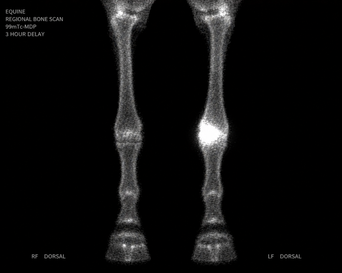
High Voltage presents with a 6-week history of intermittent right forelimb lameness that has been difficult to localize. Nerve blocks have been inconclusive. Radiographs of the foot, fetlock, and carpus are all within normal limits.
A bone scintigraphy study is performed. A radiotracer (technetium-99m labeled methylene diphosphonate) is administered intravenously. The tracer is taken up by osteoblasts — bone cells actively involved in remodeling. Gamma radiation emitted by the tracer is detected by a gamma camera to produce the image.
The scintigraphy image shows a focal region of markedly increased tracer uptake in the right proximal metacarpal region — a finding that was not visible on any radiographic view.
Equine forelimb nuclear scintigraphy
Which explanation best accounts for why the scintigraphy image reveals a finding that the normal radiographs did not — and correctly identifies what the focal increased uptake represents?
a. The scintigraphy image shows greater bone density than radiography because the radiotracer accumulates in calcium-rich bone, and denser bone produces a stronger signal — the hotspot represents an area of mineralization not yet detectable on radiography.
b. The scintigraphy image has higher spatial resolution than radiography, allowing it to detect subtle structural abnormalities in cortical bone that are too small to be visible on standard projections.
c. Scintigraphy detects physiologic activity rather than anatomy — the radiotracer accumulates in areas of increased osteoblastic turnover, producing increased signal before structural changes are visible on radiography. The hotspot indicates a region of elevated bone metabolic activity at the proximal metacarpus, identifying the source of lameness that structural imaging cannot yet show.
d. Scintigraphy uses a stronger radiation source than radiography, allowing deeper penetration into the limb and detection of lesions obscured by overlying soft tissue on standard radiographic projections.
c. Correct.
This is the fundamental principle of nuclear medicine imaging: the tracer is a biologically active molecule that distributes based on physiologic processes, not anatomic density. Technetium-99m MDP accumulates where osteoblastic activity is elevated — areas of active bone remodeling, stress response, inflammation, or neoplasia. The gamma camera detects the radiation emitted by the tracer and maps its distribution. Because metabolic changes precede structural changes, scintigraphy can identify the source of lameness days to weeks before a fracture line, periosteal reaction, or other structural change becomes visible on radiography. This temporal advantage over structural imaging is its primary clinical value.
Mochi presents with a 2-month history of weight loss, polyphagia, and tachycardia. Serum total T4 is elevated. Hyperthyroidism is confirmed biochemically.
The clinician wants to determine whether one or both thyroid lobes are affected before selecting treatment — radioactive iodine therapy requires knowing the distribution of functional thyroid tissue.
Ultrasound of the neck shows a mildly enlarged left thyroid lobe. The right lobe appears normal in size.
The clinician orders thyroid scintigraphy using technetium-99m pertechnetate, which is taken up by thyroid follicular cells based on their metabolic activity.
Which explanation best accounts for why thyroid scintigraphy provides information that ultrasound alone cannot — and correctly identifies what the scan is measuring that ultrasound does not?
a. Ultrasound shows thyroid size and structure but cannot determine whether tissue is functionally active — scintigraphy measures tracer uptake based on metabolic activity of follicular cells, directly showing which tissue is producing excess thyroid hormone rather than which tissue is enlarged.
b. Ultrasound has lower resolution than scintigraphy for evaluating small neck structures — scintigraphy provides better anatomic detail of the thyroid because the radiotracer enhances structural boundaries between the gland and adjacent tissues.
c. Ultrasound cannot image the thyroid because it is located posterior to the tracheal cartilage, which creates acoustic shadowing. Scintigraphy can image structures behind bone and cartilage because gamma radiation penetrates these barriers.
d. Ultrasound cannot detect thyroid hormone — scintigraphy measures the concentration of T4 within the thyroid gland directly, providing a spatial map of hormone content that predicts which lobe is overproducing.
a. Correct.
This is the core distinction between structural and functional imaging for this clinical question. Ultrasound can show that the left lobe is enlarged — but enlargement does not always correlate with function. A lobe could be enlarged but non-functional (cystic, fibrotic, or adenomatous with suppressed activity), or functional tissue could be present without enlargement (ectopic thyroid tissue, or a lobe that is hyperactive without yet being structurally enlarged). Scintigraphy measures tracer uptake, which directly reflects follicular cell activity — the physiologic process responsible for hormone production. For treatment planning, knowing which tissue is functionally active is the critical question, not which tissue is largest.
The scintigraphy result is now being interpreted. The image shows:
• Left thyroid lobe: markedly increased tracer uptake (bright hotspot)
• Right thyroid lobe: very low to absent tracer uptake
• Salivary glands: symmetric faint uptake bilaterally (expected normal finding)
The clinician presents four possible interpretations of this pattern to the team.
Feline Thyroid scintigraphy
Which interpretation of this scintigraphic pattern is most consistent with the clinical presentation and correctly applies the principle that tracer uptake reflects metabolic activity?
a. Both thyroid lobes are diseased — the left lobe is hyperactive (producing excess hormone) and the right lobe has been destroyed by the disease process, explaining the absent uptake.
b. The left lobe is anatomically enlarged, causing it to take up more tracer simply due to greater tissue mass — the right lobe appears absent because it is smaller and its signal is below the detection threshold, not because it is functionally different.
c. The scintigraphy result is non-diagnostic because asymmetric uptake could represent either a normal variant or disease — additional imaging with MRI would be needed to determine which lobe is abnormal.
d. The left lobe has increased metabolic activity results in high tracer uptake. The right lobe shows suppressed uptake because excess T4 from the left lobe has suppressed pituitary TSH, withdrawing the normal trophic stimulus from the right lobe and reducing its functional activity to below detectable levels.
d. Correct.
This is the physiologically accurate interpretation of this scintigraphic pattern. In cats with unilateral thyroid adenoma, the affected lobe produces T4 autonomously — independent of TSH regulation. The resulting excess T4 suppresses pituitary TSH secretion via negative feedback. Without TSH stimulation, the normal contralateral lobe reduces its follicular cell activity, resulting in very low tracer uptake. The right lobe is anatomically present and structurally normal — it is functionally suppressed, not destroyed. This pattern — unilateral hotspot with contralateral suppression — is the classic scintigraphic appearance of unilateral functional adenoma in cats and directly informs the treatment approach.
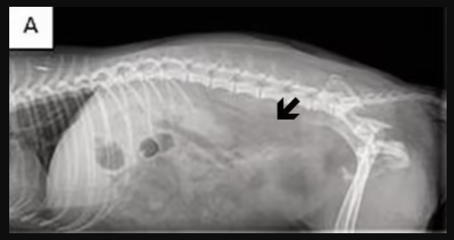
Barnaby presents with a 3-week history of regurgitation immediately after eating. The owner reports food and liquid coming back up without apparent effort, shortly after swallowing.
Physical examination is unremarkable. Thoracic radiographs show a mildly widened esophageal shadow in the caudal thorax. The clinician suspects megaesophagus or an esophageal motility disorder.
The clinical question: is there a structural obstruction, or is there a functional problem with esophageal peristalsis?
A standard lateral thoracic radiograph with barium administered orally shows barium retained in a dilated esophagus — but cannot show whether esophageal contractions are occurring.
An esophageal fluoroscopy study with barium is then performed.
Barnaby:
Which explanation best accounts for why esophageal fluoroscopy answers the clinical question when the standard thoracic radiograph with barium could not — and correctly identifies what fluoroscopy specifically adds?
a. Fluoroscopy uses higher radiation energy than standard radiography, allowing it to penetrate the esophageal wall more effectively and visualize subtle mucosal abnormalities that standard barium radiographs cannot detect.
b. Fluoroscopy produces continuous X-ray images over time, allowing the clinician to observe the swallowing sequence and esophageal peristalsis as they occur — revealing whether the esophagus is propelling the barium bolus toward the stomach (functional peristalsis) or retaining it without coordinated contractions (motility disorder). A static radiograph captures only one moment and cannot show whether peristalsis is occurring.
c. Fluoroscopy uses a radiotracer that specifically labels esophageal smooth muscle, allowing metabolic activity of the muscle to be measured and functional contractions to be detected even when barium is not administered.
d. Fluoroscopy eliminates the need for contrast material by detecting natural differences in esophageal soft tissue density — the smooth muscle of a contracting esophagus attenuates X-rays differently from a relaxed esophagus, making peristalsis directly visible without barium.
b. Correct.
Fluoroscopy is distinguished from standard radiography by its continuous real-time acquisition — it generates a sequence of images during the examination rather than a single static exposure. For esophageal evaluation, this means the clinician can watch the swallowing sequence unfold: does barium progress from the pharynx to the stomach with coordinated peristaltic contractions? Does it pool in the esophagus? Does it reflux? A standard barium radiograph shows where barium is at a single moment but cannot capture motion. In Barnaby's case, the fluoroscopy reveals absent peristalsis with barium retention — a functional motility disorder that appears the same on a static radiograph as simple barium pooling from any cause.
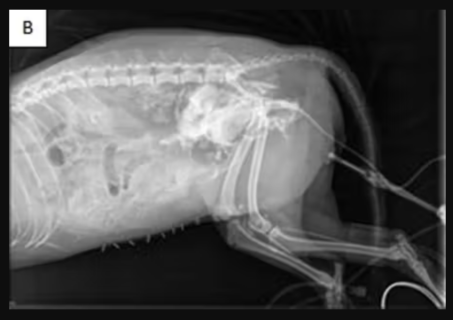
Barnaby is presented following a road traffic accident. Physical examination reveals abdominal pain and the bladder cannot be palpated in the normal location.
Urinary bladder rupture is suspected.
A standard lateral abdominal radiograph is obtained. The bladder is not visible as a distinct structure — the cranial abdomen shows increased opacity with loss of serosal detail, but no definitive bladder outline is identifiable.
The clinician explains: "Bladder and adjacent soft tissues have similar X-ray attenuation — without contrast, the bladder wall blends into surrounding structures."
A positive contrast cystogram is performed: iodinated contrast is instilled via urinary catheter into the bladder.
Barnaby:
Which explanation best accounts for why the standard radiograph could not confirm bladder rupture AND why the contrast study successfully demonstrates the injury?
a. The standard radiograph failed because the bladder is too small to be visible radiographically — contrast expands the bladder to a size large enough to be detected. The contrast also stiffens the bladder wall, making it easier to identify its outline.
b. The standard radiograph failed because X-rays cannot penetrate the fluid-filled bladder — contrast absorbs X-rays differently from fluid, allowing the beam to pass through the bladder and reveal its contents and wall.
c. The standard radiograph failed because trauma causes inflammation that scatters X-rays unpredictably, preventing formation of a clear image. Contrast reduces scatter by providing a stable high-attenuation reference structure.
d. The standard radiograph could not visualize the bladder because the bladder wall and surrounding soft tissues have nearly identical X-ray attenuation — no contrast boundary exists between them. Iodinated contrast instilled into the bladder lumen has very high X-ray attenuation (due to iodine's high atomic number), creating a large attenuation difference between the lumen and adjacent structures, making the bladder outline and any rupture site clearly visible.
d. Correct.
This is a direct application of the soft tissue contrast limitation established in Session 2A: soft tissues that have similar X-ray attenuation cannot be distinguished from each other radiographically. The bladder wall, urine, and surrounding abdominal soft tissues all attenuate X-rays at similar levels. Adding iodinated contrast to the bladder lumen introduces a material with very high X-ray attenuation — iodine has an atomic number of 53, far higher than soft tissue — creating a radiopaque lumen that contrasts sharply with the surrounding soft tissues. If the bladder is intact, the contrast fills and outlines a normal shape. If ruptured, contrast leaks beyond the bladder margins, revealing the site and extent of injury.
Three clinical questions are presented. For each one, the team must select the most appropriate initial imaging modality from the full range covered in this module and Sessions 2A and 2B: radiography, CT, ultrasound, MRI, bone scintigraphy, thyroid scintigraphy, fluoroscopy, contrast radiography.
Clinical question 1: A 9-year-old cat with weight loss and an elevated T4 — the clinician wants to know whether one or both thyroid lobes are functionally active
Clinical question 2: A 3-year-old Warmblood horse with intermittent forelimb lameness and normal radiographs — the clinician wants to identify the anatomic region of elevated bone metabolic activity before pursuing more detailed structural evaluation.
Clinical question 3: A 5-year-old Border Collie with regurgitation and suspected esophageal dysmotility — the clinician wants to observe whether esophageal peristalsis is occurring during swallowing.
Which set of modality selections correctly matches each clinical question with the appropriate imaging approach AND provides the strongest justification for each choice?
a. Question 1: thyroid scintigraphy — directly shows which lobe is functionally active by measuring tracer uptake, informing the treatment decision. Question 2: bone scintigraphy — detects regions of elevated metabolic activity before structural changes appear, localizing the lameness source for targeted structural follow-up. Question 3: esophageal fluoroscopy with barium — provides real-time dynamic imaging of the swallowing sequence, allowing direct observation of whether peristalsis is occurring.
b. (Question 1): MRI — provides superior soft tissue contrast for thyroid evaluation, allowing precise measurement of each lobe's volume and internal architecture. (Question 2): CT — cross-sectional imaging eliminates superimposition, allowing identification of subtle cortical lesions in the distal limb that radiography misses. (Question 3): CT — rapid acquisition allows evaluation of the esophagus in cross-section without requiring patient cooperation during swallowing.
c. (Question 1): ultrasound — provides real-time imaging of thyroid size and vascularity, which correlates with functional activity. (Question 2): radiography with multiple views — additional projections will identify the source of lameness if the initial study was incomplete. (Question 3): standard thoracic radiograph with barium — barium pooling in the esophagus indicates reduced motility without requiring fluoroscopy.
d. (Question 1): radiography of the neck — thyroid enlargement is visible on radiographs and correlates with functional overactivity. (Question 2): ultrasound of the distal limb — identifies fluid accumulation and soft tissue swelling at the source of lameness. (Question 3): nuclear scintigraphy of the esophagus — radiolabeled food bolus imaging directly measures esophageal transit time.
a. Correct.
All three selections match the clinical question to the modality that specifically answers it. Question 1 requires functional information (which lobe is active) not available from structural imaging — thyroid scintigraphy directly measures follicular cell activity. Question 2 requires detection of metabolic activity before structural changes appear — bone scintigraphy provides whole-body physiologic mapping that can localize the source of lameness before a fracture or lesion is radiographically visible. Question 3 requires real-time observation of a dynamic physiologic process (peristalsis) — fluoroscopy with barium contrast provides continuous temporal imaging that a static radiograph cannot. Each selection is driven by what type of information the clinical question requires.
A dog presents with suspected abdominal free fluid. Which modality is most sensitive for detecting even small volumes of free fluid, and what is the physical basis for this sensitivity?
A. Radiography — free fluid attenuates X-rays differently from soft tissue organs, producing a distinct bright opacity at the fluid margin.
B. CT — Hounsfield units precisely quantify fluid density and allow it to be distinguished from surrounding organs.
C. Ultrasonography — free fluid is anechoic, creating high natural contrast against echogenic organ surfaces, and allows real-time guided sampling.
D. MRI — free fluid appears very bright on T2-weighted sequences, providing the highest contrast of any modality.
Answer: C
Ultrasonography is the most sensitive modality for detecting abdominal free fluid. Anechoic fluid creates strong contrast against surrounding tissues and allows real-time guided sampling during the same procedure.
Which of the following correctly states why MRI is the preferred modality for evaluating a dog with suspected spinal cord compression, rather than radiography or CT without contrast?
A. MRI uses sound waves to penetrate the vertebral column and directly visualize the cord, while X-ray based modalities cannot pass through bone.
B. MRI generates contrast between the spinal cord, disc material, cerebrospinal fluid, and surrounding structures through T1 and T2 relaxation differences, which radiography and unenhanced CT cannot reliably provide.
C. MRI eliminates superimposition by acquiring cross-sectional slices, which radiography cannot do, making disc herniation visible for the first time.
D. MRI does not use ionizing radiation, making it the only safe choice for neurologic patients who require repeated imaging.
Answer: B
MRI provides superior soft tissue contrast, allowing clear differentiation between spinal cord tissue, disc material, cerebrospinal fluid, and surrounding structures. This makes it the preferred modality for evaluating spinal cord compression.
In stepwise imaging, the most advanced modality available should always be selected first to avoid missing findings and to minimize the total number of studies a patient undergoes.
A. True
B. False
Answer: B (False)
Stepwise imaging begins with the least invasive and most accessible modality capable of answering the immediate clinical question. More advanced imaging is used only when additional information is needed.
A general practice clinician is evaluating a 6-year-old Thoroughbred gelding with a three-week history of right forelimb lameness. Nerve blocks have localized pain to the foot. Radiographs of the foot are normal. Which modality and rationale best supports the next imaging step?
A. CT — to obtain cross-sectional images of the foot and eliminate superimposition from the standard radiographic projections.
B. MRI — to evaluate soft tissue structures of the foot including the deep digital flexor tendon, navicular bone marrow, and collateral ligaments, which cannot be adequately assessed by radiography.
C. Bone scintigraphy — to perform a whole-body functional survey and identify regions of elevated osteoblastic activity before ordering targeted structural imaging.
D. Repeat radiography with additional projections — to ensure all standard views of the foot have been obtained before escalating to advanced imaging.
Answer: B
Because the lameness has already been localized to the foot and radiographs are normal, MRI is the best next step to evaluate soft tissue structures and bone marrow that cannot be adequately assessed with radiography.
A clinician must select an imaging modality for a 10-year-old cat with suspected intracranial disease. The cat is in poor body condition and has an elevated anesthetic risk. Which practical factor most directly limits the modality options, and what is its implication?
A. Cost — MRI and CT are expensive, so radiography should be used regardless of its diagnostic limitations for intracranial evaluation.
B. Anesthesia requirement — both CT and MRI require general anesthesia in veterinary patients, which carries heightened risk in this patient; this must be weighed against the diagnostic benefit of cross-sectional imaging.
C. Radiation dose — CT delivers higher radiation than radiography, so ultrasound should be substituted as the primary intracranial modality.
D. Availability — nuclear scintigraphy should be pursued instead because it does not require anesthesia and is available at most referral centers.
Answer: B
Both CT and MRI typically require general anesthesia in veterinary patients. In a patient with increased anesthetic risk, the potential diagnostic benefit must be carefully weighed against that risk.
Which of the following questions should a clinician ask before ordering any imaging study, according to the stepwise imaging framework?
A. Which modality is the most advanced and therefore most likely to detect any abnormality present?
B. What will this study tell me that I do not already know, and how will that information change what I do for this patient?
C. Which modality delivers the lowest radiation dose, regardless of whether it can answer the clinical question?
D. Has the patient had this modality before, and if so, is a different modality required to avoid repetition?
Answer: B
The guiding principles of stepwise imaging are determining what new information the study will provide and how that information will influence patient management. Imaging should be purposeful and clinically relevant.
Which statement correctly describes how a standard projection is formed — and identifies the consequence of this formation process for anatomic structures at different depths within the patient?
a. A single X-ray beam passes through the patient in a straight line from tube to detector, and every structure along the beam path contributes its attenuation to the same point on the detector — producing a 2D image in which structures at all depths are superimposed in the same image plane.
b. The X-ray tube rotates around the patient during a projection radiograph, acquiring images from multiple angles — these are combined into a single 2D image in which depth information from each angle is preserved and structures can be separated by their distance from the detector.
c. Projection radiography detects echo reflections from tissue interfaces as the X-ray beam interacts with acoustic impedance differences in the patient — structures at different depths are separated by the time delay of returning echoes.
d. The radiographic detector records the radiotracer signal emitted by metabolically active tissues during a projection exposure — structures at different depths emit different amounts of radiation based on their metabolic activity, allowing them to be distinguished on the final image.
a. Correct.
Projection radiography uses a stationary X-ray tube and a single exposure. The beam travels from tube through patient to detector, passing through every structure in its path simultaneously. Each structure attenuates the beam by an amount determined by its composition and thickness. The detector records the cumulative result of all these attenuations at each detector point. Structures at the skin surface and structures deep within the patient both contribute to the same detector point if they lie along the same beam path. This is why structures at different depths and different lateral positions are superimposed in the final image: all depth information is collapsed into a 2D plane.
Which statement correctly explains how CT produces cross-sectional images that eliminate the superimposition present in projection radiography?
a. CT uses a higher-energy X-ray beam that penetrates tissue more deeply than standard radiography, allowing the detector to record the exact depth at which each structure is located from a single exposure.
b. CT uses a stationary X-ray tube and a flat detector array that records the depth of each attenuating structure electronically — software then reconstructs this depth information into cross-sectional slices from the single exposure.
c. CT rotates the X-ray tube around the patient, acquiring hundreds of projections from different angles. Mathematical reconstruction algorithms use the angular diversity of these projections to calculate the attenuation value at each specific point in the cross-sectional plane, producing an image in which each pixel represents a specific 3D location rather than a cumulative beam-path attenuation.
d. CT uses ultrasound waves rather than X-rays, allowing the scanner to measure the acoustic impedance of each tissue at a specific depth through echo return timing — this produces cross-sectional images without superimposition because sound waves return from each depth at a different time.
c. Correct.
CT eliminates superimposition through acquisition geometry, not through a different type of detector or higher beam energy. The X-ray tube rotates around the patient (typically 360 degrees per rotation), acquiring a projection at each angle. From any single projection angle, structures are still superimposed along the beam path. But by acquiring projections from all angles around the patient, the reconstruction algorithm can determine: given all these different projections, what attenuation value at each specific spatial location is consistent with all the data? The result is an image in which each pixel represents the attenuation at a known 3D location — structures are separated by their actual position, not combined along a beam path.
Which statement correctly describes the mechanism of bone scintigraphy — and identifies the specific advantage it provides over radiography and CT when evaluating a horse with poorly localized lameness and normal radiographs?
a. Bone scintigraphy produces cross-sectional images of the skeleton by rotating the gamma camera around the patient and reconstructing images from multiple projections — this eliminates superimposition and allows detection of subtle cortical fractures not visible on 2D radiographs.
b. A radiotracer that accumulates in areas of elevated osteoblastic activity is administered intravenously. The tracer emits gamma radiation as it decays, which is detected by a gamma camera to produce a map of tracer distribution throughout the skeleton. Metabolically active regions — where bone is remodeling, responding to stress, or reacting to injury — show elevated uptake before structural changes are visible on radiography or CT.
c. Bone scintigraphy uses sound waves directed at the skeletal surface to detect acoustic impedance differences between normal and abnormal bone — diseased bone reflects sound differently, producing a detectable signal that identifies the site of pathology before structural changes are visible.
d. Bone scintigraphy measures differences in hydrogen proton relaxation between normal and diseased bone, producing signal intensity differences on the image that identify areas of pathology before structural abnormalities appear on radiographs.
b. Correct.
Bone scintigraphy is a functional imaging technique. The radiotracer (typically technetium-99m MDP) is taken up by osteoblasts — cells actively involved in bone formation and remodeling. The gamma camera passively detects gamma radiation emitted by the tracer from within the patient and maps its distribution. Regions with elevated metabolic activity (stress fracture, periosteal reaction, bone edema, active remodeling) accumulate more tracer and appear as focal hotspots. Because these metabolic changes precede visible structural changes, scintigraphy can identify the source of lameness before radiography or CT show any abnormality. This temporal advantage over structural modalities is the defining justification for scintigraphy in the scenario of normal radiographs with unlocalized lameness.
Which statement correctly explains why free abdominal fluid causes loss of organ margin visibility on abdominal radiographs — and correctly identifies the physical property responsible?
a. Free abdominal fluid causes loss of organ margin visibility because fluid completely absorbs X-rays, creating a uniformly opaque region that prevents the beam from reaching deeper structures — structures behind the fluid cannot be imaged.
b. Free abdominal fluid causes loss of organ margins because fluid scatters X-rays in all directions as the beam passes through it, degrading image resolution and preventing fine structural detail from being captured on the detector.
c. Free abdominal fluid causes loss of organ margins because the fluid physically compresses the organs, flattening their surfaces and eliminating the curved margins that are normally visible on radiographs.
d. Free abdominal fluid occupies the space normally filled by fat, which provides contrast between organs on abdominal radiographs because fat has lower X-ray attenuation than soft tissue. Fluid has the same X-ray attenuation as soft tissue organs — when fluid surrounds organs, there is no attenuation difference at the organ surface, eliminating the visible boundary.
d. Correct.
Normal abdominal radiographic serosal detail depends on fat providing a lower-attenuation interface around organs: the fat-to-organ boundary produces a detectable attenuation step that appears as a visible margin. Free fluid displaces this fat and directly contacts the organ surface. Since fluid and soft tissue organs attenuate X-rays similarly, there is no attenuation difference at the organ-fluid interface and no visible boundary on the radiograph. This is the same soft tissue contrast limitation established in Session 2A: soft tissues of similar composition cannot be distinguished radiographically. Free fluid is a soft tissue opacity that eliminates the fat-opacity interfaces that normally provide organ visibility.
Which statement correctly explains why abdominal ultrasound can clearly visualize individual organ margins when they are surrounded by free fluid — identifying the acoustic property responsible for this contrast advantage?
a. Free abdominal produces minimal echo return because sound waves transmit through it with very few acoustic impedance interfaces to reflect them. Organ capsules and surfaces produce echoes at acoustic impedance differences. The anechoic fluid provides a high-contrast dark background against which the echogenic organ surfaces are clearly visible.
b. Free abdominal fluid increases X-ray transmission around organs, reducing attenuation differences between the fluid and organ surfaces and making each organ appear as a distinct high-contrast structure on the ultrasound image.
c. Free abdominal fluid acts as an acoustic lens that focuses the ultrasound beam directly onto the organ surfaces, increasing the intensity of returning echoes from organ margins and making them appear brighter and more distinct than in a non-effusive abdomen.
d. Free abdominal fluid emits its own low-level ultrasound signal as it moves within the abdomen, creating a background hum that the ultrasound machine subtracts to reveal the higher-amplitude signals from organ surfaces beneath.
a. Correct.
This is the acoustic mechanism that makes ultrasound superior to radiography for abdominal organ visualization in the presence of effusion. Free fluid is acoustically homogeneous: it contains no internal interfaces to reflect sound, so sound waves transmit through it without returning echoes, producing an anechoic (uniformly dark) region on the image. Organ capsules and parenchymal surfaces create acoustic impedance differences that reflect sound back toward the transducer, appearing as echogenic (bright) structures. The contrast between the anechoic fluid and the echogenic organ surfaces is naturally high — much higher than the contrast between fluid and soft tissue on a radiograph. The more fluid present, the more clearly individual organs are outlined.
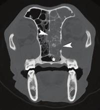
Barnaby presents with a 6-week history of left-sided nasal discharge and epistaxis.
A lateral skull radiograph is obtained. The image shows a region of mixed opacity in the nasal cavity — some increased soft-tissue opacity and some preserved turbinate architecture. The finding is visible but cannot be attributed to the left or right nasal passage, and its depth within the nasal cavity cannot be assessed.
A student says: "I can see something abnormal in the nasal region but I cannot tell which side it is on."
The clinician confirms this observation is correct and asks the student to explain why.
Which explanation correctly accounts for why the lateral skull radiograph can detect the nasal opacity change but cannot determine whether it is on the left or right side — and identifies the physical property of radiography responsible?
a. The radiograph cannot lateralize the finding because digital radiography lacks sufficient resolution for the nasal cavity — higher-resolution film radiography would separate left and right nasal structures.
b. The radiograph cannot lateralize because nasal disease in dogs is always bilateral — unilateral pathology does not produce detectable opacity changes on lateral skull projections.
c. Radiography produces a 2D projection image by passing the X-ray beam through all structures simultaneously along its path — everything in the beam is combined into a single image plane. In the lateral projection the beam passes through both left and right nasal passages, superimposing them. There is no mechanism to separate structures by depth or laterality in a single projection.
d. The radiograph cannot lateralize because it uses non-ionizing radiation that scatters unpredictably through the nasal turbinates, preventing accurate spatial attribution of the opacity signal.
c. Correct.
This is the fundamental consequence of projection imaging. The X-ray beam travels in a straight line from tube through patient to detector, and every structure in its path contributes to the same point on the detector. In the lateral skull projection the beam traverses both nasal passages from one side to the other — a finding in the left passage and a finding in the right passage produce overlapping signals at the same detector location. There is no way to assign depth or laterality to any finding from a single projection. This is why the finding is detectable (something changed the attenuation) but not localizable (cannot determine where within the 3D nasal anatomy it originates).
The lateral skull radiograph has confirmed a nasal opacity change that cannot be lateralized or characterized in depth.
Two next-step options are available:
• Option 1: VD skull radiograph — available in-house immediately, low additional cost, no anesthesia required. Will separate left and right nasal structures and allow lateralization of the finding.
• Option 2: CT nasal cavity — requires referral to a specialist center, general anesthesia, significantly higher cost (~10-15x the VD radiograph). CT acquires cross-sectional slices by rotating the X-ray beam around the patient, reconstructing attenuation at specific 3D locations — eliminating superimposition and providing precise localization, extent, and bone involvement.
The owner has been informed of the nasal finding and is awaiting a recommendation.
Which recommendation most appropriately justifies the next imaging step — correctly weighing what information each option provides against its practical implications?
a. Obtain the VD skull radiograph as the immediate next step. It will lateralize the finding at low cost without anesthesia, answering the specific question the lateral view could not. If the VD confirms significant unilateral disease, CT is then clearly justified for staging and treatment planning.
b. Proceed directly to CT without a VD radiograph. CT provides all information the VD would provide plus cross-sectional detail, so the VD is an unnecessary step that adds radiation exposure without adding information CT would not already provide.
c. Obtain the VD radiograph only and discharge with a treatment trial. The VD will fully characterize the nasal disease including mass extent, bone involvement, and soft tissue detail, making CT unnecessary if the VD is normal.
d. Obtain MRI instead of CT — MRI provides superior soft tissue contrast using non-ionizing radiation, making it safer and more informative for nasal mass characterization than CT.
a. Correct.
A VD projection directs the beam dorsoventrally, separating left and right nasal structures in the image plane and directly addressing the lateralization limitation identified in Part 1A. This is inexpensive, immediately available, and requires no anesthesia. If the VD confirms significant disease, CT is then warranted: it provides cross-sectional localization, bone involvement assessment, and soft tissue detail required for treatment planning. This stepwise approach matches the level of modality to the level of question. CT is not wrong — but without knowing which side is affected, it is premature as an immediate first step.
High Voltage presents with 5 weeks of intermittent right forelimb lameness that has resisted localization. Multiple diagnostic nerve blocks have been inconclusive. Radiographs of the foot, fetlock, and carpus are within normal limits.
The clinician considers three imaging options for the next step:
• Bone scintigraphy — radiotracer maps osteoblastic metabolic activity throughout the skeleton; requires specialized nuclear medicine facility; horse is briefly radioactive post-study
• CT of the distal limb — cross-sectional imaging of bone and soft tissue; requires referral and general anesthesia; targets the region of highest suspicion
• Ultrasound of the distal limb — real-time evaluation of tendons and ligaments; available immediately; non-ionizing
The primary clinical question: in which anatomic region of the limb is the lameness source located?
Which explanation best justifies bone scintigraphy as the most appropriate next imaging step for this specific clinical question — and correctly identifies the key advantage scintigraphy provides over CT and ultrasound in this scenario?
a. Bone scintigraphy is appropriate because it is the only non-ionizing modality that evaluates bone — CT uses ionizing radiation and carries a higher dose risk than scintigraphy for a young performance horse.
b. Bone scintigraphy is appropriate because it provides higher spatial resolution than CT for detecting subtle cortical stress fractures in the distal equine limb.
c. Bone scintigraphy is appropriate because the whole-body survey allows assessment of soft tissue tendon and ligament injuries throughout all four limbs simultaneously, a capability that CT and ultrasound cannot match in a single study.
d. Bone scintigraphy detects physiologic activity rather than anatomy — radiotracer accumulates where osteoblastic activity is elevated, identifying regions of bone remodeling before structural changes appear on radiography or CT. With normal radiographs and an unlocalized lameness, functional imaging provides a whole-body physiologic map that identifies which skeletal region is metabolically active, enabling targeted structural follow-up.
d. Correct.
This is the scenario where functional imaging specifically outperforms structural imaging. Radiographs are normal, meaning no structural changes are yet detectable. CT would provide excellent cross-sectional bone detail — but which region to scan? Without localization, CT targets a clinical guess and may miss the lesion entirely if the wrong area is chosen. Ultrasound evaluates soft tissue structures but not bone metabolic activity. Bone scintigraphy provides a whole-body physiologic map: regions of stress fracture, early periosteal reaction, focal bone edema, or active remodeling produce elevated tracer uptake before becoming structurally visible. This temporal advantage over structural modalities is the defining justification for scintigraphy when all structural imaging is normal and the lameness source remains unlocalized.
Mochi presents with 10 days of progressive abdominal distension, reduced appetite, and mild lethargy.
Two imaging studies are performed and reviewed side by side:
• Image 1: lateral abdominal radiograph — increased uniform soft-tissue opacity throughout the abdomen, loss of serosal detail, no distinct organ margins identifiable except a partial hepatic silhouette.
• Image 2: abdominal ultrasound — a large volume of anechoic fluid surrounds the organs. Individual organ margins are clearly visible against the fluid. Liver, spleen, and kidneys identifiable as distinct structures.
Which explanation best accounts for why the ultrasound provides superior characterization of the abdominal contents compared with the radiograph — and correctly identifies the physical mechanisms responsible for each finding?
a. The ultrasound is superior because it uses higher-energy radiation than radiography, allowing better penetration through the fluid and improved differentiation of soft tissue structures with similar X-ray attenuation.
b. The radiograph shows loss of serosal detail because free fluid has the same X-ray attenuation as surrounding soft tissue organs, eliminating the contrast boundaries between them. Ultrasound shows individual organs clearly because anechoic fluid provides a high-contrast background against echogenic organ surfaces — a contrast advantage arising from acoustic impedance differences that radiography cannot achieve for soft tissues surrounded by fluid.
c. The radiograph is non-diagnostic because radiography can only image gas opacity structures — soft tissue organs produce no radiographic signal. Ultrasound images soft tissue because it detects metabolic activity rather than physical properties.
d. The radiograph cannot image abdominal organs in cats because feline patients are too small for radiographic resolution. Ultrasound provides adequate resolution for small patients.
b. Correct.
This explains both findings using the correct physical mechanism for each modality. On the radiograph: free fluid attenuates X-rays similarly to abdominal soft tissue organs (both are soft-tissue opacity). When fluid replaces the normal fat interfaces between organs, the contrast boundaries disappear, producing the ground-glass opacity and serosal detail loss. On ultrasound: free fluid is anechoic (minimal echo return from homogeneous fluid) while organ capsules produce echoes at acoustic impedance differences. The anechoic fluid provides a naturally high-contrast background against which echogenic organ surfaces are clearly visible. This acoustic contrast advantage is inherent to the physics of ultrasound and cannot be replicated by radiography for soft tissue structures embedded in fluid.
Two clinical questions now require imaging-guided answers:
• Question 1: What is the character of the effusion (transudate, exudate, hemorrhage, chyle)? Can the fluid be safely sampled?
• Question 2: What is the full extent of the liver mass — does it involve the hepatic vasculature or adjacent structures? Is there evidence of additional lesions?
Available modalities and key properties:
• Ultrasound (already performed): real-time, non-ionizing, fluid and soft tissue, available in-house, low cost, allows guided procedures
• CT: cross-sectional, ionizing, bone and soft tissue with contrast, moderate cost, referral required, general anesthesia required
• MRI: cross-sectional, non-ionizing, superior soft tissue contrast, high cost, referral required, general anesthesia required, limited availability in this region
• Scintigraphy: functional/metabolic, ionizing, whole-body, specialized center only, high cost
• Radiography (already performed): 2D projection, ionizing, limited soft tissue contrast, available in-house, low cost
The owner has expressed that cost is a consideration but is willing to proceed if clinical benefit is clearly explained.
Which imaging plan most appropriately addresses both clinical questions — and provides the strongest integrated justification for each modality selected and each modality excluded?
a. Proceed directly to CT for both questions. CT provides cross-sectional imaging of the hepatic mass with vascular detail and can characterize peritoneal fluid distribution simultaneously. This eliminates the need for any additional steps and provides the most complete information in one study.
b. Perform MRI instead of CT for Question 2. MRI provides superior soft tissue contrast for hepatic mass characterization without ionizing radiation, making it safer and more informative for this clinical scenario.
c. Perform ultrasound-guided abdominocentesis to characterize the effusion (Question 1) — real-time ultrasound guidance allows safe sampling. If fluid analysis and ultrasound findings support a significant hepatic mass requiring staging, refer for CT to characterize its full extent and vascular involvement (Question 2). MRI is not indicated at this stage given cost and limited availability when CT provides equivalent information for abdominal evaluation. Scintigraphy has no role — the question is structural extent, not metabolic activity.
d. Continue with ultrasound examination only for both questions. Ultrasound can sample the fluid under real-time guidance and fully characterize the hepatic mass extent including vascular involvement. CT and MRI are not needed because ultrasound provides equivalent information at lower cost without anesthesia.
c. Correct.
This plan sequences the steps appropriately and justifies each modality selection and exclusion explicitly. Question 1 is best answered by abdominocentesis with ultrasound guidance: real-time capability makes sampling safe, results are rapid, and fluid analysis directly characterizes the effusion. This may also change the subsequent plan — a simple transudate from cardiac disease requires different follow-up than hemorrhage from a ruptured hepatic mass. Question 2 requires cross-sectional imaging if staging is warranted: CT provides adequate soft tissue contrast for hepatic mass characterization, vascular involvement assessment, and evaluation for additional lesions — at lower cost, better availability, and with equivalent diagnostic yield compared with MRI for this indication. MRI is excluded because CT provides equivalent information for abdominal evaluation without the additional cost and availability barriers. Scintigraphy is excluded because the question is structural extent, not metabolic activity.
A veterinary student is about to assist with a radiographic study. According to the ALARA principle, which of the following best describes what ALARA requires?
A. Staying below the annual regulatory dose limit of 50 mSv per year for radiation workers.
B. Reducing radiation exposure to the lowest level reasonably achievable in every imaging encounter, regardless of whether dose limits are being approached.
C. Wearing lead personal protective equipment during every radiographic exposure, regardless of distance from the beam.
D. Limiting the number of radiographic studies performed per patient to reduce cumulative dose.
Answer: B
ALARA requires reducing radiation exposure to the lowest level reasonably achievable during every imaging encounter. It is an ongoing safety principle, not simply a requirement to stay below regulatory dose limits.
A radiographic suite is 3 meters wide. A technician standing 0.5 meters from the X-ray tube moves to 1 meter away during an exposure. By approximately how much does the radiation intensity at the technician's position change?
A. It decreases by half — intensity is inversely proportional to distance.
B. It decreases to one-quarter — intensity is inversely proportional to the square of the distance.
C. It decreases by one-third — intensity decreases linearly with distance.
D. It does not change significantly — distance has minimal effect at the short ranges typical of a radiographic suite.
Answer: B
The inverse square law states that radiation intensity decreases with the square of the distance from the source. Doubling the distance reduces exposure to one-quarter of its original intensity.
The static magnetic field of a superconducting MRI scanner turns off automatically between patient scans, so MRI suite safety screening only needs to occur when the scanner is actively acquiring images.
A. True
B. False
Answer: B (False)
Superconducting MRI magnets are always on. Safety screening must occur every time a person or object enters the MRI environment, regardless of whether a scan is actively being performed.
A dog is scheduled for an abdominal CT with intravenous iodinated contrast. Pre-scan bloodwork reveals elevated creatinine consistent with reduced renal function. Which contrast safety concern is most directly raised by this finding?
A. Ferromagnetic hazard — iodinated contrast contains metal ions that may interact with the CT magnetic field.
B. Contrast-induced nephropathy — iodinated contrast agents are filtered by the kidneys and can cause acute deterioration of renal function in patients with pre-existing renal impairment.
C. Barium aspiration — iodinated agents carry a risk of aspiration pneumonia if administered to a patient with compromised renal function.
D. Gadolinium retention — iodinated contrast agents may accumulate in brain tissue in renally impaired patients.
Answer: B
Iodinated contrast agents are excreted by the kidneys. Patients with impaired renal function are at increased risk for contrast-induced nephropathy and require careful evaluation before contrast administration.
A veterinary student observes that a staff member is about to bring a standard steel oxygen cylinder into the MRI suite to support a patient under anesthesia. What is the correct action for the student?
A. Allow it — oxygen cylinders are classified as MRI Conditional and are safe under standard clinical field strengths.
B. Say nothing — correcting a staff member is outside the student's professional scope.
C. Immediately communicate the safety concern to the supervising clinician or MRI safety officer — standard steel oxygen cylinders are ferromagnetic and pose a projectile hazard in the MRI environment.
D. Ask the staff member to move the cylinder closer to the wall to reduce the risk of it being attracted to the magnet.
Answer: C
Steel oxygen cylinders are ferromagnetic and can become dangerous projectiles in the MRI environment. Students have a professional responsibility to immediately communicate identified safety concerns.
A dyspneic (trouble breathing) cat requires thoracic radiographs to guide immediate management. The owner asks about the risk of the procedure. Which statement best reflects the professional responsibilities involved?
A. The student should explain the risks to the owner, since they are present and the supervising clinician is busy.
B. Sedation risk-benefit assessment and client communication are the supervising veterinarian's responsibilities; the student may assist with positioning and apply PPE but may not make sedation decisions or communicate clinical findings to the owner independently.
C. Because the cat is dyspneic, sedation is contraindicated and radiographs should not be obtained until the cat is stable.
D. Radiographic positioning risk is low enough that sedation risk-benefit assessment is not required for a routine thoracic radiograph.
Answer: B
Clinical decision-making, risk assessment, and communication with clients are the supervising veterinarian's responsibilities. Students assist with procedures and communicate observations through the supervising clinician.
A dosimetry badge worn outside a lead apron at collar level overestimates occupational radiation dose, so personnel who wear lead aprons consistently can disregard their badge readings.
A. True
B. False
Answer: B (False)
Dosimetry badges monitor exposure to unshielded areas such as the head, neck, and thyroid. Badge readings remain important for regulatory monitoring and occupational safety, even when lead aprons are worn consistently.
Which statement correctly identifies the three components of the ALARA radiation protection triad — and correctly states the physical principle underlying one of them?
a. Time, distance, shielding — increasing distance reduces exposure by attenuation of the beam through air, which absorbs a fixed proportion of X-ray energy per meter traveled.
b. Time, distance, shielding — increasing distance reduces exposure through the inverse square law: doubling the distance from a point source reduces intensity to one-quarter of the original value.
c. Time, technique, shielding — reducing exposure time decreases occupational dose proportionally, and optimizing radiographic technique eliminates the need for distance-based protection.
d. Distance, shielding, collimation — tightening beam collimation is the primary occupational exposure reduction strategy because it eliminates the primary beam reaching personnel outside the collimated field.
b. Correct.
ALARA stands for As Low As Reasonably Achievable and is implemented through three strategies: reducing time of exposure, increasing distance from the source, and using shielding. Distance follows the inverse square law: intensity is proportional to 1/d², so doubling the distance multiplies intensity by (1/2)² = 1/4. This geometric relationship makes distance an extremely powerful protective strategy: small increases in distance produce large reductions in exposure intensity.
Which statement correctly explains how lead shielding and beam collimation each reduce occupational radiation exposure — identifying the distinct physical mechanism responsible for each?
a. Lead shielding reflects scatter radiation back toward the X-ray tube, preventing it from reaching personnel. Beam collimation increases beam energy, which reduces the number of exposures needed to produce a diagnostic image.
b. Lead shielding eliminates all scatter radiation reaching the covered body part through complete photon absorption. Beam collimation eliminates all scatter radiation generated in the patient by restricting the beam to zero width at the detector.
c. Lead shielding and beam collimation both work through the same mechanism: they reduce the time the patient is exposed to the primary beam, which proportionally reduces scatter reaching personnel.
d. Lead shielding attenuates scatter radiation reaching the covered body part by absorbing a proportion of incident photons — high atomic number and density of lead make it effective at diagnostic X-ray energies. Beam collimation reduces the volume of patient tissue irradiated, which reduces total scatter generated at the source. The two mechanisms are distinct and complementary.
d. Correct.
Lead shielding works through attenuation: lead has atomic number 82 and high physical density, properties that make it effective at absorbing X-ray photons at diagnostic energies. Typical lead aprons and gloves attenuate 90–95% of scatter. Collimation works through source reduction: restricting the primary beam to the area of clinical interest means less patient tissue is irradiated, which means less total scatter is generated in the patient and therefore less scatter reaches personnel. These are distinct mechanisms — shielding addresses scatter after it is generated; collimation reduces how much scatter is generated in the first place. Combining both addresses the problem from two directions.
Which statement correctly identifies the radiation classification of diagnostic ultrasound — and correctly describes what the mechanical index measures?
a. Ultrasound is non-ionizing — it uses sound waves rather than electromagnetic radiation. The mechanical index is a measure of acoustic pressure amplitude; higher mechanical index settings increase the potential for tissue cavitation effects.
b. Ultrasound is ionizing at higher power settings — the high-frequency sound waves carry sufficient energy to ionize tissue atoms when the mechanical index exceeds 1.0. Below mechanical index 1.0, ultrasound is non-ionizing and safe for all patients.
c. Ultrasound is non-ionizing; the mechanical index measures the total energy deposited in tissue per unit time during an ultrasound examination and determines the cumulative occupational exposure limit for sonographers performing repeated studies.
d. Ultrasound is ionizing because high-frequency sound waves carry photon energies capable of breaking molecular bonds in biological tissue, similar to the mechanism of ultraviolet radiation. The mechanical index measures the ionizing dose delivered per examination.
a. Correct.
Ultrasound uses high-frequency sound waves — a form of mechanical (pressure) energy rather than electromagnetic radiation. It is therefore non-ionizing: it does not carry sufficient energy per photon to ionize atoms or break chemical bonds. The mechanical index (MI) is defined as the peak negative acoustic pressure divided by the square root of the ultrasound frequency. It is a measure of the mechanical stress the sound wave exerts on tissue. At high MI values, dissolved gases in tissue can form and collapse rapidly (cavitation), potentially causing cell damage. Diagnostic ultrasound systems display the MI and are designed to operate below established safety thresholds (typically MI <1.9 for soft tissue).
Which statement correctly explains why ferromagnetic objects are dangerous in an MRI environment — and correctly identifies a critical property of the MRI magnetic field that makes this hazard present at all times?
a. Ferromagnetic objects become dangerous in MRI because the scanner emits strong ionizing radiation during image acquisition that interacts with metal objects, causing them to heat rapidly and burn adjacent tissue. This hazard is present only during active scanning.
b. Ferromagnetic objects are dangerous because the scanner emits pulsed radiofrequency energy that causes metal objects to resonate at their natural frequency, shattering them into fragments that can injure personnel. This hazard is present only during image acquisition when radiofrequency pulses are active.
c. Ferromagnetic objects experience a powerful attractive force from the MRI static magnetic field — a force that increases rapidly as the object approaches the bore. The static magnetic field is always on in superconducting MRI systems, even when no scan is in progress, making the projectile hazard continuous rather than intermittent.
d. Ferromagnetic objects are dangerous in MRI because the magnetic field magnetizes them permanently, causing them to interfere with future radiographic examinations by creating artifacts. This is a long-term equipment concern rather than an immediate safety hazard.
c. Correct.
Ferromagnetic materials (iron, cobalt, nickel, and many steel alloys) are strongly attracted to magnetic fields. The MRI scanner's static field exerts a translational force on ferromagnetic objects that scales with the field gradient — force increases dramatically as the object enters the fringe field and approaches the bore. This can accelerate a ferromagnetic object to high velocity in a fraction of a second, creating a life-threatening projectile. Critically, most clinical MRI scanners use superconducting magnets that maintain the magnetic field continuously — the field does not turn off between scans, overnight, or during patient changeover. Screening must occur at the controlled access boundary before any entry, at any time.
Which statement correctly explains why sedation carries increased risk in a dyspneic patient — and correctly identifies who has professional authority to make the sedation decision?
a. Sedation is dangerous in dyspneic patients because sedatives cause tachycardia and hypertension, which increase myocardial oxygen demand and can precipitate cardiac arrest in patients with underlying cardiac disease. The decision to sedate belongs to the owner, who must provide informed consent.
b. Sedation carries increased risk in dyspneic patients because sedatives and anesthetic agents reduce respiratory drive and can impair the patient's ability to compensate for underlying respiratory disease. The decision to sedate is a clinical judgment that belongs to the supervising veterinarian, not to students or veterinary technicians.
c. Sedation is safe in dyspneic patients as long as oxygen supplementation is provided during the procedure — supplemental oxygen fully compensates for any reduction in respiratory drive caused by sedative agents. The student may administer mild sedation independently if the veterinarian is temporarily unavailable.
d. Sedation is dangerous in dyspneic patients primarily because sedatives reduce heart rate, causing hypotension and cardiovascular collapse in patients with pulmonary hypertension. Students who identify this risk are authorized to refuse sedation administration on patient safety grounds.
b. Correct.
Respiratory drive — the physiological stimulus to breathe based on CO2 and O2 levels — is suppressed by most sedative and anesthetic agents. In a healthy patient with normal respiratory reserve, this suppression is easily managed. In a dyspneic patient who is already breathing at maximum compensatory capacity, sedative-induced reduction in drive can tip the patient into respiratory failure. Airway muscle tone may also be reduced, worsening any dynamic obstruction. The clinical judgment of whether this risk is outweighed by the benefit of diagnostic imaging requires veterinary assessment of the specific patient. Students and technicians do not have the clinical authority or training to make independent sedation decisions.
A veterinary student is manually restraining Barnaby during thoracic radiography. The student is positioned close to the primary beam.
Three exposure-reduction options are available but none are currently being used:
• A movable lead barrier is positioned 1 metre from the table
• A lead apron is hanging adjacent to the table
• The beam collimation field extends well beyond the area of interest
The supervising technician asks: "What should you do first to most effectively reduce your exposure right now?"
Context: radiation exposure intensity follows the inverse square law — doubling the distance from a point source reduces intensity to one-quarter. Occupational dose limits are established by regulatory bodies; personnel are required to keep cumulative exposure as low as reasonably achievable (ALARA) regardless of whether dose limits are being approached.

Which action would most effectively reduce the student's occupational radiation exposure immediately — and correctly reflects the prioritization of ALARA strategies when multiple options are simultaneously available?
a. Wear the lead apron — shielding provides the most reliable protection because it physically blocks radiation regardless of positioning changes, and aprons attenuate the majority of scatter radiation reaching the torso.
b. Tighten beam collimation to the area of interest — reducing the field size decreases the volume of irradiated tissue and therefore reduces scatter radiation reaching personnel, eliminating the need for positioning changes.
c. Request sedation for Barnaby so that manual restraint is no longer required — eliminating manual restraint removes the student from the immediate vicinity of the primary beam entirely and removes all occupational exposure for the student.
d. Step behind the movable lead barrier — increasing distance from the primary beam source substantially reduces exposure intensity through the inverse square law, and the barrier provides additional shielding. This combined effect of distance plus shielding provides greater protection than either a lead apron alone or collimation adjustment alone.
d. Correct.
Stepping behind the movable barrier achieves two protective effects simultaneously: (1) increased distance from the primary beam reduces intensity by the inverse square law — moving 1 metre farther reduces intensity to approximately one-quarter of the previous value; (2) the lead barrier provides additional attenuation of residual scatter. This combined distance-plus-shielding effect is more effective than wearing a lead apron while remaining adjacent to the table (shielding only) or tightening collimation without repositioning (scatter reduction only). In ALARA prioritization, maximizing distance is the highest-impact immediate strategy when it is available.
The clinical situation has changed: Barnaby cannot be sedated at this time due to cardiovascular concerns, and a second person is not available to operate the exposure switch from a distance.
Manual restraint by the student is genuinely required.
The student cannot step behind the barrier during the exposure. Two protective measures remain available:
• Lead gloves are available for the student's hands, which will be positioned at the edge of the collimated field
• The collimation field can be tightened to precisely the area of clinical interest
The technician asks: "Given that you must restrain, what should you do to minimize your exposure as much as possible?"
Which combination of actions best reflects ALARA-consistent practice for the student who must perform manual restraint — and correctly explains how each action reduces occupational exposure?
a. Wear the lead gloves AND tighten beam collimation to the area of interest — lead gloves attenuate scatter reaching the hands positioned near the field edge, while tightening collimation reduces the volume of irradiated patient tissue and therefore reduces scatter generated at the source.
b. Wear the lead gloves only — collimation adjustments are the radiographer's responsibility and should not be altered by personnel performing restraint, as unauthorized adjustments to technique settings may compromise image quality.
c. Increase the exposure time (mAs) to allow the patient to be repositioned during a longer exposure — longer exposures allow more positioning adjustments and reduce the number of repeat exposures needed.
d. Rotate the student with another team member between exposures — alternating personnel distributes cumulative dose across multiple individuals, eliminating occupational exposure risk for any single person.
a. Correct.
When manual restraint cannot be avoided, combining available protective measures is the correct ALARA approach. Lead gloves attenuate scatter radiation reaching the hands — which are the closest part of the student to the primary field and therefore the highest-dose body part in this scenario. Tightening collimation to the area of interest reduces the volume of irradiated tissue, which reduces the total amount of scatter generated in the patient. Less scatter generated means less scatter reaching the student's hands and body. The combination of shielding (lead gloves) and reduced scatter generation (collimation) provides more protection than either measure alone.
A pregnant radiographer is assisting with imaging Mochi for suspected abdominal effusion.
Two modalities are available for the initial evaluation:
• Abdominal radiography — requires the technician to position Mochi and potentially restrain her during exposure
• Abdominal ultrasound — requires the technician to hold the probe in contact with Mochi's abdomen during image acquisition
The clinical question is detection and characterization of abdominal fluid.
Context: for the fetus, there is no established safe threshold for ionizing radiation exposure — even low doses carry a theoretical risk. Guidelines generally recommend minimizing pregnant personnel exposure to ionizing radiation where alternatives exist. Ultrasound uses high-frequency sound waves — it does not use ionizing radiation. The mechanical index (MI) is a measure of acoustic pressure amplitude; high MI settings can cause tissue cavitation. Diagnostic ultrasound operates at MI levels well below the threshold for clinically significant effects.

Which explanation best justifies abdominal ultrasound as the most appropriate modality for the pregnant technician in this scenario — and correctly addresses the safety profile of ultrasound compared with the available alternative?
a. Ultrasound is appropriate because it requires shorter examination time than radiography — the reduced examination time minimizes the technician's proximity to the patient and therefore reduces any occupational exposure risk.
b. Ultrasound is appropriate because it produces superior image quality for abdominal fluid detection and therefore reduces the need for repeat examinations — fewer examinations means less total occupational radiation exposure over time.
c. Ultrasound uses non-ionizing sound waves rather than ionizing radiation — personnel performing ultrasound examinations are not exposed to ionizing radiation during image acquisition. This eliminates the fetal radiation exposure risk associated with radiographic positioning. Ultrasound itself operates at mechanical index levels well below the threshold for clinically significant biological effects in diagnostic applications.
d. Ultrasound is appropriate because the lead apron worn during radiography does not protect the fetus adequately — lead aprons attenuate less than 50% of diagnostic X-ray energies, making the fetal dose from radiographic positioning unacceptably high.
c. Correct.
This answer correctly identifies the primary justification (non-ionizing vs ionizing) and introduces the mechanical index nuance accurately. Ultrasound uses high-frequency sound waves — no ionizing radiation is emitted during image acquisition, so personnel performing ultrasound examinations receive no ionizing radiation dose. The fetal radiation risk from radiographic positioning (even with protective measures) is therefore eliminated when ultrasound is used. The mechanical index context is important: ultrasound is not completely without biological interaction — acoustic energy can cause tissue effects at sufficiently high MI settings — but diagnostic applications operate at MI levels (typically <1.9) well below clinically significant thresholds for soft tissue. This makes diagnostic ultrasound effectively safe for both the patient and the operator in routine practice.
A technician is preparing Barnaby for an MRI examination of the spine. As the technician enters the MRI scanner room, they are carrying a portable oxygen cylinder to support Barnaby's anesthetic monitoring.
The MRI scanner is active — the magnetic field is always on, even when no scan is in progress.
Magnetic hazard warning signs are posted at the suite entrance. A controlled access boundary is marked on the floor.
The technician did not stop at the screening checkpoint before entering.

Which statement best identifies the primary safety failure in this scenario AND correctly explains the mechanism responsible for the associated hazard?
a. The primary failure is not wearing a dosimetry badge before entering the MRI suite — MRI scanners emit ionizing radiation during image acquisition that accumulates as occupational dose and must be monitored.
b. The primary failure is not screening the oxygen cylinder for ferromagnetic properties before entering the MRI field — the strong static magnetic field can exert a powerful attractive force on ferromagnetic objects, accelerating them toward the magnet bore at high velocity and creating a life-threatening projectile hazard.
c. The primary failure is not pre-medicating Barnaby with antihistamines before MRI contrast administration — gadolinium-based contrast agents commonly used in MRI cause allergic reactions in dogs that require prior prophylaxis.
d. The primary failure is positioning the anesthetic monitoring equipment too close to the scanner bore — electrical equipment near the bore can interfere with image quality through radiofrequency emission, creating artifact that compromises diagnostic accuracy.
b. Correct.
Standard portable oxygen cylinders are ferromagnetic (steel construction). The MRI magnetic field — which is always present, not just during scanning — exerts a powerful attractive force on ferromagnetic objects that increases dramatically as the object approaches the magnet bore (the force scales with field gradient). A ferromagnetic oxygen cylinder entering the fringe field can be rapidly accelerated toward the bore, becoming a high-velocity projectile. This is the most immediately life-threatening MRI safety failure: objects weighing several kilograms can reach the bore in fractions of a second. All equipment must be screened as MRI-safe or MRI-conditional before crossing into the controlled access zone.
A dog presents with moderate-to-severe dyspnea. Physical examination reveals rapid shallow breathing and increased respiratory effort. The owner reports a 2-day history of worsening breathing difficulty.
Thoracic radiographs are indicated to evaluate for pleural effusion, pneumothorax, or pulmonary disease.
The dog becomes increasingly stressed during positioning attempts. Image quality is compromised by patient movement.
A veterinary student suggests: "We should sedate the dog so we can get a diagnostic image — the owner says they want to know what is wrong."
The supervising clinician is considering the suggestion.
Context: sedation in a dyspneic patient carries risk — sedatives and anesthetic agents can reduce respiratory drive, cause airway relaxation, and compromise the ability to compensate for underlying respiratory disease. However, a diagnostic thoracic radiograph may be essential for immediate management decisions. Dyspneic patients are also at risk from stress during restraint and positioning.

Which response best reflects the most appropriate clinical and professional approach to this situation — and correctly integrates patient safety, diagnostic necessity, client communication, and team ethics?
a. Proceed with sedation immediately because a diagnostic radiograph is required for treatment and the owner has consented to imaging — owner consent authorizes the clinical team to use whatever means are necessary to obtain a diagnostic study.
b. Refuse sedation under any circumstances because respiratory disease is an absolute contraindication to all sedation — any sedative agent administered to a dyspneic patient will cause respiratory arrest.
c. Stabilize the patient first where possible — provide oxygen supplementation, minimize stress, and allow rest before attempting positioning. The decision to sedate should be made by the supervising veterinarian after assessing the urgency of imaging versus the risk of sedation-induced respiratory compromise in this patient, and the decision and its rationale should be communicated clearly to the owner.
d. The student should administer mild sedation independently to improve positioning before the supervising clinician returns — mild agents carry low risk and delay in obtaining a diagnostic image may compromise patient care.
c. Correct.
This response applies risk-benefit reasoning appropriately and assigns communication responsibility correctly. Stabilization (oxygen, stress reduction, rest) may improve the patient's tolerance for positioning and may allow adequate radiographs without sedation. If sedation is required, the decision belongs to the supervising veterinarian, not the student, because it requires clinical assessment of sedation risk versus diagnostic urgency in this specific patient. The owner must be informed of the sedation decision and its rationale — this is both an ethical and a legal requirement. The student's role is to flag the concern, not to make or refuse the sedation decision unilaterally.
Standard radiographic evaluation of most body regions in dogs and cats require:
A. Only the number of views needed to make a diagnosis
B. At least two orthogonal projections
C. A single projection
D. Oblique projections only
B. At least two orthogonal projections
Which imaging modality does not use X-rays to produce an image?
A. CT
B. Radiography
C. Fluoroscopy
D. Nuclear scintigraphy
D. Nuclear scintigraphy
Which statement about MRI safety is correct?
A. MRI magnets are always on; metal objects must never enter the MRI room.
B. MRI magnets are only on while a patient is being scanned, it is safe to enter the room when a patient is not being imaged.
C. It is never safe for people to enter the MRI room
D. Metal objects are only dangerous when they are near the patient.
A. MRI magnets are always on; metal objects must never enter the MRI room.
Session 5A
A student begins evaluating a thoracic radiograph and immediately notices a large opacity in the left lung field. They describe this finding and write their interpretation, then stop. Which error does the systematic framework specifically exist to prevent in this scenario?
A. Premature closure — the student applied interpretation before completing description.
B. Satisfaction of search — stopping evaluation after finding the first abnormality, without completing systematic review of the rest of the image.
C. Modality misidentification — the student failed to confirm the image was a radiograph before interpreting it.
D. Technical quality error — the student did not assess whether the image was adequately exposed before describing the finding.
Answer: B
Satisfaction of search occurs when a clinician stops looking after identifying one abnormality. The systematic framework requires a complete region-by-region review to ensure additional findings are not missed.
Session 5A
A radiograph is labeled with a patient name and study date. Before beginning interpretation, the student checks the label against the clinical record and finds a discrepancy — the label matches a different patient. What is the correct next action?
A. Interpret the image as labeled, then flag the discrepancy in the medical record as a note.
B. Interpret the image based on what the anatomic appearance suggests about the patient's species and size, ignoring the label.
C. Stop evaluation, report the label-image mismatch to the supervising clinician, and do not interpret the image until it is traced to the correct patient.
D. Compare the image to the patient's previous radiographs to determine which label is correct, then proceed.
Answer: C
A label-image mismatch is a patient safety issue. Interpretation should stop immediately until the study can be correctly matched to the appropriate patient.