Anatomy and Physiology Exam 3 Lecture - The Skeletal System
 Call Kai
 Learn
Learn Practice Test
 Spaced Repetition
 Match
Match Flashcards
 Knowt Play
1/30

There's no tags or description
Looks like no tags are added yet.
Name | Mastery | Learn | Test | Matching | Spaced | Call with Kai | Chat |
|---|
No analytics yet
Send a link to your students to track their progress
31 Terms
Regions of the skeleton
Axial Skeleton - Forms the central axis; skull, vertebral column, ribs, sternum, and sacrum (at the base of the spine and wedged between the hip bones of the pelvis)
Appendicular Skeleton - Includes the limbs and girdles (bones in the appendicular axis that connect your limbs to your central axis)
Pectoral girdle - upper extremity (shoulder)
Pelvic girdle - lower extremity (pelvis)
Major Skull Cavities
Cranial Cavity - Contains the brain
Orbital Cavity - Contains the eyes
Nasal Cavity - Contains the nasal passages
Oral Cavity - Contains the mouth and tongue
Middle Ear Cavities - Contains the ear bones

Bone Feature - Fossa (Depression)
A shallow, basin-like depression in a bone surface.
Cranial Fossa
The purpose of the Cranial Fossa is to form the floor of the cranial cavity, supporting, compartmentalizing, and protecting the distinct regions of the brain.
Anterior Fossa (purple circle) - the shallowest and most superior depression, housing the frontal lobes and olfactory pathways.
Middle Fossa (pink circle) - cradles the central skull base, accommodating the temporal lobes and the pituitary gland.
Posterior Fossa (blue circle) - The deepest and the largest, located at the base and back of the skull, containing the cerebellum and brainstem, including the passage way where the brain transitions into the spinal cord.

Bone Feature - Articulation (where two bones meet that form a joint) - Condyle
A large, smooth, rounded articular projection that forms joints with other bones, often covered with cartilage.

Bone Feature (Articulation) - Facet
A small, flat, smooth surface on a bone where it articulates with another bone.
Bone Feature (Articulation) - Head
A prominent, round epiphysis (end of a long bone) supported by a narrow neck.
Ex: Rib
Bone Feature (Projection/Process) - Epicondyle
A rough, non-articular bony raised or projected area located on or above a condyle that anchor muscles, ligaments, and tendons.
Ex: medial epicondyle of the femur

Bone Features (Projection/Process) - Trochanter
A very large, blunt, irregularly shaped projection found only on the femur
You can feel it as it is the prominent bony bump on the upper, outer side of your hip and thigh

Bone Features (Projection/Process) - Tubercle
A small rounded and slightly roughened projection or process that serves as an attachment point for muscles, ligaments, and tendons.

Bone Feature (Projection/Process) - Tuberosity
A large, rounded, and roughened bump on a bone that serves as an attachment point for muscles, tendons, and ligaments.

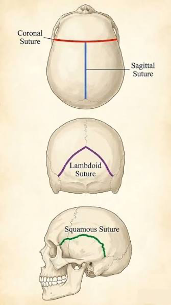
Bone Feature (openings) - Fissure
A narrow slit-like opening in a bone (or between separate bones) that typically allows blood vessels and nerves to pass through.
Ex: Coronal Suture, Squamous Suture, Lambdoid Suture
Bone Feature (Openings) - Foreman and Canal
Foreman - a round passageway or hole
Canal (Meatus) - a tube-like passageway or canal through a bone
Sesamoid Bones
Definition - special types of short bones formed within tendons where there is considerable pressure or physical stress
Function - Protect tendons from wear and stress, acting like pulleys to improve leverage, reduce friction, and absorb weight-bearing pressure.
The patella (kneecap) is the largest sesamoid bone in the human body, sitting at the front of the kneecap, inside the quadriceps tendon
There are two pea-sized sesamoid bones that lie beneath of the big toe, helping with weight-bearing and pushing off during walking
Small sesamoid bones are commonly found near the thumb joints and in the wrists.

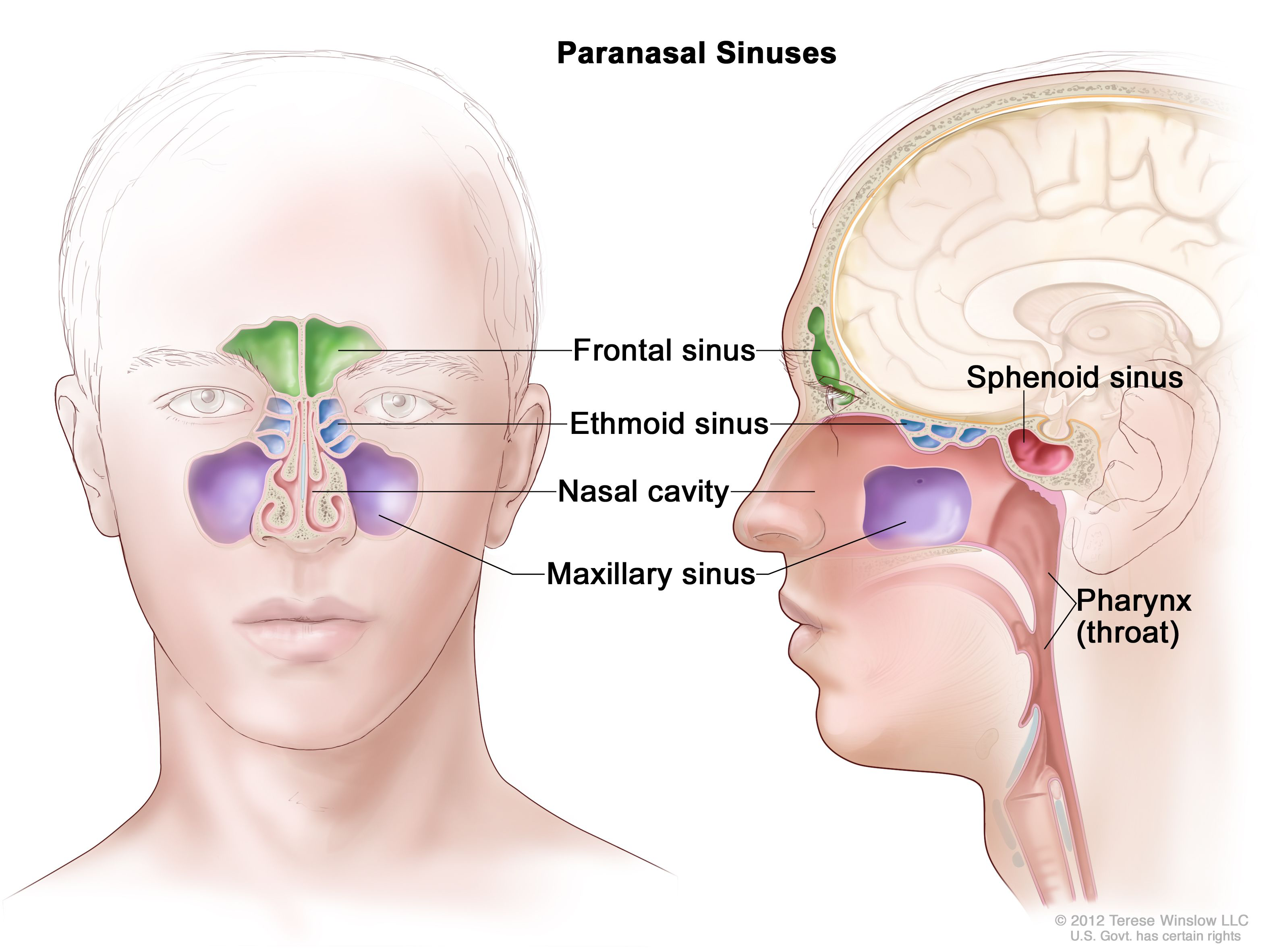
Paranasal Sinuses
Four pairs of air filled cavities located in the bones of your face and skull that surround the nasal cavity and produce mucus to drain in to the nose. They lighten the skull and resonate the voice.

Paranasal Sinuses - Frontal Sinuses
Located in the forehead area above your eyebrows

Paranasal Sinuses - Ethmoid Sinuses
Positioned in the small spaces between your eyes, behind the bridge of the nose

Paranasal Sinuses - Sphenoid
Located deep inside the skull behind the eyes and nose

Paranasal Sinuses - Maxillary
The largest pair, located in your cheekbones below your eyes
Human Vertebrae
Composed of 33 vertebrae during early development and is divided into 5 distinct regional types.
Cervical Vertebrae
7 Vertebrae (C1 - C7)
Located in the neck region, the smallest vertebrae and feature a triangular Vertebral Foreman for blood vessels and Intervertebral Foreman for spinal nerves
The first two (C1 atlas and C2 axis) are specialized to support the movement of the skull
C1 - nod your head “yes” movement
C2 - Odontoid process, allows rotation of head “no” movement
Thoracic Vertebrae
12 Vertebrae: T1 - T12
Located in the upper back and more massive than cervical but smaller than lumbar
Spinous process pointed and angled downward
Superior and Inferior costal facets and transverse processes for articulation of the ribs
Lumbar Vertebrae
5 Vertebrae: L1 - L5
Thick, stout body and blunt, squarish spinous process
Located in the lower back, the largest and strongest moveable bones designed to support most of the body weight.
Sacral Vertebrae - Sacrum
5 separate sacral vertebrae (S1 - S5) fused by age 26 that form a single, triangular bone called the sacrum, which articulates with the pelvic girdle
Anterior View = Smooth and Concave
Posterior View = Rough
Coccygeal Vertebrae - Coccyx
4 Vertebrae (Co1 - Co4) that fuse by age of 30
Provides attachment site for muscles of pelvic floor
Cornua - Hornlike projections on Co1 for ligaments to attach from coccyx to sacrum
Represents the terminal end of the spinal cord
Fractured by fall or during childbirth
Rib Cage
The average person has 12 pairs of ribs (24 individual ribs in total) which attach anteriorly (at the front) in three distinct ways
True Ribs (1-7) attach directly to the sternum (breastbone) through their own individual pieces of costal cartilage
False Ribs (8-12) attach indirectly to the sternum, their cartilage fuses together and joins the coastal cartilage of the 7th rib rather than connecting to the sternum on its own
Ribs 11 and 12 are floating, not attached to sternum at all (no anterior attachment, only anchor posteriorly to the spine)
Bony Structure of the Shoulder
Humerus - The proximal bone of the arm. It’s rounded head articulates with the scapula to form the main ball-and-socket shoulder joint.
Scapula (Shoulder Blade) - A flat, triangular bone located on your upper back (posterolateral chest wall) over ribs 1 - 7. It features important attachment sites for multiple shoulder muscles.
Clavicle (Collarbone) - An S shaped long bone running horizontally between the sternum and scapula. It acts as a strut to hold the arm away from the trunk, transmitting forces from the upper limb to the axial skeleton.

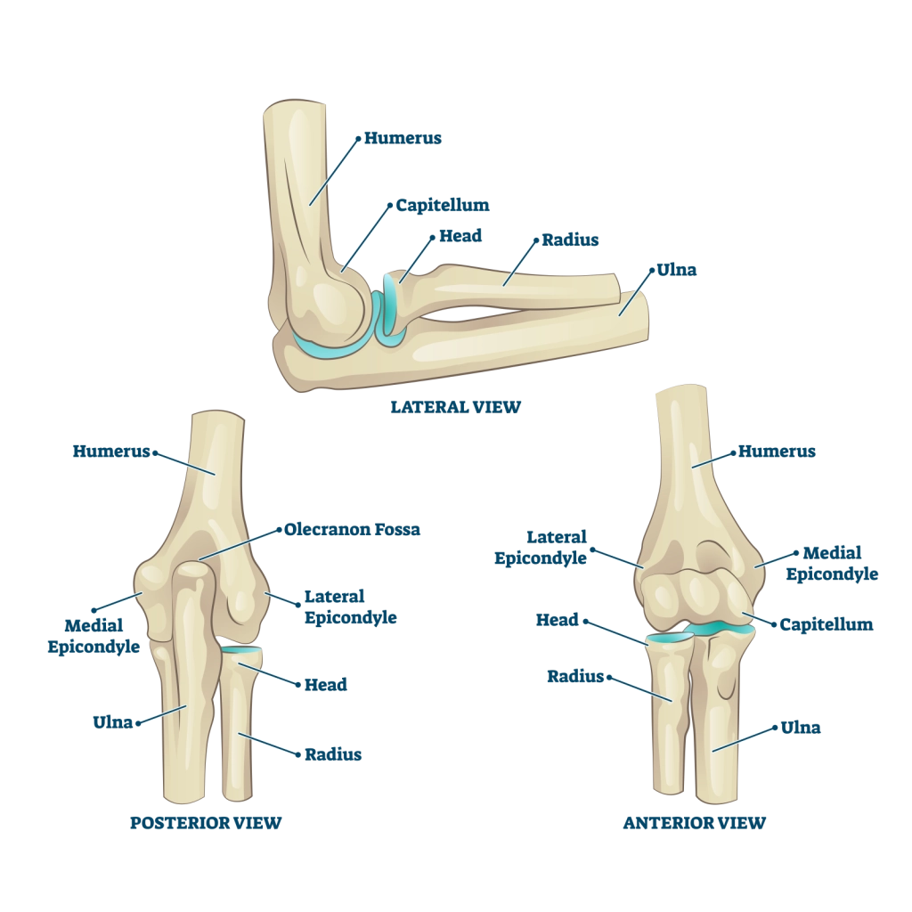
Bony Structures of the Elbow
Humerus - The distal end of the upper arm expands to form distinct condyles, articular surfaces, and fossae that meet the forearm bones.
Radius - The lateral bone of the forearm (thumb side), its disc-shaped, round head articulates with both the humerus and ulna, allowing for forearm rotation.
Radial tuberosity for biceps muscle
Ulna - The medial bone of the forearm (pinky side), its u-shaped and forms the main hinge joint with the humerus and forms the bony part of the elbow
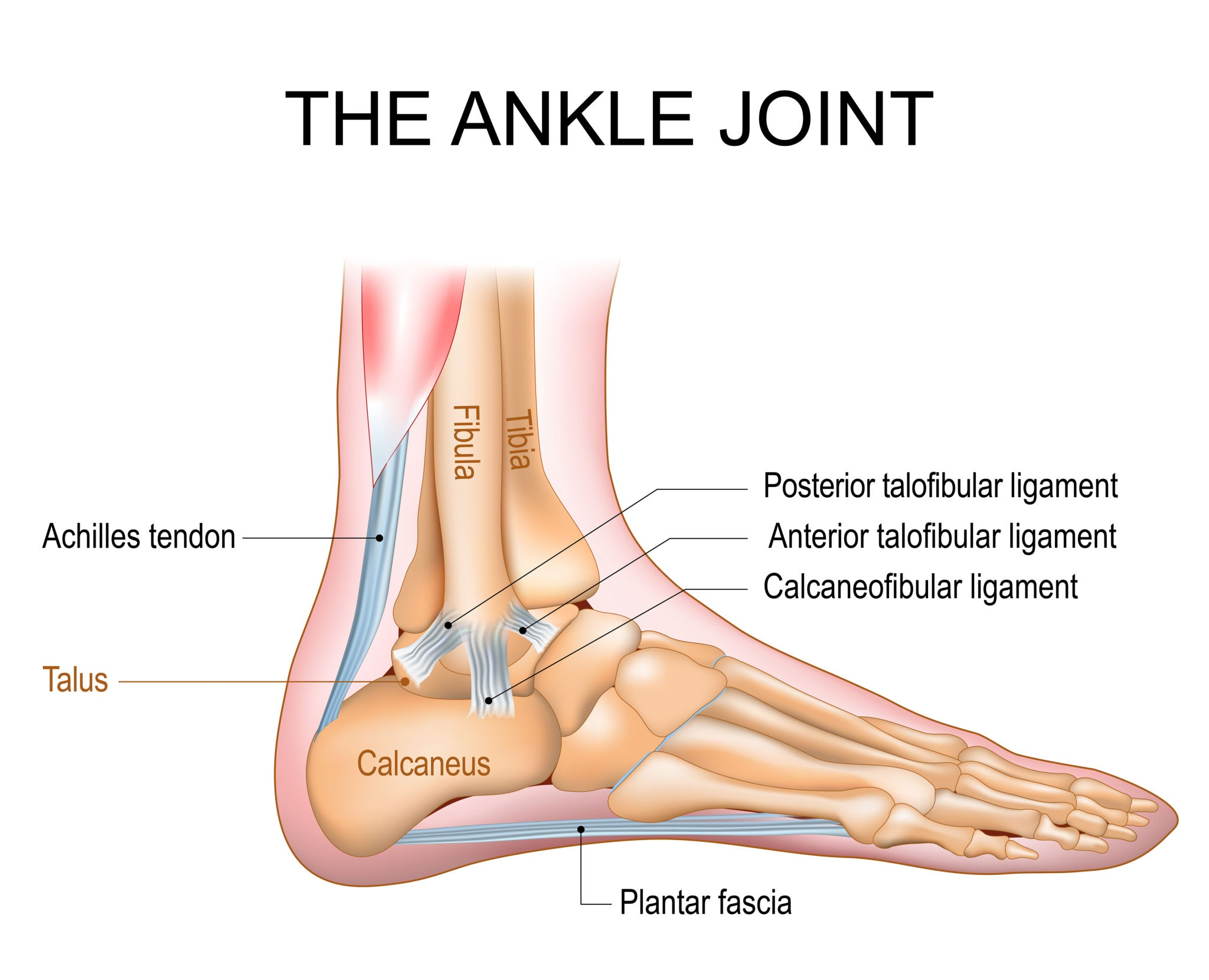
Bony Features of the Ankle Joint
Tibia (Shin bone) - The larger, weight bearing medial bone the lower leg. Its distal end forms the roof and inner wall of the ankle joint.
Fibula (Calf bone) - The slender, lateral bone of the lower leg. It does not bear body weight directly, but it forms the outer stabilization wall of the ankle joint.
Talus (Ankle bone) - A rounded tarsal bone of the foot. It sits beneath the tibia and fibula, but above the heel bone connecting your leg to your foot, helping carrying your body weight.
Pelvis - What axial and appendicular bones form the pelvis?
Axial Bones - Sacrum and Coccyx
Appendicular - Ilium, Ischium, Pubis
Ilium - Superior, flared portion that forms the prominent hip crest (iliac crest). Greater sciatic notch contains the sciatic nerve.
Ischium - Posterior and inferior portion of the pelvis that includes the ischium tuberosity that supports body weight when sitting
Pubis - the anterior portion that curves medially to join the opposite pubic bone at the pubic symphysis.
Female Pelvis is less massive, more shallow, and larger pubic arch than male

Functions of the arches of the foot
Sole of the foot does not normally rest on the ground. If it does its called pes planus
3 springy arches absorb stress of walking
Medial Longitudinal Arch - Runs along the inner side of the foot from heel to hallux, main shock absorber
Lateral Longitudinal Arch - Extends along the outer side of the foot from heel to little toe, providing balance and stability
Transverse Arch - Extends across the middle of the foot, distributes weight across the forefoot.
Arches are held together by short, strong ligaments