ANAT 5010 - unit 15
1/95
There's no tags or description
Looks like no tags are added yet.
Name | Mastery | Learn | Test | Matching | Spaced | Call with Kai |
|---|
No analytics yet
Send a link to your students to track their progress
96 Terms
thoracic wall
external boundary of thoracic cavity
- ribs 1-12
- sternum
- costal cartilages
- vertebrae TV1-12
- intercostal muscles
- intercostal neurovasculature
what makes up the thoracic wall?
- TV1
- rib 1
- manubrium
what are the boundaries of the superior thoracic aperture?
- TV12
- rib 12
- anterior tips of ribs 1-12
- costal cartilages of ribs 11-12
- xiphoid process
what are the boundaries of the inferior thoracic aperture?
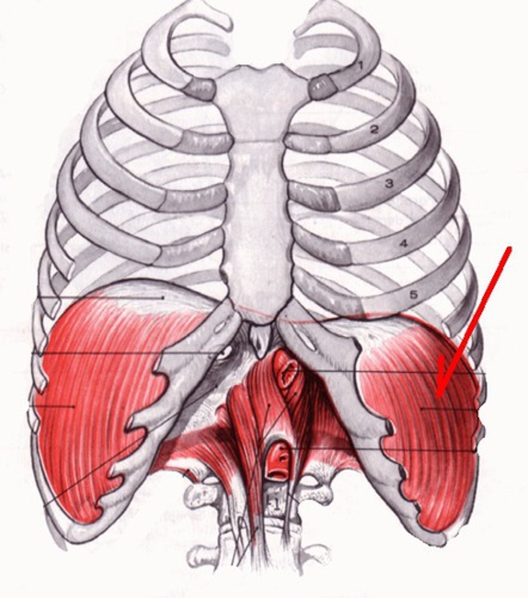
diaphragm
flat, skeletal muscle; floor of thoracic cavity; roof of abdominal cavity
manubrium
most superior bone of sternum; articulates with clavicles at clavicular notches; articulates with ribs 1-2; articulates with body of sternum at sternal angle
TV4-5
what vertebral level is the sternal angle at?
body of sternum
middle bone of sternum; articulates with ribs 2-7; articulates with xiphoid process at xiphisternal junction
TV9
what vertebral level is the xiphisternal junction at?
xiphoid bone
inferior bone of sternum; articulates with rib 7
head of rib
articulates with TV bodies
neck of rib
area between head and tubercle of rib
tubercle
rough patch with facet for transverse process of TV
angle of rib
curve; most posterior point of rib cage
shaft
body; central; lateral
costal groove
inferior; houses neurovasculature that passes through the ribs
scalene tubercle
divides grooves for subclavian vein and artery on the superior surface of rib 1; attachment point for anterior scalene muscle
costovertebral joint
between head of rib and bodies of two TV
costotransverse joint
between tubercle of rub and transverse process of same level TV
costal cartilages
attach to anterior ends of each rib via costochondral joints
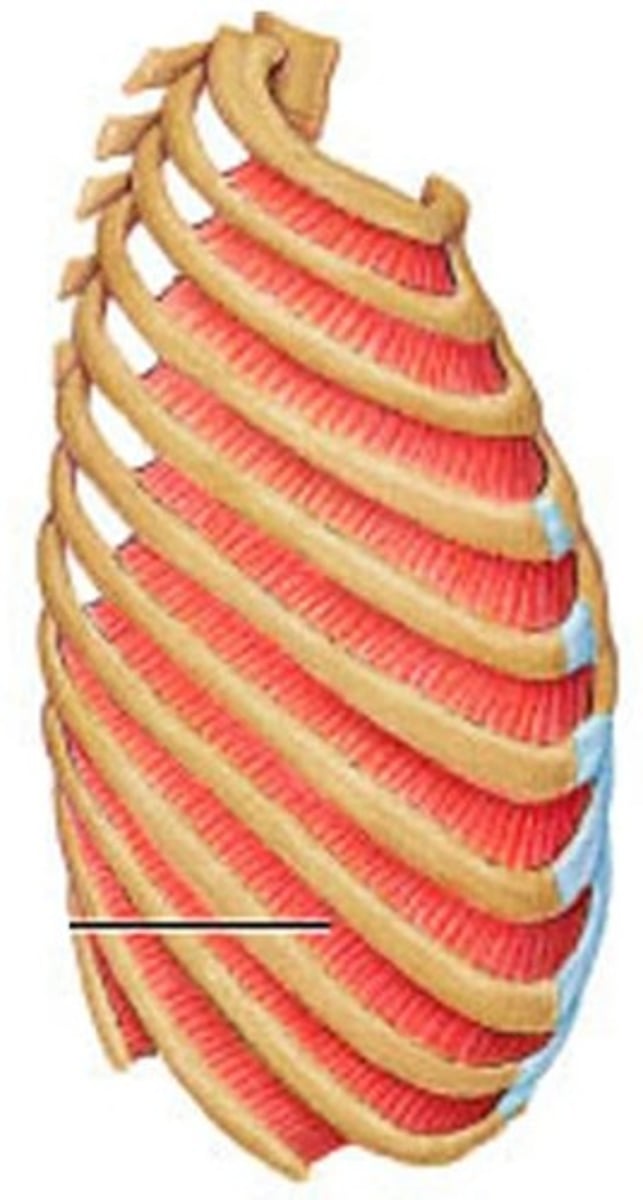
costal arch
defined by margin of cartilages of ribs 7-10
joint 1
what rib joint is fibrocartilage, immobile, and articulates with the manubrium?
joint 2
what rib joint is synovial and articulates with the sternal angle?
joint 3-6
what rib joint is synovial and articulates with the body of the sternum?
joint 7
what rib joint is synovial and articulates with the xiphisternal junction?
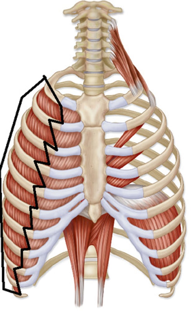
true ribs (1-7)
cartilages attach directly to the sternum
false ribs (8-10)
cartilages attach indirectly to the sternum via other cartilages
floating ribs (11-12)
cartilages do not attach to sternum
origin: lower border of superior rib
insertion: upper border of inferior rib
action: elevate rib; assist in respiration
innervation: intercostal nerves
what is the o,i,a,in of the external intercostals?
origin: upper border of inferior rib
insertion: lower border of superior rib
action: depress ribs; assist in expiration
innervation: intercostal nerves
what is the o,i,a,in of the internal intercostals?
innermost intercostals
fibers of internal intercostals separated by the neurovascular bundle (V A N)
anterior intercostal membrane
fibrous continuation of external intercostal muscles
posterior intercostal membrane
fibrous continuation of internal intercostal muscles
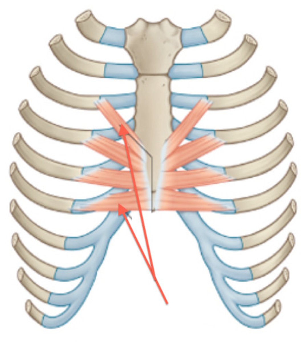
origin: body of sternum, xiphoid process, costal cartilages
insertion: internal surfaces of costal cartilages of ribs 2-6
action: depress ribs; assist in expiration
innervation: intercostal nerves
what is the o,i,a,in of the transversus thoracis?
mixed
are the intercostal nerves mixed, motor, or sensory?
T1 VPR
atypical intercostal nerve that contributes to the brachial plexus
T2 VPR
atypical intercostal nerve; intercostobrachial nerve formed from lateral cutaneous branch of VPR; contributes to medial brachial cutaneous nerve
T12 VPR
atypical intercostal nerve; subcostal; abdominal
internal thoracic artery
branch of subclavian artery; travels in thoracic wall superficial to the transversus thoracis muscle; supplies anterior ICSs 1-6
- superior epigastric artery
- musculophrenic artery
what are the terminal branches of the internal thoracic artery?
superior epigastric artery
medial branch of the internal thoracic artery; courses inferiorly to the anterior abdominal wall
musculophrenic artery
lateral branch of the internal thoracic artery; courses along the costal margin
interal thoracic
what are the anterior ICS 1-6 arteries a branch of?
musculophrenic
what are the anterior ICS 7-11 arteries a branch of?
highest intercostal artery
what are the posterior ICS 1-2 arteries a branch of?
descending thoracic aorta
what are the posterior ICS 3-11 arteries a branch of?
anterior intercostal veins
drain into internal thoracic or musculophrenic veins
posterior intercostal veins
drains into azygos system of veins
mediastinum
midline space between lungs; divided into superior and inferior at sternal angle
- anterior mediastinum
- middle mediastinum
- posterior mediastinum
what are the divisions of the inferior mediastinum?
- thymus
- brachiocephalic veins and superior vena cava
- aortic arch and branches
- trachea
- esophagus
- thoracic duct
- phrenic nerves
- vagus nerves
what are the contents of the superior mediastinum?
thymus gland
primary lymphatic organ; begins involution into fatty tissue after 1 year old; bi lobed; anterior to major blood vessels
brachiocephalic veins
formed from subclavian vein and internal jugular vein
superior vena cava
where do the brachiocephalic veins drain into?
- brachiocephalic trunk
- left common carotid
- left subclavian
what are the branches of the aortic arch?
ligamentum artertiosum
connects inferior surface of aortic arch to pulmonary trunk; landmark for left recurrent laryngeal nerve
phrenic nerves
cross anterior to subclavian artery; cross anterior to root of lungs; run between mediastinal pleura (external) and pericardium (internal)
vagus nerves
cross anterior to subclavian artery; cross posterior to root of lungs; give off laryngeal branches
recurrent laryngeal nerves
branches of vagus nerves; mixed sensory and motor
right recurrent laryngeal nerve
loops under right subclavian artery
left recurrent laryngeal nerve
loops under aortic arch lateral/posterior to ligamentum arteriosum
- fat
- connective tissue
- inferior 1/2 of thymus
- lymph nodes
what are the contents of the anterior mediastinum?
- heart
- pericardium
- roots of great vessels
- primary bronchi
what are the contents of the middle mediastinum?
pericardium
membrane that surrounds the heart
serous membrane
double layered membrane with thin cavity of fluid between the layers
fibrous pericardium
external layer of serous membrane; dense CT
serous parietal pericardium
fuses to internal side of fibrous layer; external to serous fluid
serous visceral pericardium
adheres to heart surface; "epicardium"
phrenic nerve
what provides sensory innervation to the pericardium?
cardiac tamponade
compression of heart due to fluid accumulation in pericardium
- ascending aorta
- pulmonary trunk (pulmonary arteries)
- pulmonary veins
- superior vena cava
- inferior vena cava
what all makes up the roots of the great vessels?
descending thoracic aorta
inferior continuation of aortic arch at TV4; passes through diaphragm at TV12
abdominal aora
continuation of the descending thoracic aorta
- 3rd-11th posterior intercostal arteries
- subcostal artery
- left bronchial artery
- esophageal artery
- mediastinal artery
- pericardial artery
- superior phrenic artery
what are the branches of the descending thoracic aorta?
azygos system of veins
handles drainage of posterior thoracic wall
azygos vein
drains superiorly into superior vena cava; right of median plane
hemiazygos vein
drains into azygos vein; drains superiorly then crosses vertebral body at TV9
accessory hemiazygos vein
drains into azygos vein; drains inferiorly then crosses vertebral body at TV8
thoracic duct
largest lymphatic vessel in the body; extends superiorly from cisterna chyli at LV1-2; passes through superior mediastinum; empties into left venous angle
esophagus
flattened muscular tube; inferior continuation of pharynx at CV6; terminates at TV11; posterior to trachea and anterior to and right of the aorta
inferior thyroid artery
what is the origin of arterial branches to the upper portion of the esophagus?
- esophageal artery
- bronchial artery
- mediastinal artery
what is the origin of arterial branches to the middle portion of the esophagus?
esophageal branch of left gastric artery
what is the origin of arterial branches to the lower portion of the esophagus?
inferior thyroid vein to the SVC
what drains the upper portion of the esophagus?
azygos system to the SVC
what drains the middle portion of the esophagus?
esophageal branch of left gastric vein to the hepatic portal vein
what drains the lower portion of the esophagus?
esophageal plexus
supplies smooth muscle of esophagus; contains postganglionic sympathetic fibers from the cardiopulmonary splanchnic nerves; contains preganglionic parasympathetic fibers from the vagus nerve
anterior vagal trunk
continuation of the left vagus nerve
posterior vagal trunk
continuation of the right vagus nerve
thoracic sympathetic trunk
continuous with that of cervical and lumbar regions; lies near head and neck of ribs
lateral
where are white communicating rami located?
medial
where are gray communicating rami located?
thoracic splanchnic nerves
preganglionic sympathetic fibers; branch off thoracic ganglia medially without synapsing; innervate targets in abdomen
greater splanchnic nerve
branches from T5-T9 ganglia
lesser splanchnic nerve
branches from T10-T11 ganglia
least splanchnic nerve
branches from T12 ganglia