The Conducting System & Heart
 Call Kai
 Learn
Learn Practice Test
 Spaced Repetition
 Match
Match Flashcards
 Knowt Play
1/24

There's no tags or description
Looks like no tags are added yet.
Name | Mastery | Learn | Test | Matching | Spaced | Call with Kai | Chat |
|---|
No analytics yet
Send a link to your students to track their progress
25 Terms

What is #1
SA Node

What is #2
AV Node

What is #3
AV Bundle, Bundle of His

What is #4
Purkinje fibers
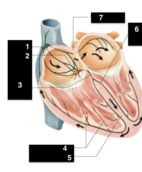
What is #5
Interventricular Septum
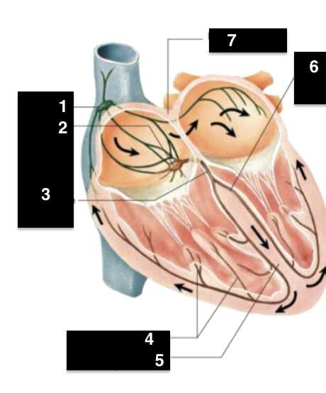
What is #6
Left bundle branch
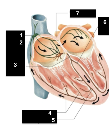
What is #7
Interatrial Septum
Conduction Pathway
SA Node → Atria → AV Node → Bundle of His → Right and Left Bundle Branches → Purkinje Fibers
Epicardium
outermost layer of heart
Myocardium
Cardiac Muscle, responsible for pumping action of heart
Endocardium
Inner most layer
The cardiac cycle
the period between the start of one heartbeat and the beginning of the next
Systole
Contraction of a chamber
Diastole
Relaxation of a chamber
Phases of the cardiac cycle
Atrial systole → atrial diastole → ventricular systole → Ventricular diastole
Ausculation
listening to heart sound via stethescope
S1
“lub” caused by the closing of the AV valves
S2
“dub” caused by the closing of the semilunar valves
S3
a faint sound associated with blood flowing into the ventricles
S4
faint sound associated with atrial contraction
Heart Murmur
abnormal sound consisting of a clicking, rushing, or gurgling sound heard before, between, or after normal heartbeat
Equipment Needed to take pulse
Inflatable Cuff & Pump, Pressure Gauge, Stethoscope, sphygmomanometer
Locations to take Pulse
1) Wrist: Radial Pulse
2) Neck: Carotid Pulse
3) Inner Elbow: Brachial Pulse
What creates a pulse
The artery walls expanding and contracting as blood flows through