Hilary building a phenotype
 Call Kai
 Learn
Learn Practice Test
 Spaced Repetition
 Match
Match Flashcards
 Knowt Play
1/145

There's no tags or description
Looks like no tags are added yet.
Name | Mastery | Learn | Test | Matching | Spaced | Call with Kai | Chat |
|---|
No analytics yet
Send a link to your students to track their progress
146 Terms
how do enzymes speed up reactions?
they lower the activation energy for the conversion to the transition state because:
the active site is more complementary to the transition state than the substrate, meaning the substrate is under strain and the transition state is stabilised (lower energy level than when free)
they position the substrate into the most favourable steric arrangement, close to the involved residues in the active site (by weak, short-range noncovalent bonds = close fit with substrate)
what are the steps of dna replication? what are the differences between prokaryotes and eukaryotes?
an initiator protein recognises and binds an A/T rich (weakly bonded) region in the DNA, causing separation by torsional stress
this allows the helicase ring to be opened + closed around one strand by loader proteins
ATP hydrolysis causes a conformational change in helicase pulling the strand through, acting as a wedge
SSBs prevent re-annealing and secondary structure folding
short ~11nt RNA primers are produced (no exonuclease site, so can start from scratch without proofreading unlike DNA polymerase)
a beta clamp ensures DNA polymerase doesn’t fall off (esp. leading strand), attached by a clamp loader
DNA polymerase adds dNTPs 5’ → 3’ (in Okazaki fragments on lagging strand, trombone model), driven by the release of pyrophosphate and facilitated by 3 magnesium ion cofactors
the primers are removed and the strand elongated until they can be joined by DNA ligase
topoisomerases remove DNA positive supercoils from helicase unwinding
in prokaryotes: DnaA initiator, DnaC loader, DnaB helicase, DnaG primase, pol-III primarily, pol-I degrades primers- theta shape, no histones so 100kb/min
in eukaryotes: ORC initiator, CDC + CDT loaders, MCM helicase (mini chromosome maintenance), pol alpha starts using primase subunit, then swapped for pol delta/epsilon, primers removed by RNAase H1 + flap endonuclease, telomerases used- multi origin, histones so 2kb/min
what are the different ways that cells can regulate metabolic pathways? mention the examples of each
by changing the amounts of enzymes present by gene expression eg. the lac operon- this is a slow response
by controlling the activity of pre-existing enzymes by:
mass action- increasing the substrate concentration increases enzyme activity, but this method has a limit because all enzymes have a Vmax that can’t be exceeded
negative feedback loops and allosteric regulation- the end product of a reaction controls the enzyme activity by allosteric inhibition eg. regulation of ATCase by CTP, and of PFK by PEP/citrate
spatial organisations- changing from diffuse to localised enzyme distributions increases enzyme activity by increasing the local concentration and reducing metabolite diffusion eg. purinosomes in the IMP production pathway
post-translational modifications- PTMs change the properties of enzymes eg. phosphorylation of pyruvate decarboxylase by kinases
how is CTP production regulated?
aspartate is converted into cytosine triphosphate (CTP, a dNTP for DNA replication) by aspartate transcarbamoylase (ATCase)
CTP over-production is prevented by end-product inhibition because CTP is an allosteric inhibitor of ATCase
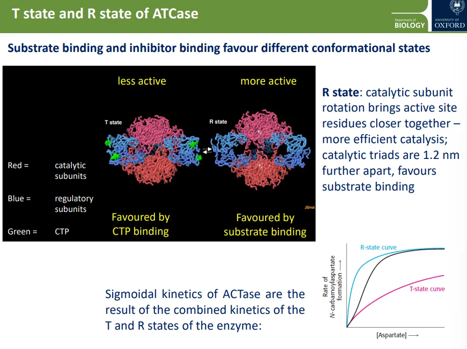
ATCase has allosteric kinetics (sigmoidal) instead of Michaelis-Menten (hyperbolic), because:
when the aspartate substrate binds to one of the 6 active sites, it causes a conformational change by the rotation of the 2 catalytic trimers- this converts the enzyme into a relaxed, R, state, favouring further substrate binding (cooperative binding)
if CTP binds to one of the 3 regulatory dimers, it will favour the tense, T, state conformation so that the trimers are tightly bound together- this makes it harder for aspartate to bind
this causes sigmoidal kinetics, which allows for a fast response within a narrow substrate concentration window
how is PFK activity regulated in E. coli vs humans?
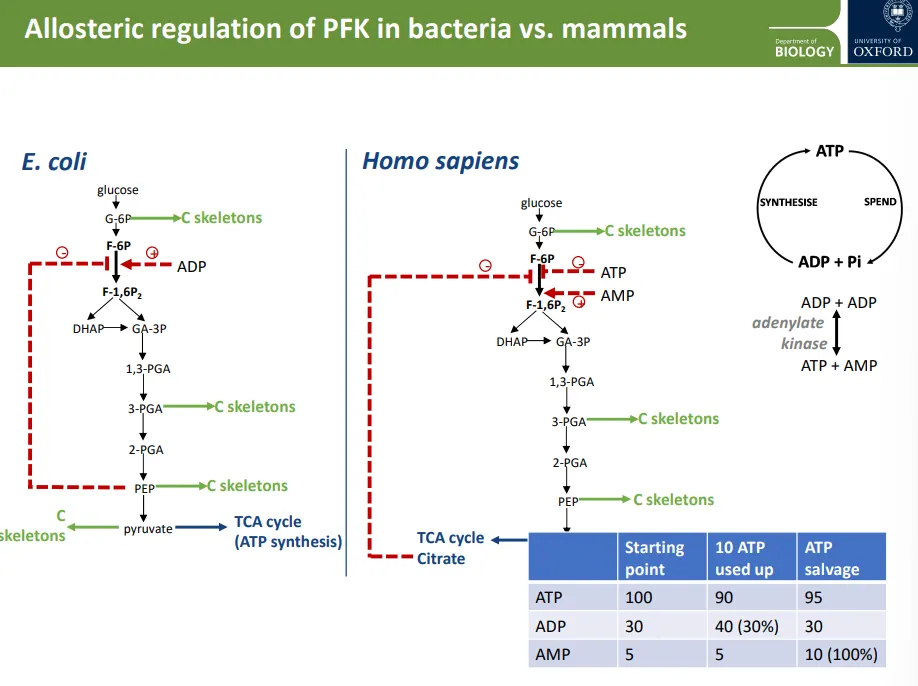
phosphofructokinase (PFK) is an enzyme used early in the glycolysis pathway
in E.coli:
if PEP (the last step of glycolysis before pyruvate) concentrations are high, it starts to allosterically inhibit the PFK enzyme, so carbon skeletons can be removed from the glycolysis pathway at glucose 6-phosphate for biosynthesis, because enough pyruvate is being produced for the TCA cycle
if ATP synthesis is slow, ADP builds up, which promotes PFK activity so that more pyruvate is produced for the TCA cycle and ATP synthesis
in humans:
the PFK enzyme is inhibited by citrate from the TCA cycle instead of PEP
it is activated by AMP, not ADP, and is inhibited by ATP
this is because in humans, adenylate kinase converts ADP into ATP and AMP, for ATP salvage
the PFK enzyme can be a lot more sensitive to an increase in AMP, because if we relied on ADP as a signal, it wouldn’t change enough (table)
how is IMP production regulated?
inosine monophosphate (IMP) is used to produce purine nucleotides
the 6 enzymes in the IMP production pathway are controlled by changing their spatial localisation
when purines are highly available, the enzymes are widely distributed
when there is a shortage of purines, the enzymes come together in biocondensates (localised regions) called purinosomes
enzyme activity increases in purinosomes compared to the diffuse form, because the local concentration of enzyme increases and there is less opportunity for the metabolites to diffuse away
how is PDC activity regulated?
pyruvate decarboxylase (PDC) converts pyruvate into acetyl CoA for the TCA cycle
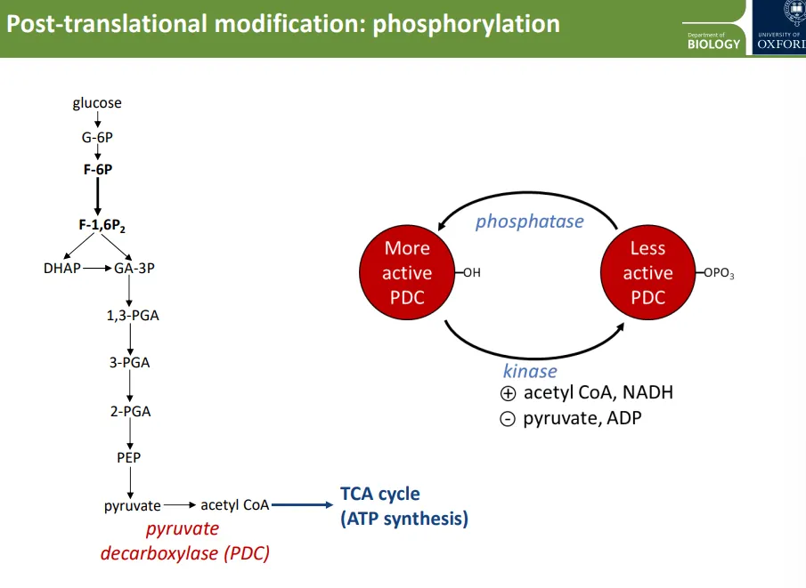
PDC is controlled by post-translational modification (phosphorylation by kinase enzymes and dephosphorylation by phosphatases)
PDC is more active when dephosphorylated, and less active when phosphorylated
the kinase enzyme is promoted by acetyl CoA and NADH, and inhibited by pyruvate and ADP, so:
when PDC isn’t active enough (high pyruvate + ADP), kinase is inhibited, so the enzyme is dephosphorylated and more active, to produce acetyl CoA for the TCA cycle
when PDC is too active (high acetyl CoA and ATP), kinase is promoted, so the enzyme is phosphorylated and less active, so the TCA cycle decreases
what is the energy source used to polymerise polypeptides, starch and cellulose?
polymerisation is not a thermodynamically favourable reaction, so it requires coupling to a more favourable one
to make polypeptides, one GTP is hydrolysed for each aa. added
to make starch, two ATP are used for each glucose monomer added (one is hydrolysed directly, the second is used to make ADP-glucose, which then gets hydrolysed)
to make cellulose, one UTP is used for each glucose monomer added, by making UDP-glucose
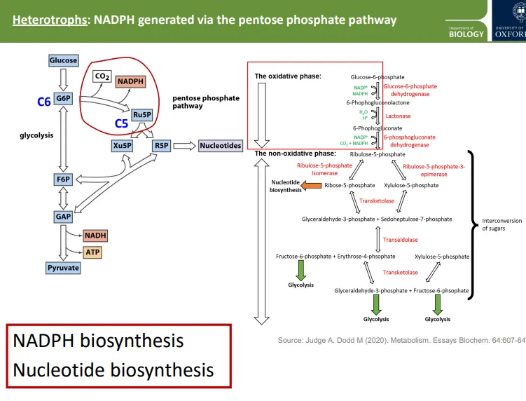
what is the pentose phosphate pathway used for?
photosynthesis can produce some NADPH, but in non-photosynthetic organisms and the non-photosynthetic tissues of plants, the pentose phosphate pathway is used to meet NADPH needs (for biosynthesis)
glucose can be diverted from glycolysis into the pentose phosphate pathway instead
this process produces NADPH, CO2 and other compounds, which get converted so that they can then enter glycolysis, or be used to biosynthesise nucleotides
how is glutamate biosynthesised?
bioavailable nitrates are converted into ammonium ions by nitrate and nitrite reductase, using NADPH (produced by the pentose phosphate pathway)
these react with a-ketoglutarate (same as 2-oxoglutarate), which is produced in the TCA cycle, catalysed by glutamate dehydrogenase, to make the amino acid glutamate using NAD(P)H
the production of most other amino acids involves transamination by the reverse of this reaction
the starting molecules to produce all amino acids come from the TCA cycle, pentose phosphate pathway and glycolysis
mammals have lost many amino acid biosynthesis enzymes, so essential amino acids must be gained from diet
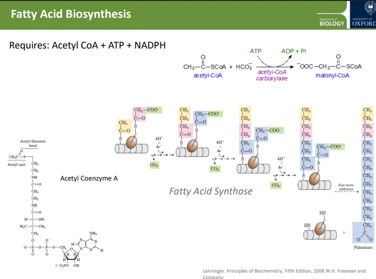
how are fatty acids biosynthesised?
when there is sufficient glucose in the cell, acetyl-CoA is drawn away from the TCA cycle to biosynthesise fatty acids
acetyl-CoA is converted into malonyl-CoA, using ATP, catalysed by acetyl-CoA carboxylase
an acetyl coA and a malonyl-CoA (continually replaced) bind to the active site of fatty acid synthase at 2 cysteine residues
in each reaction, 2 carbons from the malonyl-CoA are added to the emerging chain (which starts from the acetyl-CoA), using protons and electrons and producing CO2
why is glycolysis the most simple process it can be?
glycolysis has to act within many constraints:
glucose can easily diffuse out of the cell, so the addition of negatively charged phosphates must be done early in the pathway to reduce loss of substrate
glycolysis must proceed via a route that avoids the production of highly reactive intermediates that would be toxic to the cell
glycolysis must generate a range of intermediate molecules that provide specific carbon skeletons that can be used in other biosynthesis pathways eg. amino acids
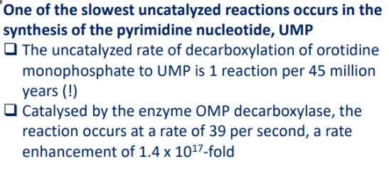
what’s an example of a reaction that is greatly sped up by enzymes?
in the synthesis of pyrimidine (UMP), orotidine monophosphate must be decarboxylated by OMP decarboxylase
the rate is enhanced by this enzyme from 1 reaction per 45 million years to 39 reactions per second
this is a 1.4 × 10¹⁷ fold increase
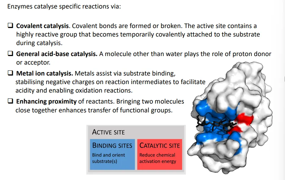
what are the four methods of enzyme catalysis?
what is a catalytic triad?
some enzymes (most commonly hydrolases and transferases) have catalytic triads, a set of three closely positioned residues
these are normally made up of an acid, a base, and a nucleophile- the acid and base polarise and activate the nucleophile to make it powerful enough for covalent catalysis (where a temporary covalent bond is formed with the substrate)
the nucleophile is normally serine or cysteine, or sometimes threonine
the base is normally histidine
the acid is normally aspartate or glutamate
what is an example of covalent and acid-base catalysis?
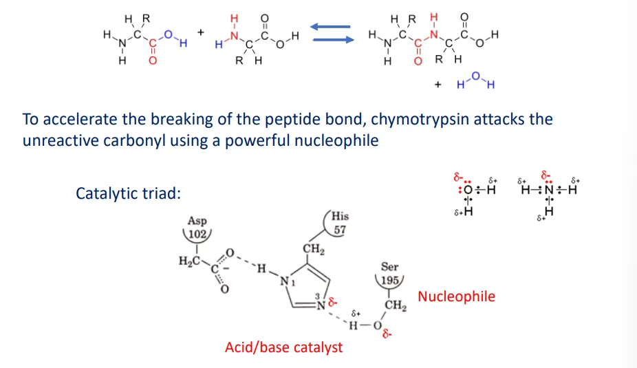
chymotrypsin, a serine protease, catalyses the hydrolysis of peptide bonds
this is done by activating a nucleophile to make it more powerful- because the nucleophile has ‘excess’ electrons it has a propensity to form new covalent bonds with the electrophile
the catalytic triad is made up of aspartate, histidine and serine
the aspartate (acidic) is hydrogen bonded to the histidine (base) to position the histidine side chain correctly, which deprotonates and activates the serine nucleophile
the activated nucleophile attacks the carbonyl in the peptide bond (producing a covalent bond) to form an intermediate (transition state)
this negative charge buildup is stabilised by an oxyanion hole (the amide groups in the backbones of glycine and serine residues)
the peptide bond breaks, but one half is still covalently bound to the enzyme as an intermediate
a water molecule attacks the carbonyl, forming a new intermediate which collapses to release the second product from the enzyme
what are the assumptions in the michaelis-menten model of enzyme kinetics and what is it?
we are measuring the initial velocity, so the reaction is irreversible as we aren’t near equilibrium
the substrate is in excess of the enzyme
the reaction is at steady state, so [ES] stays constant
the model describes how the velocity, V, changes with the substrate concentration, [S], according to the equation V = Vmax x [S] / ([S] + KM), where:
KM = Vmax / 2
KM is used as a measure of the enzymes affinity for its substrate, where a low KM means the substrate is weakly bound
![<ul><li><p>we are measuring the initial velocity, so the reaction is irreversible as we aren’t near equilibrium</p></li><li><p>the substrate is in excess of the enzyme</p></li><li><p>the reaction is at steady state, so [ES] stays constant</p></li></ul><p></p><ul><li><p>the model describes how the velocity, V, changes with the substrate concentration, [S], according to the equation <strong>V = V<sub>max</sub> x [S] / ([S] + K<sub>M</sub>)</strong>, where:</p><ul><li><p>K<sub>M</sub> = V<sub>max </sub>/ 2</p></li></ul></li></ul><p></p><ul><li><p>K<sub>M </sub>is used as a measure of the enzymes affinity for its substrate, where a low K<sub>M</sub> means the substrate is weakly bound</p></li></ul><p></p>](https://knowt-user-attachments.s3.amazonaws.com/8229f223-5c35-4500-8da3-7ebafffe2db4.png)
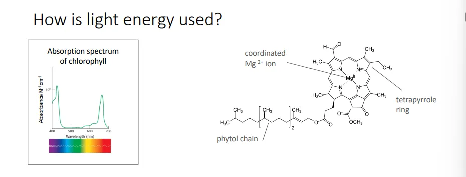
what is the structure of chlorophyll?
chlorophyll has a similar structure to heme complexes used in oxidative phosphorylation
it has a closed tetrapyrrole structure- the nitrogen atom from each pyrrole is coordinated to a central magnesium ion
they are effective photoreceptors because the pyrrole rings have alternating single and double bonds (conjugated polyenes), so the electrons aren’t as localised and restricted
this is attached to a highly hydrophobic 20-carbon phytol chain used for anchoring in the membrane
in what three ways can excited chlorophyll lose energy?
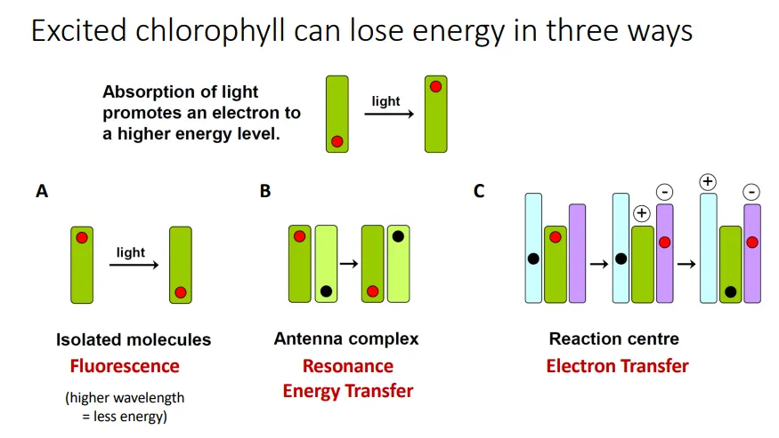
fluorescence- the energy absorbed is re-emitted as light (since some energy is lost as heat, the wavelength of the light increases ie. UV → visible) if there isn’t another chlorophyll nearby
resonance energy transfer- the energy can be passed onto an adjacent chlorophyll molecule by mechanical vibration
electron transfer- if there is a suitable electron acceptor nearby (as in reaction centres), the excited electron can be transferred, producing a charge difference through photoinduced charge separation
what is the structure of photosystems in higher plants and in cyanobacteria?
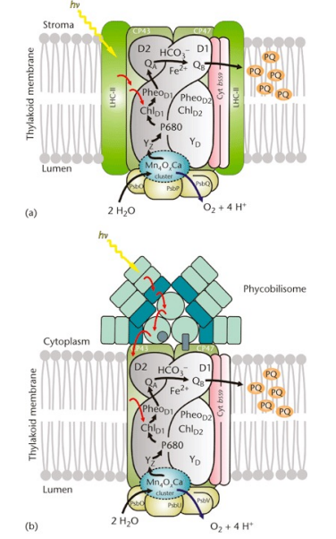
in higher plants (top picture), a ‘special pair’ of chlorophyll molecules, the reaction centre, is surrounded by an antenna complex
this complex funnels energy to the reaction centre by resonance energy transfer
this allows for photoinduced charge separation in the reaction centre by electron transfer
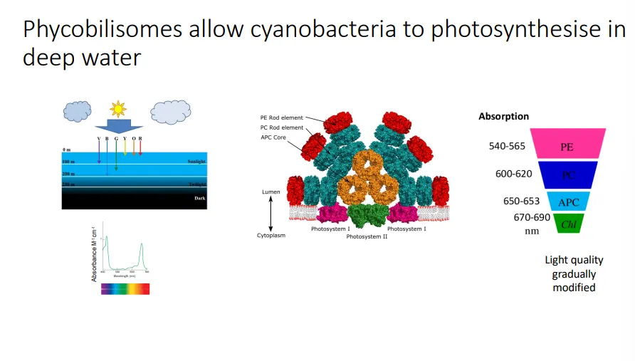
in cyanobacteria (bottom picture), the antenna complex is a structure on top of the reaction centre, called the phycobilisome
what is the phycobilisome and what is its purpose?
in cyanobacteria, the antenna complex, which is positioned on top of the reaction centre in the photosystems, is called the phycobilisome
it is made up of different pigments, not chlorophyll, called bilin chromophores- these are open-chain tetrapyrroles synthesised from heme, which are incorporated into different protein complexes: PC, PE and APC
this means the antenna complexes have different absorption properties, which allows cyanobacteria to expand their light-absorbing capacity for a deeper marine environment
water absorbs red light (long wavelength) the most, whereas blue and green (shorter wavelength) are transmitted further
the phycobilisomes specialise in absorbing shorter wavelength light (which chlorophyll can’t), and as the light is channelled towards the chlorophyll of the reaction centre, the wavelength is increased (by resonance energy transfer)
what happens at photosystem II?
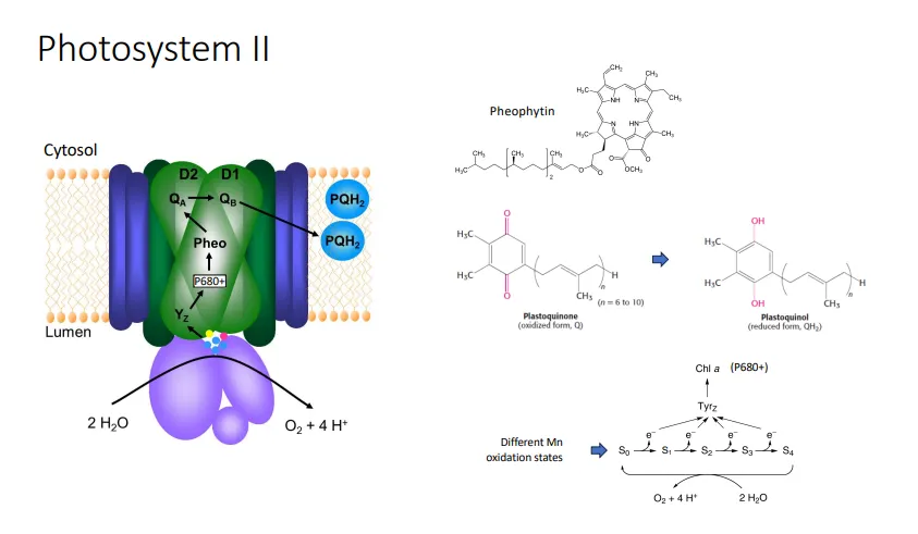
in photosystem II, an excited electron from the P680 (680nm) special chlorophyll pair is transferred to the first electron acceptor, pheophytin, to a bound plastoquinone at site QA, then to a mobile plastoquinone at site QB
the mobile plastoquinone is reduced by 2 of these electrons and the uptake of 2 protons (from the cytosol) to plastoquinol, QH2, which is used to replace the electrons lost in photosystem I
the electrons lost from photosystem II are replaced by the oxidation of water:
with each electron lost, the positively charged special pair, P680+, extracts an electron from a tyrosine residue in the water-oxidising complex (WOC)
this tyrosine radical extracts an electron from the manganese centre (can exist in multiple oxidation states)
once the manganese is oxidised 4 times, it is a strong enough oxidant to extract 4 electrons from 2 water molecules, producing oxygen and 4 protons (in the thylakoid lumen)
overall a proton gradient is created (high conc. in thylakoid lumen), which is used by ATP synthase to produce ATP used in the calvin-benson cycle to fix CO2
what happens at photosystem I?
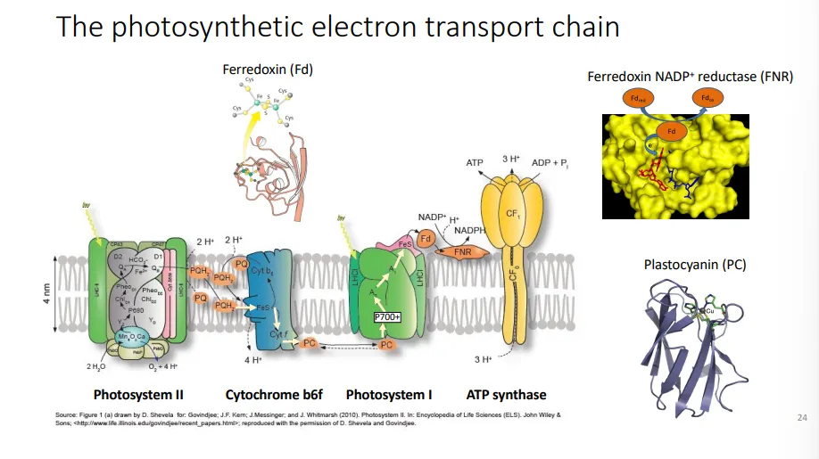
in photosystem I, an excited electron from the P700 (700nm) special chlorophyll pair is transferred to the first electron acceptor, quinone, then through a series of Fe-S clusters to ferredoxin
ferredoxin NADP+ reductase produces NADPH by transferring 1 electron from the reduced ferredoxin and 1 proton from the cytosol
the electrons lost from photosystem I are replaced by those lost from photosystem II:
the plastoquinol (QH2, reduced plastoquinone) produced at photosystem II diffuses through the membrane to the cytochrome b6f complex
this catalyses the transfer of 2 electrons from plastoquinol to plastocyanin, Pc, and releases the 2 protons into the thylakoid lumen
the reduced plastocyanin moves to photosystem I to be oxidised, replacing the electrons lost by the P700 special pair
overall a proton gradient is created (high conc. in thylakoid lumen), which is used by ATP synthase to produce ATP used in the calvin-benson cycle to fix CO2
what is the sequence of electron transfer through the two photosystems?
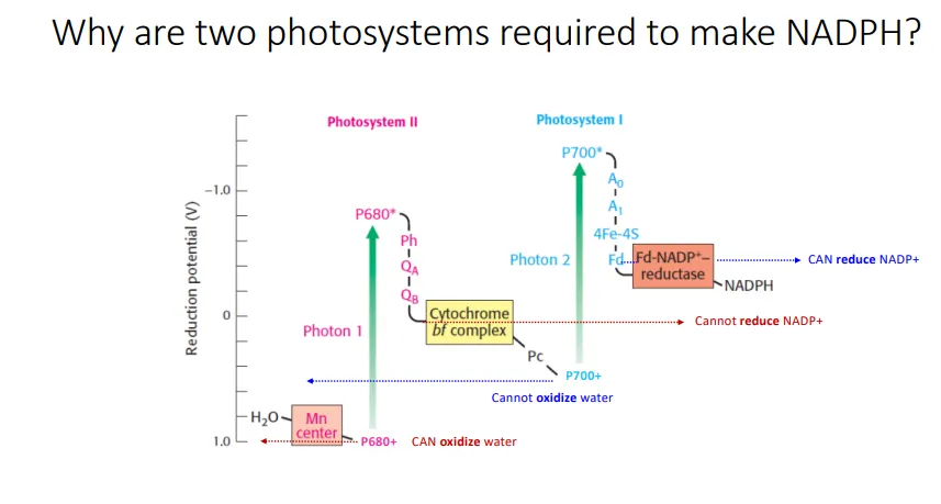
electrons are extracted from water molecules by the water-oxidising complex (manganese + tyrosine)
this replaces the electrons lost at photosystem II, which are excited by light
the excited electrons are passed through pheophytin and bound plastoquinone to a mobile plastoquinone, which is reduced to plastoquinol
the cytochrome b6f complex transfers the electrons from plastoquinol to plastocyanin
this replaces the electrons lost at photosystem I, which are excited by light
the excited electrons are passed through quinone and a series of Fe-S clusters to reduce ferredoxin
ferredoxin NADP+ reductase transfers the electrons from reduced ferredoxin to NADP+ to make NADPH
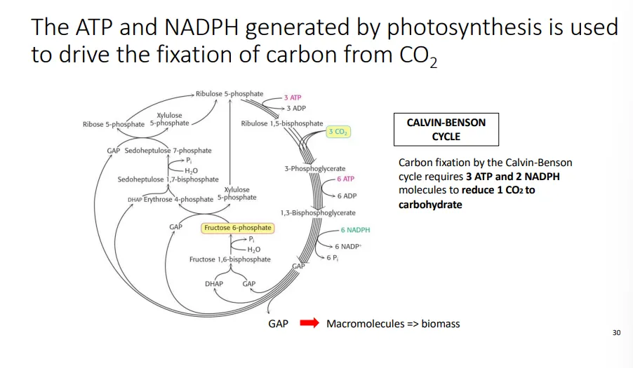
what is the basic sequence of the calvin-benson cycle?
ribulose 1,5-bisphosphate (5C) reacts with CO2 in a reaction catalysed by RuBisCo
ATP and NADPH (produced by the photosystems and ATP synthase) are used to produce glyceraldehyde 3-phosphate, GAP (3C)
1/6 of the GAP produced is the yield that can be used for biomass, while the rest is recycled back to the starting molecule in a complex cycle so as not to lose carbon
for each CO2 reduced, 3 ATP and 2 NADPH is used
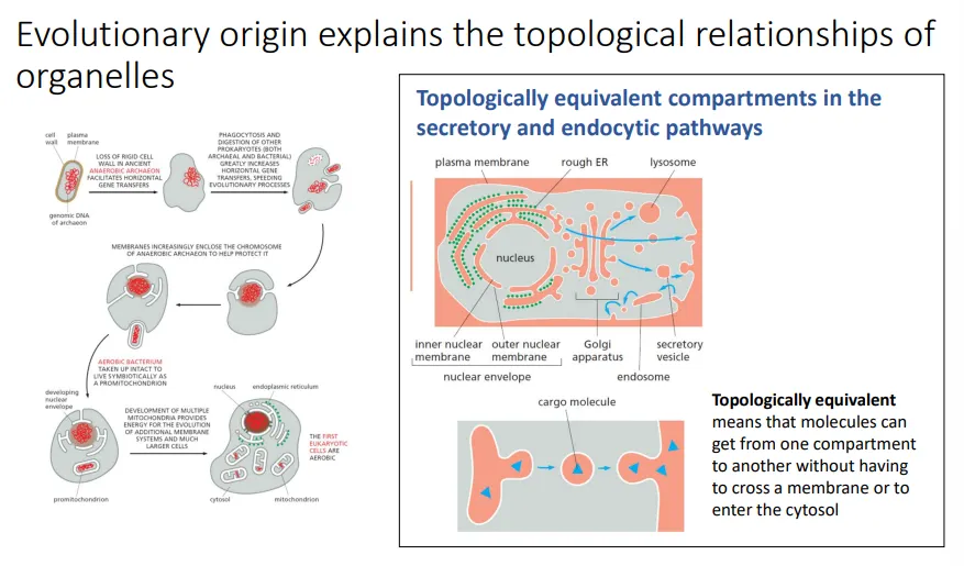
why do mitochondria and chloroplasts require a specialised transport system?
after the endosymbiotic events, huge amounts of gene transfer occurred from the endosymbionts to the nucleus so that they could be controlled and so genome efficiency could be improved
chloroplasts and mitochondria have highly reduced genomes, so they can’t produce all the proteins they need, which must be made by the nucleus
eg. human mitochondria only possess 13 protein-coding genes, all involved in oxidative phosphorylation
the other cell organelles are topologically similar, and so they can interconnect, integrate and exchange molecules easily without them crossing membranes
however, organelles of endosymbiotic origin are isolated from this vesicular traffic system because they evolved separately
this means they need their own transport system
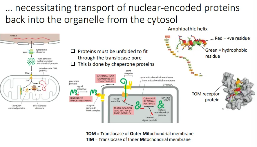
how are proteins imported into the mitochondria?
the mitochondrial proteins that must be imported from the cytosol (originating from genes in the nucleus) have a signal peptide sequence at the N-terminus
this is a recognition motif for the TOM (outer membrane) and TIM (inner membrane) translocase protein complexes
when the sequence is recognised, it triggers the transient linearisation of the precursor peptide so that it can be translocated across the double membrane into the mitochondrial matrix
then the signal peptide is cleaved off by signal peptidase to produce the mature protein
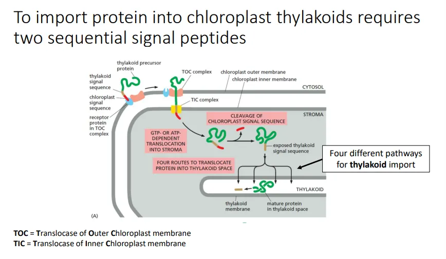
how are proteins imported into the chloroplast thylakoids?
the chloroplast proteins that must be imported from the cytosol (originating from genes in the nucleus) into the thylakoid lumen have two signal peptide sequences at the N-terminus
the first chloroplast signal sequence is a recognition motif for the TOC (outer membrane) and TIC (inner membrane) protein complexes
when the sequence is recognised, it triggers the transient linearisation of the precursor peptide so that it can be translocated across the double membrane into the chloroplast stroma
then the chloroplast signal peptide is cleaved off by signal peptidase, which exposes the second thylakoid signal sequence
this is recognised and allows the protein to be imported into the thylakoid lumen by four different pathways (then gets cleaved)
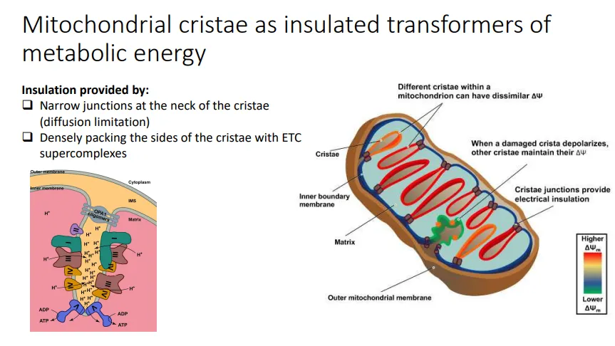
how are invaginations in mitochondria organised?
MICOS and OPA1 oligomers are protein complexes that create the junction between the cristae and the rest of the intermembrane space, at the ‘neck’
this insulates the invagination so that the proton gradients are maximised, as protons can’t diffuse into the rest of the IMS
this also means that if one cristae is damaged, the rest of the cristae don’t get depolarised, so the mitochondrion can remain functional
ATP synthase dimers are located at the tip, which results in the curvature of the membrane
how are invaginations in chloroplasts organised?
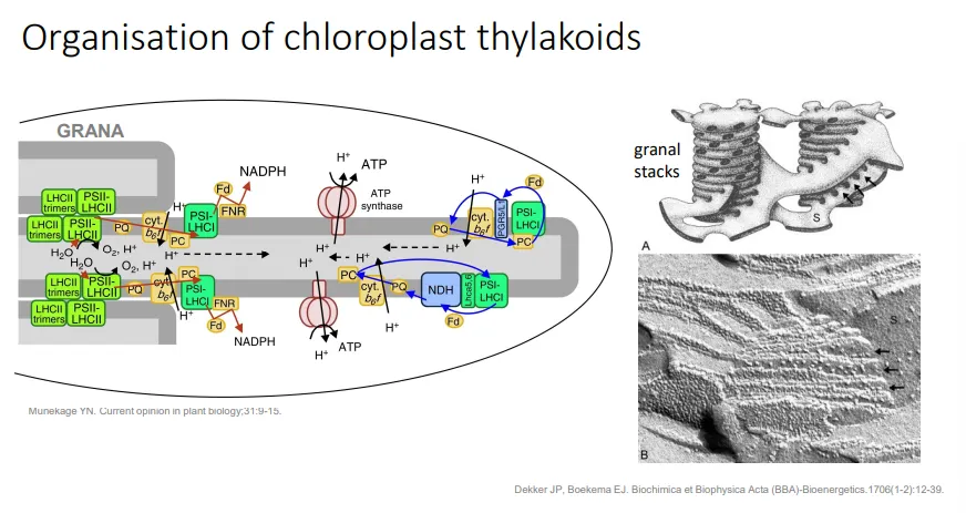
thylakoids are stacked into grana, which maximises the absorption of light
the granal stacks are connected by single thylakoids, because some sections of the thylakoid membrane must be exposed to the stroma
this allows the ETC and ATP synthase to release NADPH and ATP into the stroma for the calvin-benson cycle to occur
what are peroxisomes?
peroxisomes are organelles that metabolise fatty acids by beta-oxidation when glucose is scarce (which can go through the glycolytic pathway instead)
they use oxygen to remove electrons from the substrate, producing harmful hydrogen peroxide, which they then detoxify using catalase
the fatty acids are attached to coenzyme A, oxidised by using FAD to transfer 2 electrons and 2 protons to oxygen, and converted into acetyl CoA (which can enter the TCA cycle) by a series of enzymes
mammals evolved an electron transfer flavoprotein in their mitochondria, which is used instead of FAD and oxygen, passing the electrons directly to the ETC instead of producing hydrogen peroxide (though they still also use peroxisomes for long fatty acids)
they are also involved in detoxifying the compounds produced in photorespiration, a wasteful process where O2 competes with CO2 as a substrate for RuBisCo (as it evolved in anaerobic conditions), so that they can be used for sugar production
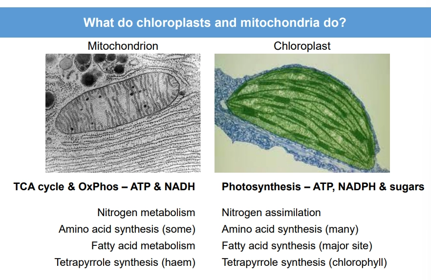
what are all the functions of mitochondria and chloroplasts?
what’s so special about ATP as an energy source?
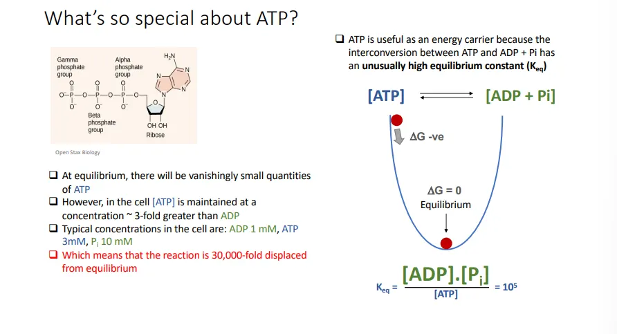
it has an unusually high equilibrium constant (conc of products/conc of reactants), so at equilibrium, there is an extremely small amount of ATP compared to ADP
however, ATP is maintained at high concentrations away from equilibrium in the cell
this greatly favours the forwards hydrolysis reaction, as the system tries to reach equilibriu,
this means the hydrolysis of ATP releases even more free energy
ATP is thermodynamically unstable, so when it breaks down it releases lots of free energy, but it is kinetically stable, so it has a slow rate of hydrolysis
this means it needs enzymes to speed up the reaction and doesn’t break down spontaneously
at the same time, the hydrolysis of ATP doesn’t release as much free energy as hydrolysing some other phosphate compounds
this intermediate free energy means the reverse condensation reaction can be driven by coupling it to the hydrolysis of other phosphate compounds
this is substrate level phosphorylation
how is ATP produced in glycolysis?
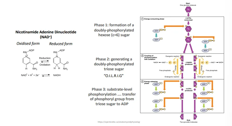
the glucose starting molecule is phosphorylated twice, at adjacent carbons, by the input of 2 ATP, to make it unstable enough to decompose into 2 3C molecules with one phosphate each
these lose two electrons and a proton, which is used to reduce the NAD+ cofactor to NADH
this reaction releases enough energy to add a free phosphate to the 3C molecule to become an unstable diphosphate molecule again
this instability then favours the release of the two phosphate groups from each 3C molecule to produce 4 ATP by substrate-level phosphorylation (coupling) and 2 pyruvate
why is fermentation necessary in anaerobic conditions?
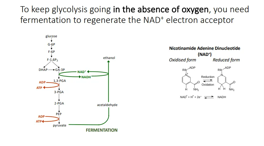
to keep the glycolysis reaction going, the NAD+ cofactor (to act as an electron acceptor) must be regenerated from NADH
in the absence of oxygen this must be done via fermentation (producing ethanol or lactic acid)
more complex organisms and aerobic organisms use the Krebs cycle to do this
what does the Krebs cycle do?
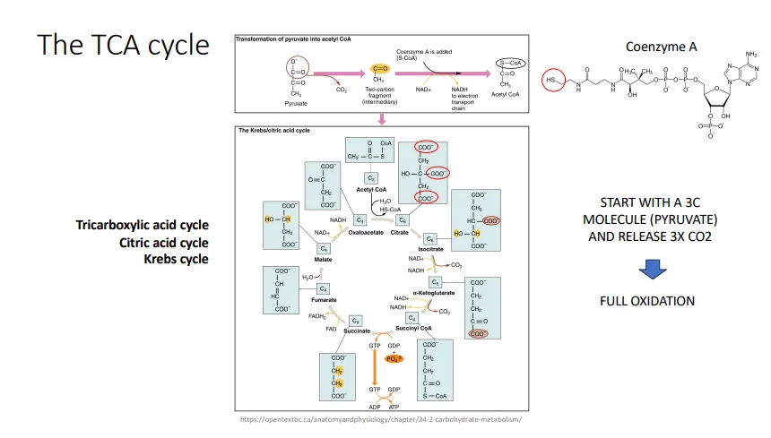
pyruvate is first oxidised to acetyl CoA, releasing 1 CO2 and 1 NADH
the Krebs cycle (citric acid/tricarboxylic acid cycle) extracts electrons from acetyl CoA to reduce NAD+ and FAD, so that they can enter the electron transport chain to produce ATP by oxidative phosphorylation (also produces 2 CO2, and 1 ATP by substrate-level phosphorylation)
this cycle is more efficient than just relying on fermentation, because it means the glucose starting molecule ends up fully oxidised to 6 CO2, so the maximum energy is released per glucose
how does the electron transport chain produce ATP?
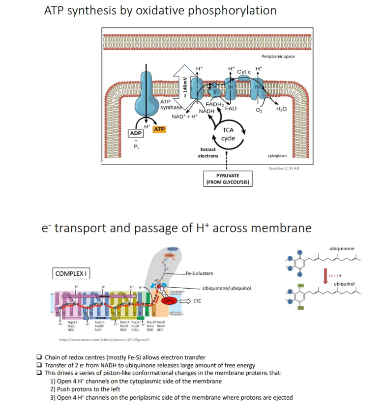
redox-active metals (in Fe-S clusters and heme complexes) are embedded, bonded to residues, in the membrane proteins of the electron transport chain
these recieve electrons from NADH and FADH2 from the Krebs cycle and glycolysis (which get oxidised, regenerating NAD+ for glycolysis)
they pass the electrons along to the ubiquinone molecule (hydrophobic) in the membrane, reducing it to ubiquinol and releasing large amounts of free energy (electrons then transferred to cytochrome c)
this drives the conformational changes in the membrane proteins, opening proton channels for protons to leave the cytoplasm
this produces an electrochemical gradient that is harnessed by ATP synthase to condense ADP to ATP, by a rotary motor mechanism (proton gradients also in alkaline smokers, origin of life)
the electrons are then used to reduce the final electron acceptor, oxygen
why is it beneficial to use proton gradients to make ATP?
flexibility- using oxidative phosphorylation means energy can be extracted from many different energy sources by extracting electrons to universal electron carriers, NAD+ and FAD (whereas just substrate-level phosphorylation is tied to specific chemistry and so is less versatile)
efficiency- the electron transport change allows more energy to be extracted from carbon sources (compared to just fermentation) by efficient regeneration of NAD+
how are proteins secreted in eukaryotes?
by the endomembrane system:
proteins with an N-terminal hydrophobic signal peptide are bound by an SRP + SRP receptor and translocated co-translationally by the ribosomes of the rough ER, either integrating into the membrane or being released into the lumen
during this process, N-linked glycans are added- short oligosaccharides (sugar chains) are coupled to the protein via a nitrogen in an asparagine residue
this is used as a signal throughout the secretion process to determine whether the protein is folded and modified correctly (controlled by chaperone proteins), as the glycan chain is truncated and edited at different points, for quality control
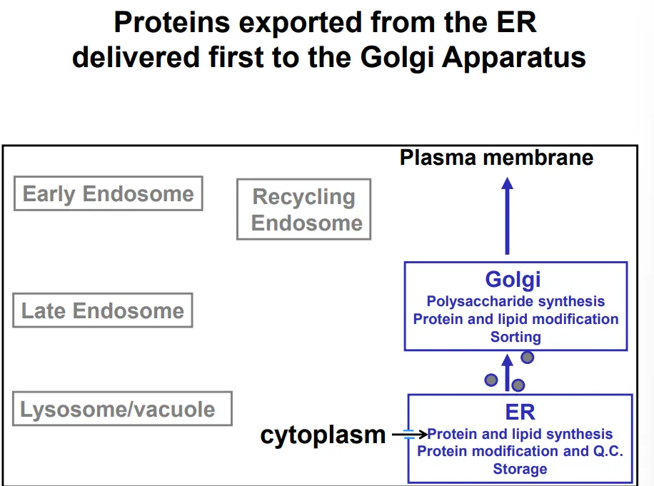
the proteins are transported from the ER to the golgi apparatus by the membrane budding into COP-II vesicles (so that they don’t need to cross membranes to be secreted)- this is also used to transport lipids produced by the smooth ER
the golgi apparatus (cis → medial → trans layers of cisternae) is a polysaccharide factory, which modifies the existing N-glycan chains for signalling and adds further polysaccharides of functional importance for glycoproteins and glycolipids
the vesicles bud off again and release the contents of the lumen (topologically equivalent to extracellular space) out of the cell, either immediately or following an external signal, in the case of regulated secretory vesicles
what is the endocytic pathway in eukaryotes?
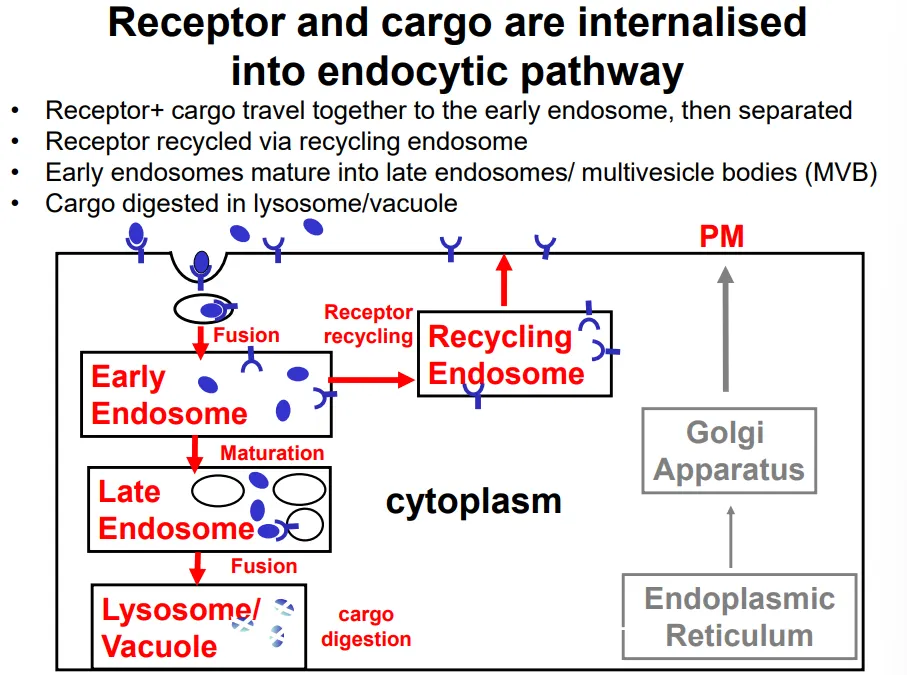
endocytosis and phagocytosis are initiated by specific receptor proteins, which form a receptor-cargo complex
clathrin-coated vesicles bud off the plasma membrane into the cell, producing an endosome
the receptor and cargo dissociate in the early endosome, so that the receptors can be re-inserted into the plasma membrane via a recycling endosome
the cargo remains in the endosome, which matures into a late endosome upon receiving additional contents (eg. enzymes) from the golgi apparatus
exisiting lysosomes fuse to this, releasing further digestive enzymes that degrade the cargo (nutrients, pathogens and signalling molecules)
during maturation, the endosome becomes increasingly acidic until it becomes a lysosome, where the pH is low enough for the digestive enzymes to function (acidic optimum so that they don’t damage the cell earlier)
how are transport vesicles formed and what proteins do each kind of vesicle use in the process?
curvature of membranes is energetically unfavourable, so to form vesicles, they must be forced into deformation by a protein coat
protein coat subunits self-assemble on the cytoplasmic side of the membrane
these are also responsible for collecting and packaging the cargo into the vesicle
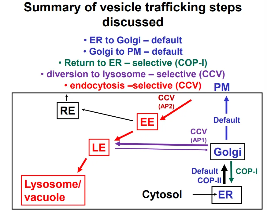
ER → golgi vesicles use COP-II coats
golgi → ER (retrieval) vesicles use COP-I coats
PM → early endosome vesicles use clathrin and adaptin 2 to form clathrin coated vesicles (AP2)
golgi → late endosome vesicles use clathrin and adaptin 1 to form clathrin coated vesicles (AP1)
how are proteins signalled to be retained or exported in the ER?
export from the ER doesn’t require a signal, it occurs by default ‘bulk flow’ (but some signals may accelerate export)
bacterial proteins that are inserted into eukaryotes (adding a signal for production in the ER, but not a signal for retention) are excreted
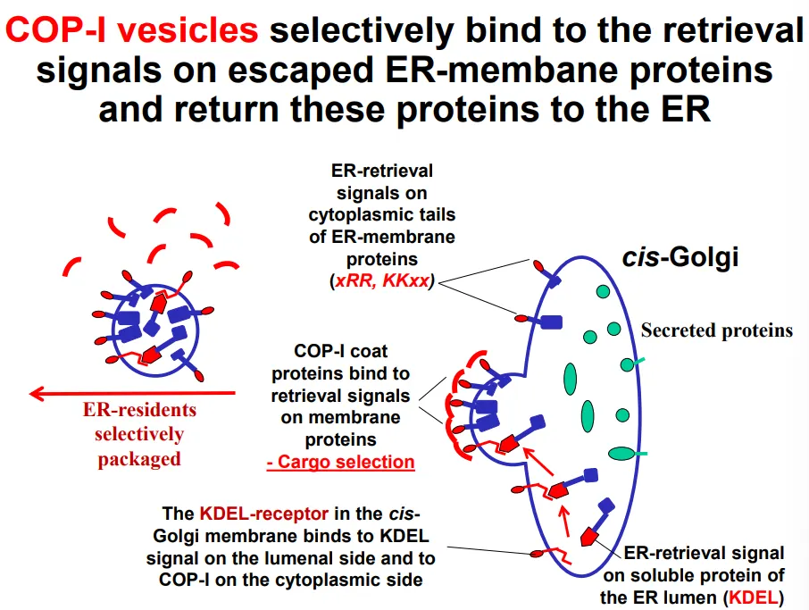
resident proteins aren’t prevented from leaving the ER, but they are continually retrieved from the golgi apparatus due to an ER-retrieval signal
these are necessary (deletion of the signal results in secretion instead of residency) and sufficient (the only sequence that is required to cause secretion)
COP-I vesicles selectively bind to retrieval signals (primary structure) for escaped ER resident proteins that are found in the cis-golgi:
diarginine (N terminus) and dilysine (C terminus) motifs on the cytoplasmic side of transmembrane ER resident proteins
KDEL receptor proteins in the cis-golgi membrane bind to the KDEL sequences (C-terminus) of luminal ER resident proteins
this is cargo selection, which then triggers vesicle formation
the KDEL receptor proteins dissociate from the cargo upon return to the ER due to pH differences between the ER and golgi lumen (the ER is less acidic, which discourages the association), so the receptors can be recycled to the golgi in COP-II vesicles
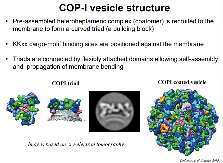
what is the structure of COP-I vesicles?
pre-assembled COP-I triads are present in the cytoplasm
these complexes have a natural curvature
multiple triads assemble to cause membrane bending, with the cargo-motif binding sites positioned against the membrane for cargo selection
these are flexibly bound together so that the size of the vesicles can change
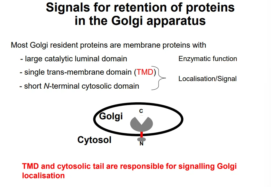
how are proteins signalled to be retained or exported in the golgi apparatus?
the large luminal domain of golgi resident proteins is functional enzymatically eg. to make polysaccharides
this is attached to a trans-membrane domain and a short N-terminal cytoplasmic domain, which act as a secondary structure signal
the different organelles in the secretion pathway have different lipid compositions, so the membrane thicknesses increase from ER → golgi → PM
this means that the TMDs of membrane proteins also increase in length, so they may be rejected if they are foreign to each organelle (eg. the hydrophobic region isn’t long enough, so charged residues clash with the fatty acid tails)
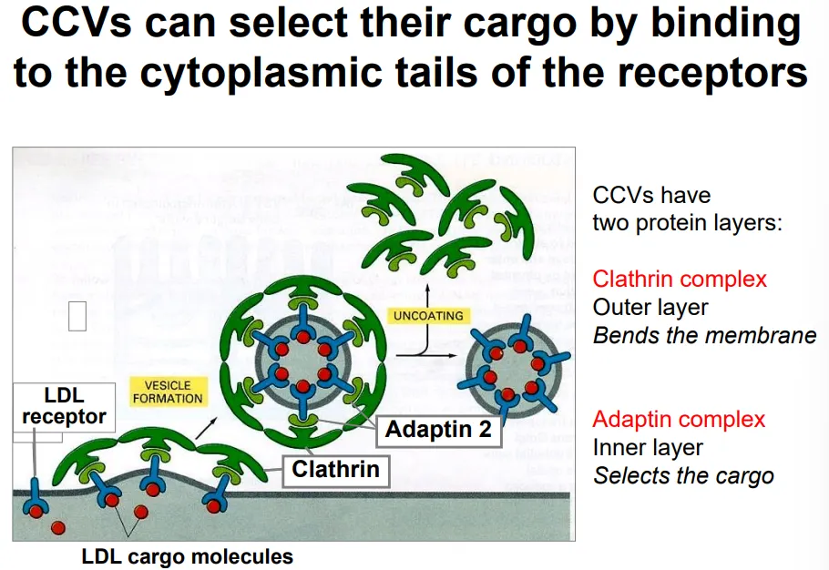
how does endocytosis occur selectively? use cholesterol as an example
cholesterol is transported through the blood as part of low density lipoprotein particles
these contain a large protein signal molecule, which binds to LDL receptors on the plasma membrane of target cells
these receptors bind to adaptin 2 complexes (cargo selection) in the cytoplasm, which in turn attach to clathrin triskelion complexes that cause membrane bending by spontaneous self-assembly, to produce a clathrin coated vesicle (CCV)
the clathrin and adaptin dissociate (using energy) from the vesicle to allow fusion to the early endosome, where the LDL receptors dissociate due to the lower pH so that they can be recycled back via recycling endosomes
how are proteins signalled to be transported from the golgi apparatus to the late endosome?
digestive hydrolases made in the ER have a signal patch in their tertiary structure, which is recognised by enzymes in the cis-golgi
these enzymes phosphorylate a mannose residue on the N-glycan chain of the protein
this modification is recognised by a mannose-6-P receptor in the trans-golgi, which binds to the glycan chain as well as adaptin 1 complexes from the cytoplasm
this causes the formation of clathrin coated vesicles, which transport the cargo to the late endosome
here the Man-6P receptor dissociates due to a decrease in pH, so that it can be recycled back to the trans-golgi
these hydrolase proteins are activated by proteases found in the lysosome, by proteolytic cleavage, so that they don’t cause damage to the cell earlier in the process
how are transport vesicles targeted to the correct destination?
the vesicle temporarily docks to a membrane by long-range tethers
here its specific v-SNARE (vesicular) proteins test their interactions with the t-SNARE (target) proteins on the membrane
if the t and v-SNARE proteins are complementary to each other, they form a very tight coiled-coil structure, which brings the vesicle close to the target membrane so that fusion can happen
if not, they won’t coil and the temporary docking will release the vesicle to try elsewhere
after fusion, NSF complexes remove the v-SNARE proteins from the target membranes, using ATP, to maintain compartment identity
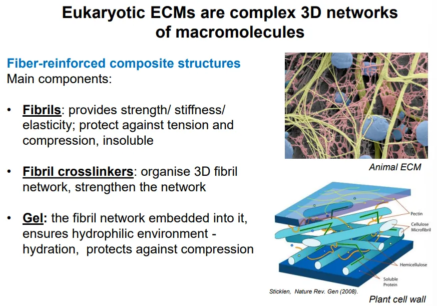
what are the different components of the animal ECM and their functions?
fibrils are made from collagen and elastin proteins:
provide strength/stiffness/elasticity
protect against tension and compression
insoluble
fibril crosslinkers are made from accessory collagen proteins:
organise 3D fibril network
strengthen the network
gel is made from glycosaminoglycans (GAGs) polysaccharide and proteoglycans (GAGs attached to a protein core):
contains the fibril network embedded
creates a hydrophilic environment = hydration
protects against compression
contains protein components (unlike the plant cell wall)
describe the structure of collagen
collagen is an insoluble fibrous protein that makes up the fibrils of the animal ECM
it is a right handed triple helix of three peptide strands made up of three amino acid repeats, every 3rd aa. being glycine
glycine is the smallest amino acid, so when the collagen forms a triple helix, the glycine is at the centre to make the most compact possible shape
rich in proline, lysine and hydroxyproline
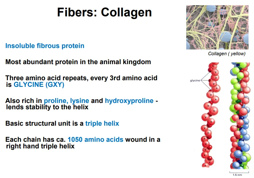
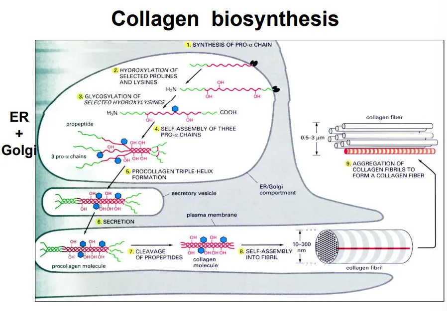
what are the steps of collagen synthesis?
ER: synthesis of pro α-chains (precursors)
ER: proline and lysine hydroxylation- vitamin C is a cofactor
ER: glycosylation of hydroxylysines
ER: self-assembly of the chains into a procollagen triple helix
golgi: N-linked glycan modifications for recognition
secretory vesicles: transfer to plasma membrane
ECM: cleavage of the ends by extracellular proteases
ECM: self-assembly into collagen fibril
ECM: aggregation of fibrils into a fibre
collagen fibres cross-link at specific, regular points, giving them a striated/banded pattern
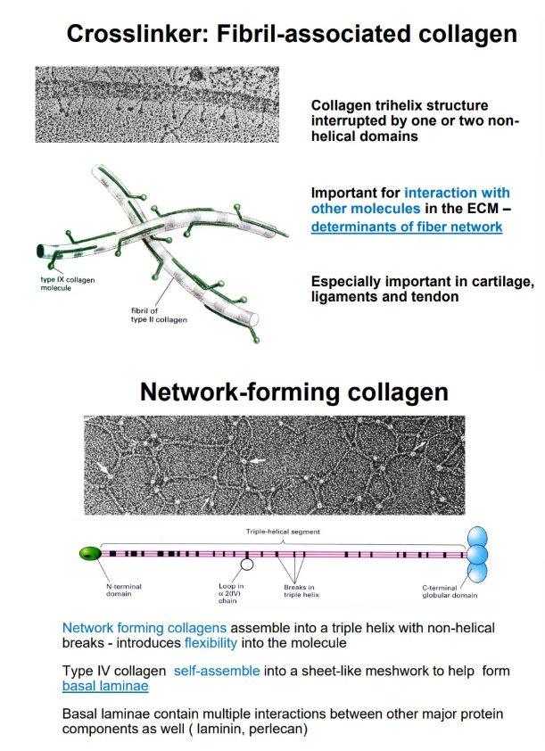
what are the two accessory forms of collagen and their structures?
crosslinker collagen has a trihelix structure interrupted by one or two hinge regions, which allow cross-linking (determines the ECM thickness)
network-forming collagen has more regular non-helical breaks, so they are very flexible and can assemble into a mesh sheet, which can be stacked by interactions between N and C globular domains to form the basal laminae (along with laminin glycoproteins)
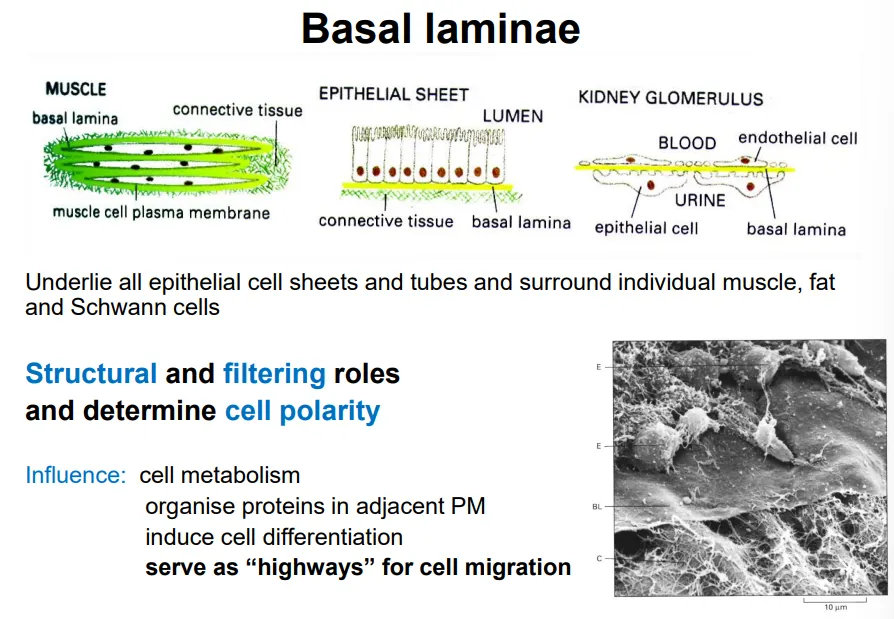
what is the basal laminae and what does it do?
the basal laminae is a specialised form of ECM that lies underneath epithelial cells and surrounds muscle, fat, and schwann cells to facilitate anchorage to connective tissue
this is primarily made up of layers of network-forming collagen sheets, with laminin glycoproteins
this has multiple regulatory roles, eg:
determining cell polarity if needed
filtering intake of molecules
organise proteins in adjacent plasma membrane
induce cell differentiation
serve as “highways” for cell migration
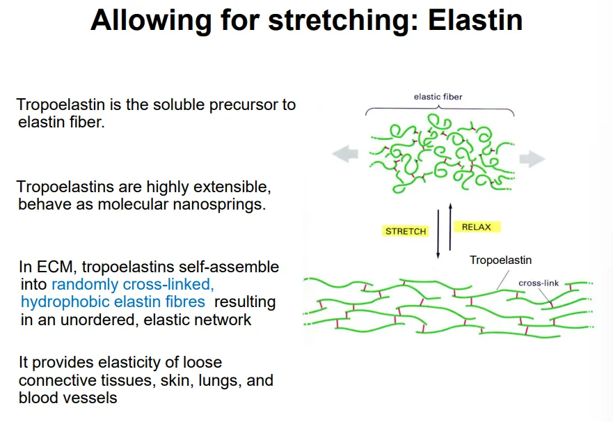
what is the structure of elastin
elastin is an insoluble protein network that forms part of the fibrils of the animal ECM
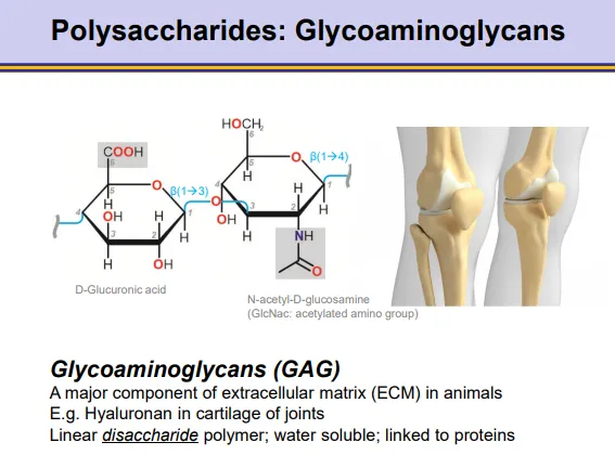
what is the general structure and properties of glycosaminoglycans?
GAGs are polymers of disaccharide monomers, which make up most of the gel in the animal ECM
one monomer is a -uronic acid, the other is an amino sugar
the most abundant is hyaluronic acid, which is glucuronic acid + N-acetylglucosamine
these are strongly hydrophilic and water-soluble
they are negatively charged due to high amounts of sulfur, which attracts sodium- this affects osmosis to give turgor pressure
they are inflexible and have a very low density
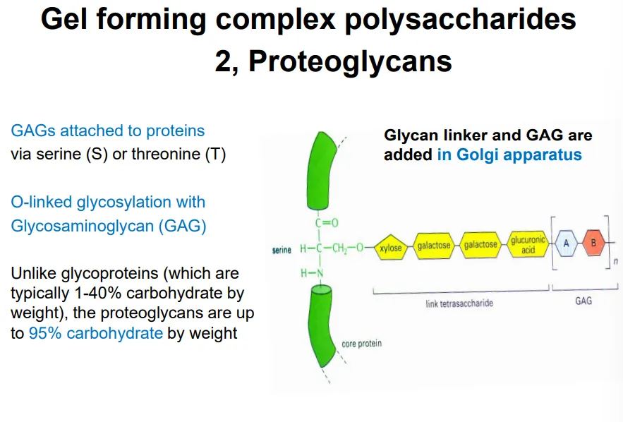
what is the general structure and properties of proteoglycans?
proteoglycans form part of the animal ECM
these are proteins highly modified by O-linked glycosylation (conjugation), with many glycosaminoglycans (GAGs) attached via serine or threonine residues (in the golgi)
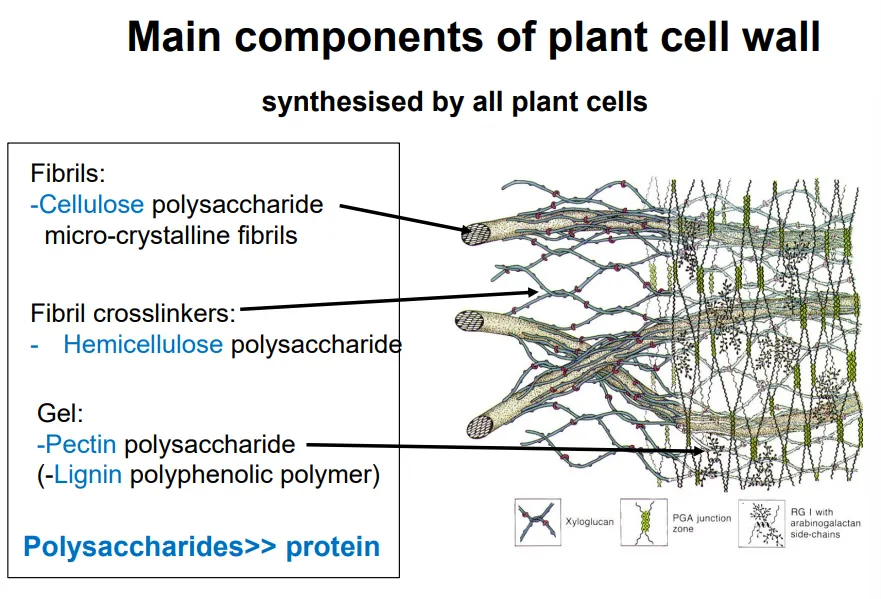
what are the different components of the plant cell wall and their functions?
very little protein content (unlike animal ECM)
hemicellulose eg. xylgoglucans
the gel is mostly made up of pectins (very complex, hydrophilic, acidic polysaccharides), with its consistency regulated by pH and Ca ions
these protect against pathogens and influence the porosity, pH, and ion balance of the cell wall
the gel can also contain lignin (polyphenol), primarily in the secondary cell wall (more rigid, produced by mature cells)
describe the organisation of cellulose fibres
cellulose polymers are produced right next to each other so that they form hydrogen bonds together
this produces a long sheet of adjacent polymer molecules, which is hydrophilic at the edges but hydrophobic on either face
these stack into microfibrils, which have a crystalline core, that can assemble into fibrils due to having a hydrophobic faces on the top and bottom and a hydrophilic faces on either side
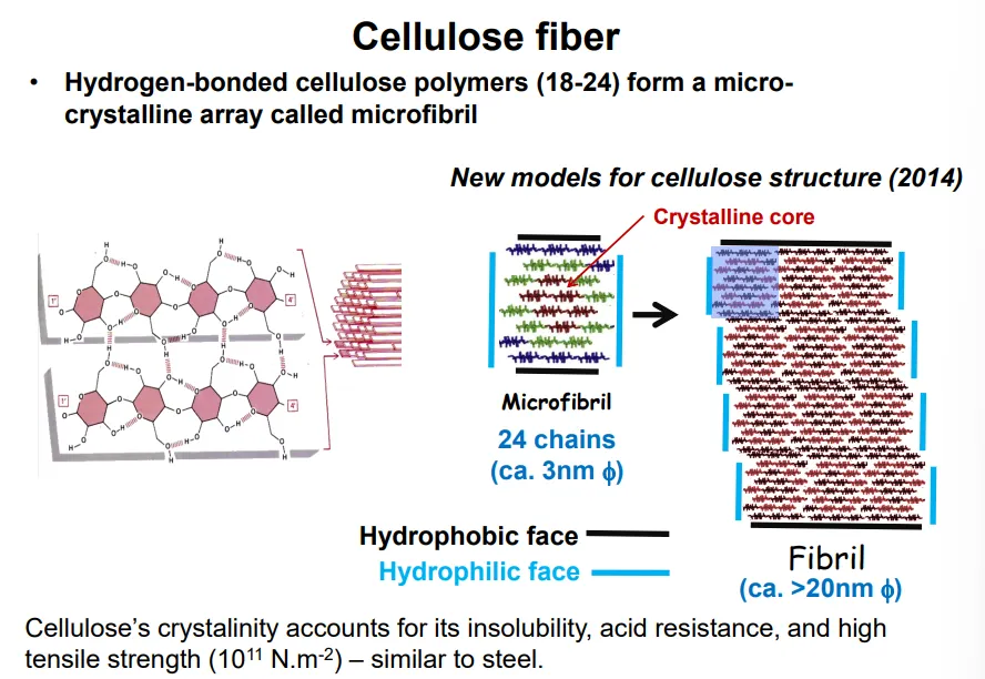
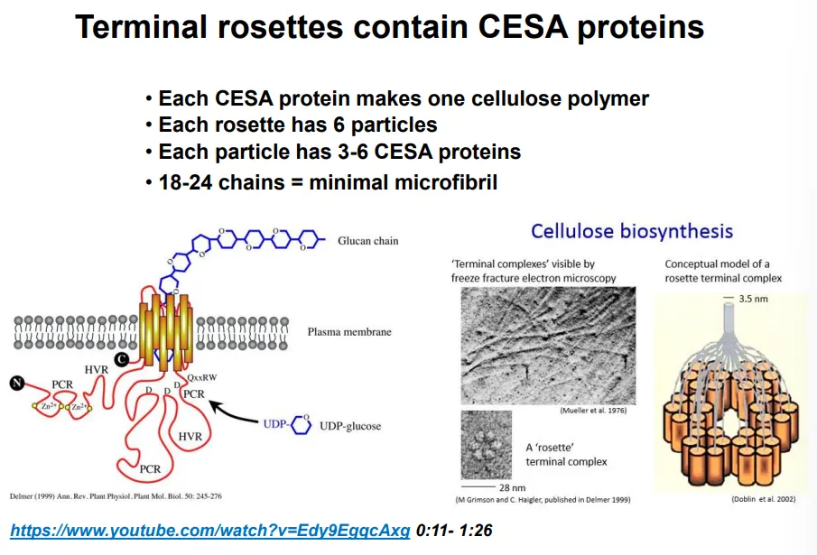
how is cellulose deposition in the plant cell wall regulated?
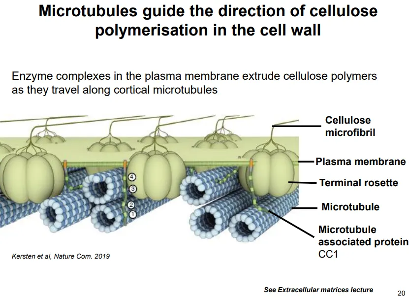
cellulose is synthesised by terminal rosette enzyme complexes in the plasma membrane (acquired by HGT from bacteria)
the terminal rosettes contain 6 particles, each of which contain 3-6 cellulose synthase A (CESA) proteins, which each make one cellulose polymer at a time
this means each rosette makes an 18-24 chain microfibril and twists them together
the cell elongates perpendicular to the orientation of the cellulose fibrils, because new material is deposited in between the fibrils
random cellulose deposition in a cell would mean it could never expand
the deposition of cellulose by terminal rosettes is directed by microtubules to occur in one direction
plant cells stop growing by depositing new layers in different orientations
this deposition is driven by the polymerisation reaction because the polymer is being pushed into an already densely packed matrix, so the terminal rosette is the part that moves instead, along the microtubules
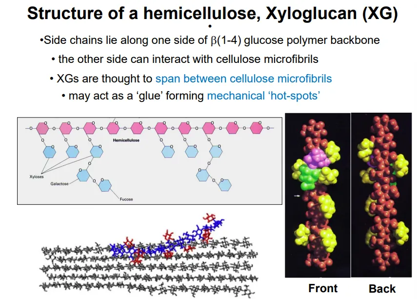
describe the general structure and function of hemicellulose, with an example
hemicellulose cross links the cellulose fibrils in the plant cell membrane
‘half’ cellulose because one side of the chain has many glucan side chains, while the other can interact with cellulose to form mechanical hot spots
eg. xyloglucan
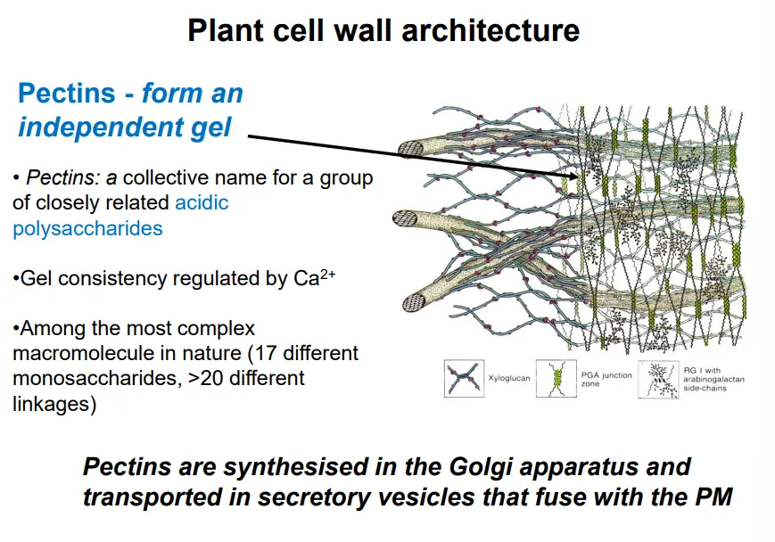
describe the composition and function of the gel in plant cell walls
the gel is mostly made up of pectins (very complex, hydrophilic, acidic polysaccharides), with its consistency regulated by pH and Ca ions
these protect against pathogens and influence the porosity, pH, and ion balance of the cell wall
the gel can also contain lignin (polyphenol), primarily in the secondary cell wall (more rigid, produced by mature cells)
how do animal cells anchor with the ECM?
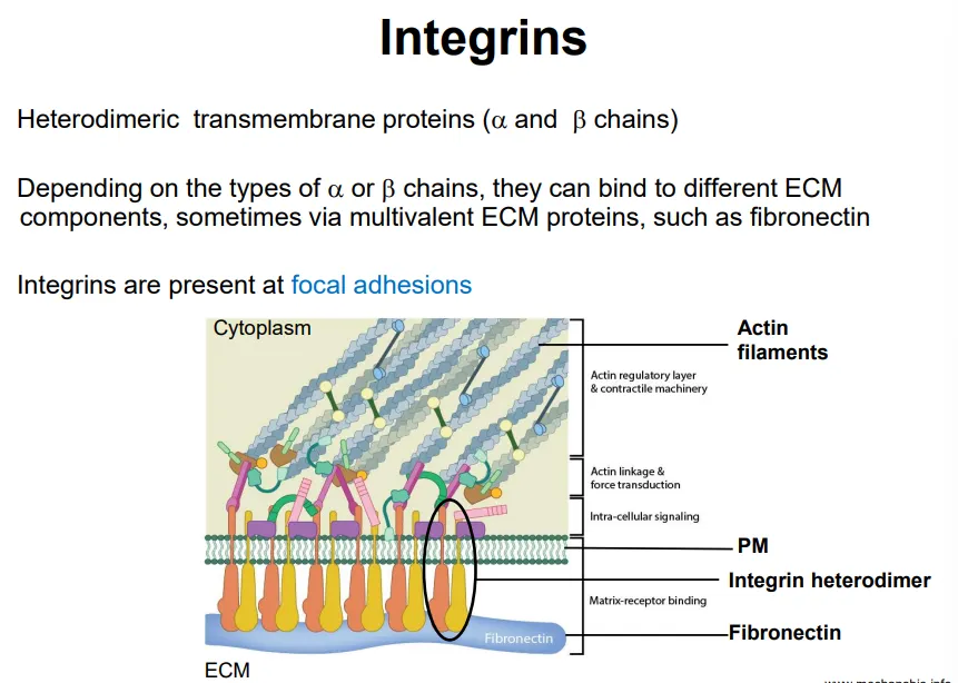
animal cells have integrins, transmembrane protein receptors that mediate cell adhesion to the ECM
these are heterodimeric with one alpha and one beta chain, which can bind to different ECM components at regions called focal adhesions
on the cytosolic side the integrin complexes bind to the cytoskeleton (for stress fibre attachment)
these focal adhesions define cell shape, mechanical and chemical signalling and control cell migration
how does the cell modify the cell wall for growth?
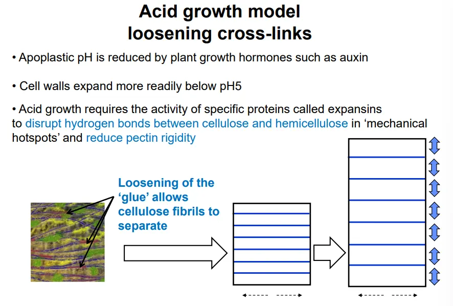
cell growth requires the loosening of the cell wall crosslinking, and the increase of turgor pressure, according to the lockhart equation: R = ɸ(P-Y)
cell expansion happens when P, pressure > Y, the yield threshold
this is determined by the wall extensibility, ɸ (determined by cross linking)
the cross linking is weakened by acidic extracellular pHs (< 5), caused by hormones eg. auxins
this triggers expansin proteins to disrupt H bonds between cellulose and hemicellulose and reduce pectin rigidity
local cell wall loosening at the apical meristem initiates new organ formation
it can also be used to detect abiotic/biotic stress (eg. pectin degradation by pathogens)
describe the 3 kinds of cytoskeleton
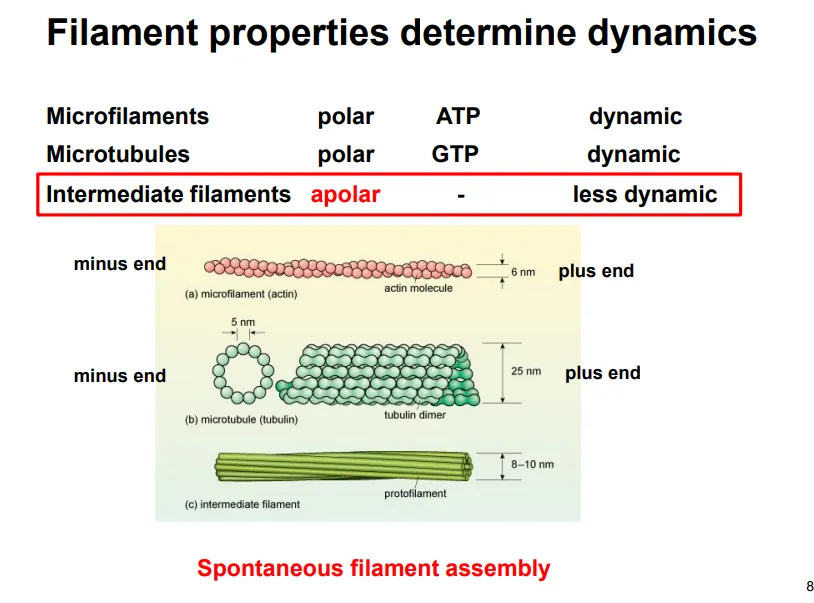
actin (micro) filaments- all eukaryotes, polar, use ATP, dynamic, 7nm
tubulin microtubules- all eukaryotes, polar, use GTP, dynamic, 25nm
intermediate filaments (multiple kinds)- only animals, apolar, don’t use ATP/GTP, less dynamic, 10nm
describe intermediate filament structure and function (give examples)
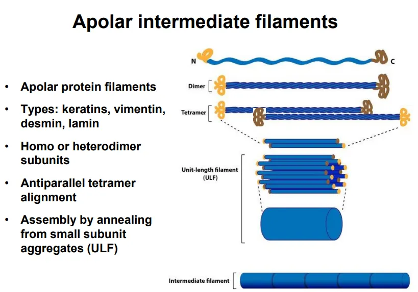
apolar (the two ends have the same properties)
long strands with globular C and N termini assemble into coiled-coil dimers
dimers assemble antiparallel into tetramers
these line up into smaller subunits, which anneal into long filaments
eg. keratin, vimentin, desmin, lamin
mainly used for structural support, determining and maintaining the cell and nucleus shape (so they aren’t needed in plants)
this is because they have a high tensile strength and are resistant to compression and bending
eg. lamins form a lattice structure which covers the inner side of the nuclear envelope as a site for chromatin anchorage
describe the structure of microtubules
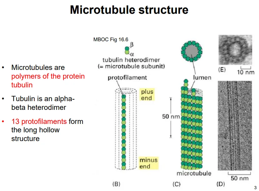
tubulin monomers are heterodimers of two subunits: alpha and beta
the heterodimers assemble end to end, so that the alpha and beta subunits alternate, into long strands called protofilaments
13 of these protofilaments assemble into a hollow cylinder
the plus end is the beta end, and the minus end is the alpha end
each of the subunits has a GTP-binding site, but only beta-tubulin can hydrolyse it- the GTP bound to alpha-tubulin lends stability and proper folding
decribe microtubule formation, polymerisation and depolymerisation
self-assembly from scratch is difficult because 13 subunits need to interact laterally, which is energetically unfavourable
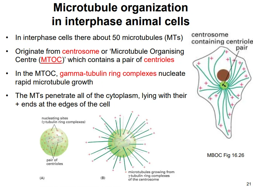
in cells gamma-tubulin ring complexes are used in initiation to act as structural templates for the alpha-tubulin of the heterodimers to bind (at the minus end)
these complexes are anchored at particular organelles
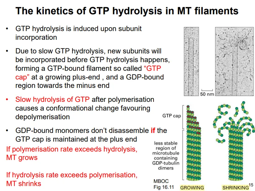
tubulin monomers, bound to GTP, are added to the plus end
the GTP provides strong lateral interactions for stability
once a monomer associates, it triggers GTP hydrolysis by the beta-tubulin subunit
this is very slow, so new subunits can incorporate before GTP hydrolysis is achieved, creating a GTP cap at the plus end
the GDP-bound heterodimer has a different conformation, which can induce dissociation if the GTP cap isn’t maintained (when the hydrolysis rate > polymerisation rate), resulting in shrinkage, and vice versa
this is dynamic instability, which can result in complete depolymerisation if there are no GTP-bound dimers to rescue the growth
what is the function and organisation of microtubules in plant cells?
in young cells microtubules radiate out from the nucleus
in larger growing cells (interphase) they associate and run parallel with the membrane
these guide cellulose deposition in the correct orientation
enzyme complexes in the plasma membrane extrude cellulose polymers as they travel along parallel membrane anchored microtubules
microtubules also act as tracks for motor proteins to move organelles and transport vesicles around the cell (along with actin) eg. the endomembrane system
the spindle fibres used in mitosis are microtubules (not from centrosome like in animals)
what is the function and organisation of microtubules in animal cells?
microtubules radiate out from the centrosome (the microtubule organising centre, MTOC), which contains a pair of centrioles- the centrosome has many gamma-tubulin ring complexes for initiation
most microtubules constantly polymerise and depolymerise out from the centrosome due to dynamic instability
this is because hitting the membrane perpendicularly causes the loss of the GTP cap and results in depolymerisation
we believe this functions to help the cell to explore and sense changes in the plasma membrane
microtubules also act as tracks for motor proteins to move organelles and transport vesicles around the cell eg. the endomembrane system
the spindle fibres used in mitosis are microtubules
(eukaryotic) cilia contain microtubules in a 9+2 formation for movement eg. sperm, and signalling/sensing eg. rod cells
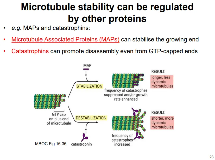
how is microtubule growth regulated?
most of the time dynamic instability isn’t observed because of protein action:
microtubule associated proteins (MAPs) stabilise the plus end to promote growth
catastrophins destabilise the plus end to promote depolymerisation
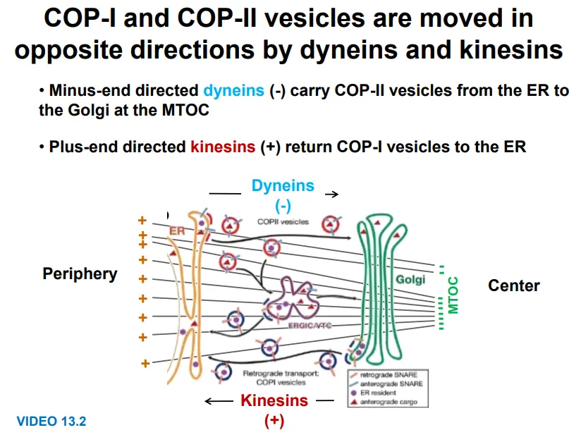
how are organelles moved around the cell by microtubules?
motor proteins bind to vesicles and organelles and move along the microtubules
kinesins move towards the plus end (outwards)- eg. for COP-I vesicles golgi → ER
these are dimers with a cargo-binding tail domain, a stalk and a two head domain, which moves along microtubules in 8nm steps, each using one ATP (binding and hydrolysis cause conformational changes for moving the heads forwards)
dyneins move towards the minus end (inwards)- eg. for COP-II vesicles ER → golgi
these are more complicated, sometimes involving accessory proteins for binding, but using ATP to make irregular movements
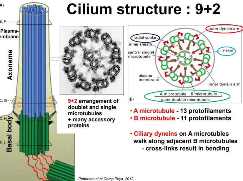
how are microtubules organised in eukaryotic cilia and how do they bend?
the basal body (originating from centrioles, but not in plants) extrudes microtubules in a 9+2 arrangement
9 doublets (13 protofilament cylindrical A microtubule + 11 protofilament attached B microtubule) are arranged in a ring, with 2 singlets in the centre
the doublets are connected by nexin proteins and have radial spokes towards the centre
the microtubules are always enclosed by the plasma membrane
ciliary dynein (a motor protein complex) attached to the A microtubules walks along the adjacent B microtubules due to ATP
this would cause the filaments to slide, but due to the nexin linking proteins and controlled activity, this causes bending
in short cilia this produces a force perpendicular to the long axis, but in long cilia this symmetric beat produces a parallel force (in a sinusoidal wave)
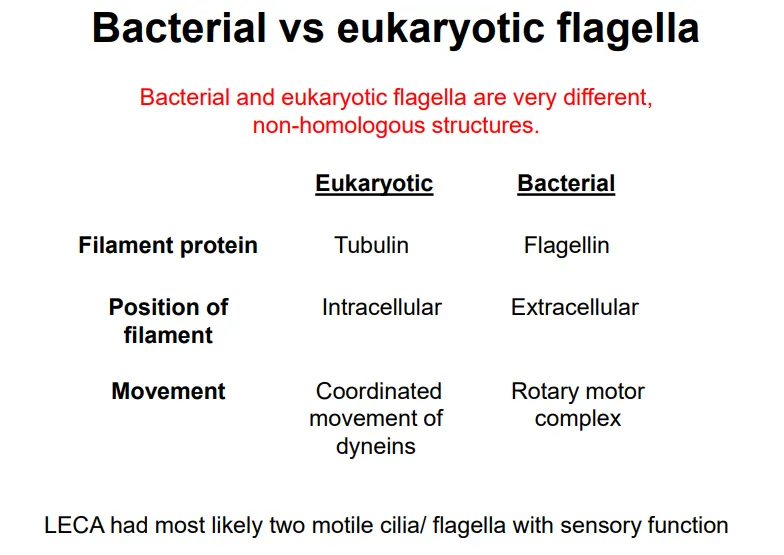
compare bacterial and eukaryotic flagellar proteins, positions and movement
describe the structure and assembly of actin filaments
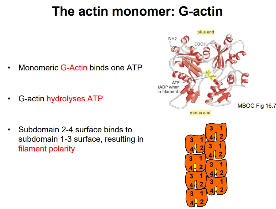
F-actin (filamentous actin) is a linear polymer of a single globular monomer, G-actin
it is a left-handed helix with 13 monomers per turn (37nm)
each G-actin binds and hydrolyses one ATP to spontaneously self-assemble (once three monomers have bound together)
one surface (2-4 subdomain) binds preferentially to the opposite surface (1-3 subdomain), so the filament is polar
polymerisation happens at the plus end, which has a greater binding affinity to ATP-bound G-actin
after addition, the G-actin monomers hydrolyse the ATP slowly, so that the minus end is made up of ADP-bound G-actin, which can dissociate (this is called treadmilling)- the middle region is ADP + Pi bound because monomers are slow to release phosphate
however the rate of filament assembly and disassembly is normally controlled by actin-binding proteins, by:
nucleation- holding the three monomers together to allow for self-assembly (energetically unfavourable otherwise)
capping- binding to the plus end to prvent further polymerisation
severing- cutting the filament to induce depolymerisation
sequestering- binding to monomers to prevent incorporation
bundling- bind to actin filaments to connect them
what are the functions of actin filaments?
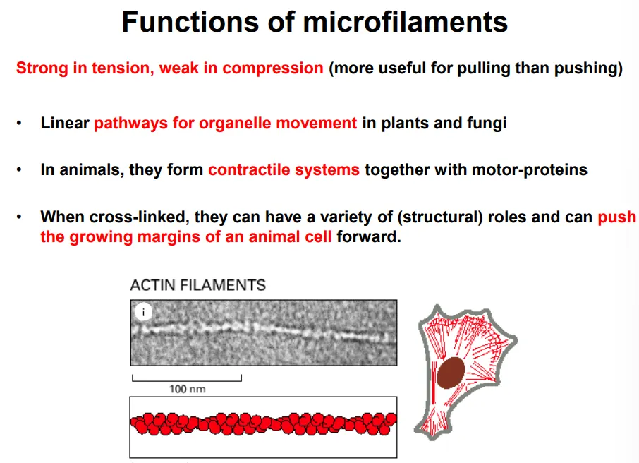
stronger in tension than compression (used for pulling)
in conjunction with myosin motor proteins, they are used for:
organelle movement in plants and fungi (by myosin motor proetins, causing cytoplasmic streaming)
changing the cell shape in contractile systems in animals, eg:
sarcomeres in muscle cells
contractile rings in cytokinesis
stress fibres anchored to focal adhesions
cell movement
how are organelles moved around the cell by actin filaments?
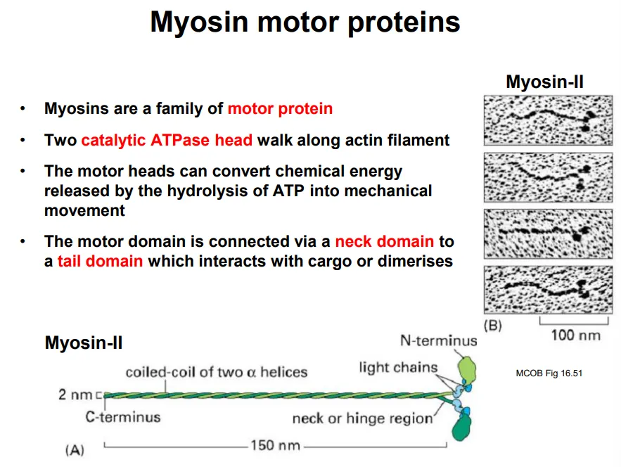
in plants and fungi, the large vacuole means organelles and vesicles must be moved around by myosin motor proteins, causing the cytoplasm to also move by cytoplasmic streaming (overcomes diffusion barrier)
two catalytic heads walk along the actin filament in steps by conformational changes from hydrolysing ATP
this is connected to a neck domain, with a tail domain on the end to bind to cargo
how are actin filaments organised in skeletal muscle cells?
in animal skeletal muscle tissue, actin and myosin filaments (myofibrils) form contractile arrays in multinucleated syncitial cells (myoblasts fused together)
muscle myosin proteins assemble into bipolar myosin filaments (thick), where the heads project out along the length in specific radial positions (43 nm repeat)
each repeating unit is a sarcomere, which is striated:
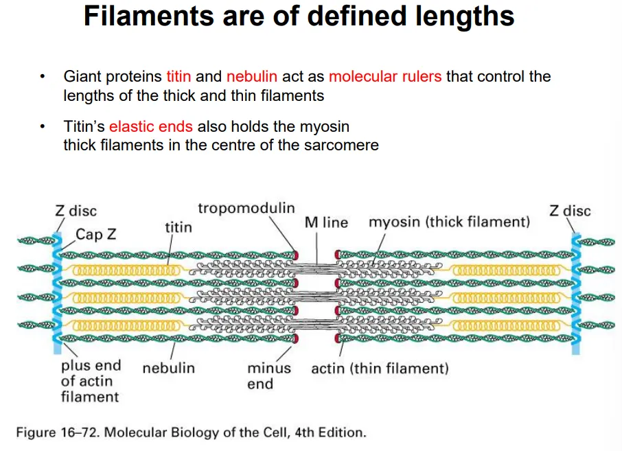
the plus ends of the actin filaments (thin) are anchored at the z disc, and the minus end is capped by tropomodulin (can’t polymerise/depolymerise)- these are all the same length due to attachment to a long nebulin protein
elastic titin proteins attach to the z disc and connect to myosin thick filaments to keep them centred along the m line
this shortens according to the sliding filament model
the thick filaments are hexagonally packed very regularly
describe how sarcomeres shorten
each sarcomere in skeletal muscle cells contracts according to the sliding filament model:
when relaxed, the titin proteins (connected to the z disc and myosin filaments) are stretched, so the actin and myosin only just overlap
when the muscle contracts, the myosin filaments walk along the actin filaments, pulling the z discs on either side towards the m line in the centre
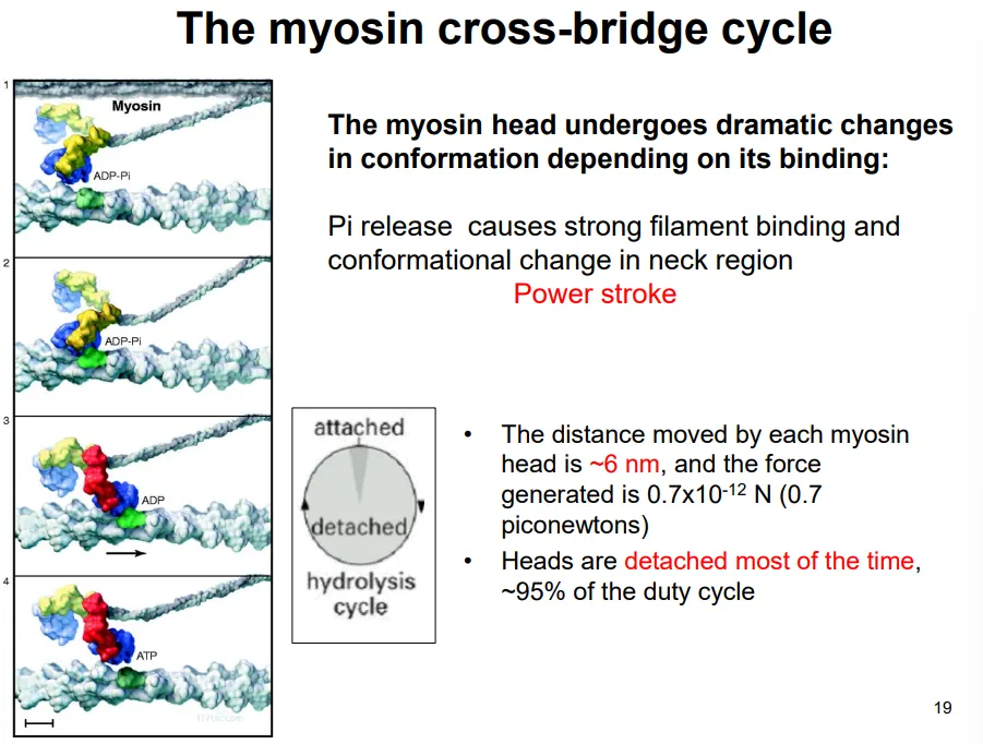
this occurs by the myosin cross-bridge cycle, driven by ATP hydrolysis:
the two myosin heads of each protein act independently
they bind to the actin and release a phosphate (from previous hydrolysis), causing the power stroke, which pulls the actin filament 6nm
the ADP dissociates, and the next ATP binds
this causes the head to detach from the actin (detached 95% of the time)
the hydrolysis of this ATP causes the head to move back to its original conformation
the spacing of the myosin heads doesn’t match up with that of the myosin binding sites of actin, and the nine radial positions of the myosin heads doesn’t match up with the hexagonal packing of actin around each thick filament
this means that not all the myosin heads can bind to actin at one time- this means there are always some heads attached to maintain tension
how is muscle contraction controlled?
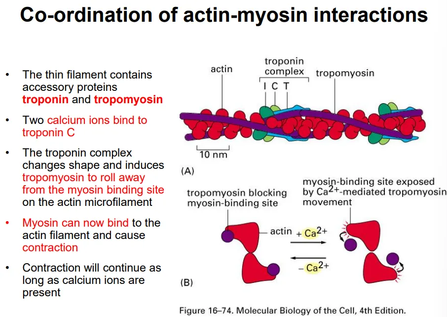
tropomyosin proteins are wrapped around the thin actin filament and cover the myosin binding sites
when an action potential arrives at the cell, it is transmitted through invaginations of the plasma membrane
this causes the voltage-gated release of calcium ions from the sarcoplasmic reticulum
two calcium ions bind to a troponin complex, which changes shape and causes the tropomyosin to detach
this allows myosin to bind to the actin filament, causing contraction by ATP hydrolysis as long as the calcium ions are present
what are the three actin filament organisations in crawling cells?
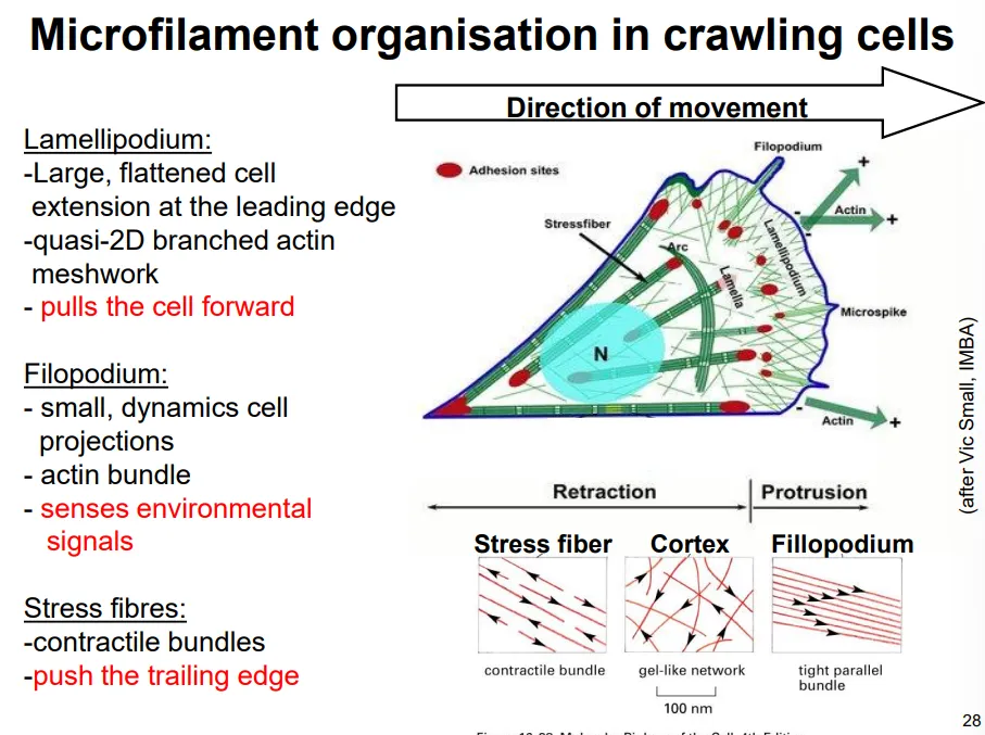
cell movement first needs filopodia to sense the environment- outgrowths of the cell containing parallel actin bundles, initiated and polymerised by formin proteins
in the lamellipodia, the actin is branched at 70 degree angles due to the binding of actin-related proteins (ARPs) that facilitate new filaments forming, by holding together a trimer
this extension occurs via a treadmilling model, where the actin filaments grow a short distance before being capped, at which point they act as a site for new filaments to be assembled, while the older end of the network is disassembled by severing proteins
new focal adhesion points will be made and stress fibres attached, which will contract (mini sarcomere) when this becomes the trailing end
this protrusion force is provided purely by actin polymerisation, not by myosin II
how are the different kinds of actin structures initiated?
GTP-binding proteins (G proteins) spatially organise actin polymerisation:
Rho stimulates stress fibre production
Rac stimulates lamellipodia extension
Cdc-42 stimulates filopodia formation
these are spatially separated around the cell in the relevant areas, to promote each actin structure
these act as molecular switches, active when bound to GTP, which they hydrolyse to become inactive
how are cytoskeletal elements interconnected?
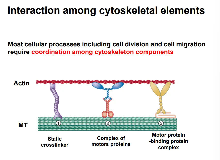
many processes require the coordination of multiple cytoskeletal components
they can be connected by different crosslinkers to coordinate them:
static crosslinkers- neither can move
motor protein complexes- both proteins can move along their elements
motor protein-binding protein complexes- one is fixed, one can slide
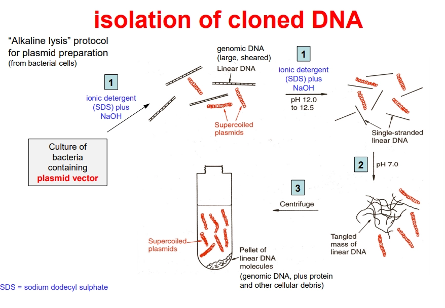
how can we isolate recombinant bacterial plasmids for sequencing?
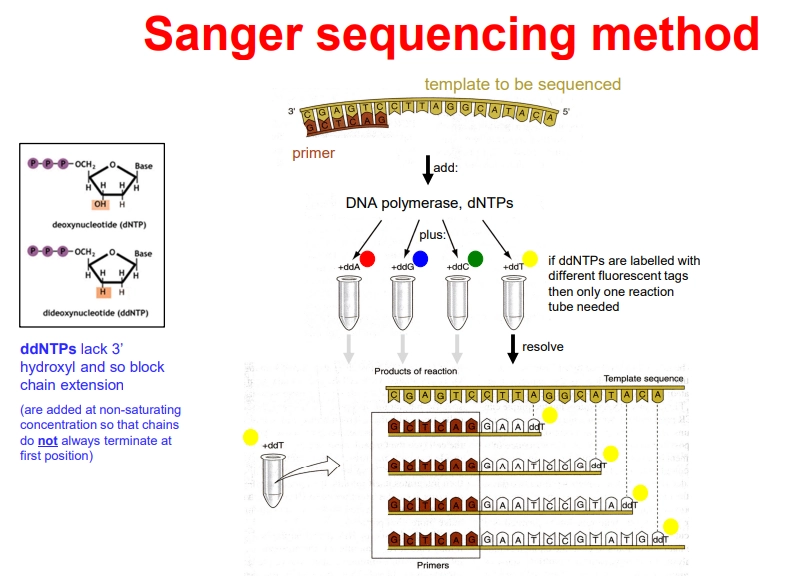
how can we sequence DNA using sanger sequencing?
PCR is now carried out in 1 tube, containing the DNA template, a buffer, DNA primers, an excess of dNTPs, DNA Taq polymerase, and differently fluorescently tagged 2’,3’-dideoxynucleotides (ddNTPs)
the primer anneals adjacent to the initiation start and DNA polymerase elongates the strand
when ddNTPs are incorporated, they terminate elongation because they have no 3’ OH to attack the next phosphate and form the phosphodiester bond
they are not added at saturating concentrations, so that termination occurs randomly at every possible position
this produces truncated strands with fluorescent tags that can be automatically detected using laser detection to produce a chromatogram
originally, the reaction had to be carried out in 4 different tubes, one for each ddNTP, because the fragments were separated by electrophoresis and detected using a radioactive primer
this process is limited to ~900 nucleotide sequences due to smaller relative fragment size differences, but is still used for small projects due to convenience and accuracy
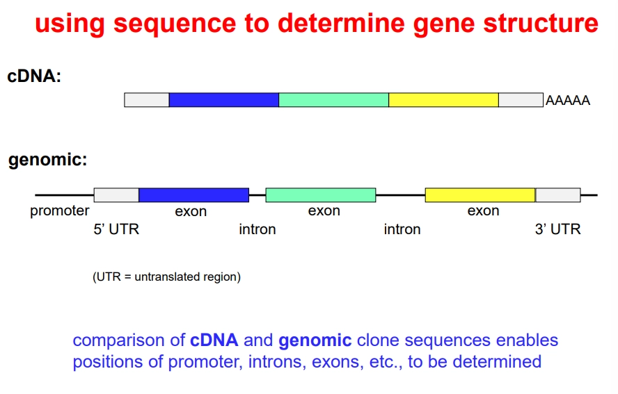
how can we use DNA sequencing to determine gene structure?
sequencing the cDNA and genomic clones can be used to determine the position of exons, introns and UTRs by comparison
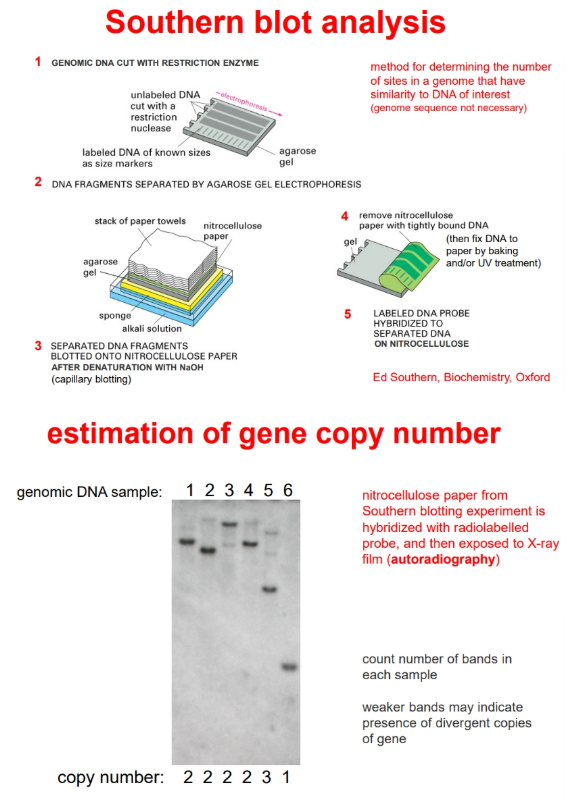
how can we estimate the number of copies of a gene that are present in a chromosome?
by southern blotting
when the probes are added to the nitrocellulose they will hybridise to any fragment that contains the gene of interest
this will reveal separate bands due to differing total fragment sizes (after radioactive/fluorescent/enzymatic detection)
the bands can be counted to determine the number of copies of the gene in the chromosome
weaker bands indicates divergent copies of the genes
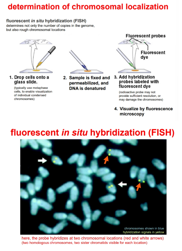
the copy number and rough chromosomal location can be determined by fluorescent in situ hybridisation (FISH)
how can we determine chromosome localisation of a gene?
by fluorescent in situ hybridisation (FISH)
how can we determine when and where a gene is expressed?
to determine what tissues produce a gene (including at different points of development/under different conditions), we can use:
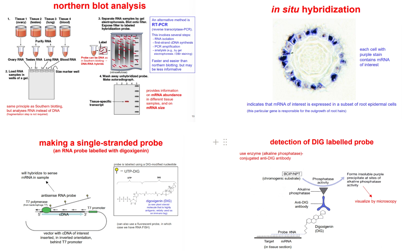
northern blotting (derived from southern blotting, but using the mRNA instead of fragmented chromosomal DNA, radioactive detection is used instead of fluorescence)
reverse transcriptase-PCR (RT-PCR) and electrophoresis
to determine what cells within a tissue express a gene, we can use:
in situ hybridisation of a single-stranded probe (more sensitivity, because it won’t re-anneal to itself after denaturation) to fixed thin sections of tissue
to produce the probes, the cDNA for our gene of interest is inserted into a plasmid vector in an inverted orientation, such that transcription of the vector produces an antisense RNA probe (no sense probe strand present = no re-annealing)
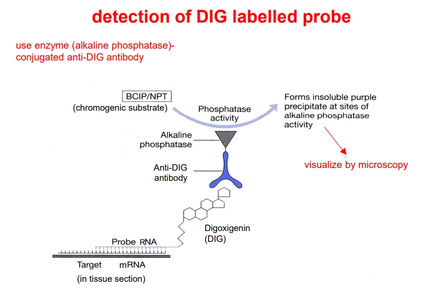
during transcription of this probe, UTP-DIG is incorporated (UTP bound to digoxigenin)- this is detected in the tissue section using an antibody attached to alkaline phosphatase, which converts a substrate from colourless to purple (detected under a microscope)
this isn’t the same as detecting where the protein itself is produced, as genes are often regulated post-transcriptionally
how can we produce single-stranded RNA probes?
these are used during in situ hybridisation (to determine what cells within a tissue express a certain gene)
the cDNA for our gene of interest is inserted into a plasmid vector in an inverted orientation, such that transcription of the vector produces an antisense RNA probe
this will hybridise to the sense mRNA, without the need for denaturation, like with a double-stranded probe (absenc2e of sense probe strand prevents the probe re-annealing to itself, decreasing noise)
during transcription of this probe, UTP-DIG is incorporated (UTP bound to digoxigenin)
this can be detected using an anti-DIG antibody conjugated to alkaline phosphatase, which can convert a colourless chromogenic substrate into a purple precipitate which can be seen through a microscope
how can we determine when and where a protein is produced?
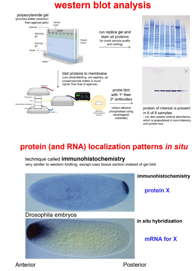
to determine what tissues accumulate a protein, we can use western blotting (derived from southern blotting, but using the protein instead of DNA, polyacrylamide gel instead of agarose, and electroblotting instead of capillary blotting)
to determine where a protein accumulates within a cell/tissue, we can use immunohistochemistry (similar to in situ hybridisation)
detection requires antibodies for the protein of interest:
a primary antibody detects the protein- this is produced by immunising an animal using the bacteria-produced protein (with a hexahistidine tag attached for purification)
a secondary antibody, conjugated to alkaline phosphatase, detects the primary antibody (this allows for signal amplification/sensitivity and use of a single reagent for multiple protein targets)
alkaline phosphatase converts a colourless chromogenic substrate to a purple precipitate (detected under a microscope)
how can DNA be cut and joined for genetic modification?
DNA can be cut using restriction endonuclease enzymes
these cut at palindromic recognition sites that are 4, 6 or 8 base pairs long
this can result in blunt or cohesive/sticky ends
eg. EcoRI
two DNA molecules with complementary sticky ends can be joined back together
the bases will form hydrogen bonds with each other, and then DNA ligase can be used to catalyse the phosphodiester bond formation
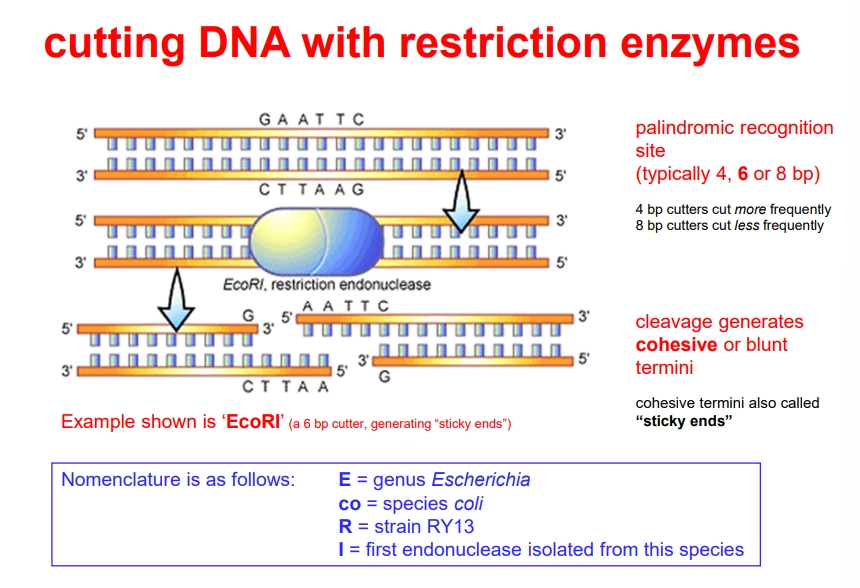
how can we monitor the recombination of DNA?
gel electrophoresis through agarose gel separates fragments of DNA by size (heavier = slower)
intercalating agents must be used to visualise the DNA itself eg. ethidium bromide binds to DNA and fluoresces
the DNA sample (cut DNA size is compared to uncut DNA and to recombinated DNA) is compared to a ladder of DNA fragments of known sizes
how can we identify whether a gene of interest has been recombinated?
by creating and screening genomic libraries:
necessary if we’re interested in the whole transcription unit or intergenic regions
contains all the DNA sequences in a cell, highly repetitively
sample DNA is fragmented before cloning, using restriction enzymes or random shearing
this is done to produce the maximum possible number of large (so we can capture the whole unit), overlapping fragments
by creating and screening cDNA libraries:
this must be used if we are interested in only the mRNA sequence of intron-containing DNA
the inserts are much smaller and need not be fragmented because these introns have been removed by reverse transcription of mRNA
not all genes may be included, dependent on the degree of gene expression for each mRNA in the cell
the DNA must be labelled with a probe
a nitrocellulose (sticks to DNA) membrane can be used to replica-plate the DNA
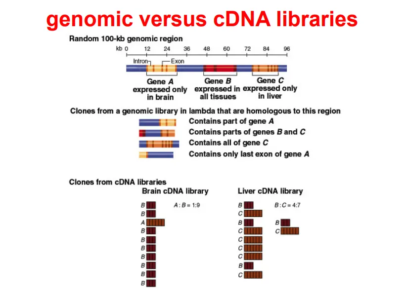
how do we choose the vector used for genetic modification?
in order of increasing insert size capacity:
plasmid- can be used for small/cDNA fragments
phage
cosmid (plasmid with cos sites, can be packaged into phages)
BAC (bacterial artificial chromosome, based on F plasmid)
YAC (yeast artificial chromosome)- can be used for much larger inserts
what are the key features of a plasmid vector?
an origin of replication, so that it will be expressed in a host cell
selectable marker(s), which can be used to identify successful plasmid uptake eg. ampicillin resistance
a multiple cloning site (MCS), where the fragment is inserted- the location of this site is important, as it can be used for insertional inactivation:
blue-white selection can be used for plasmids where the MCS is in the lacZ gene, because when beta-galactosidase is produced correctly, it converts the colourless X-gal into a blue product, so successful recombination can be measured
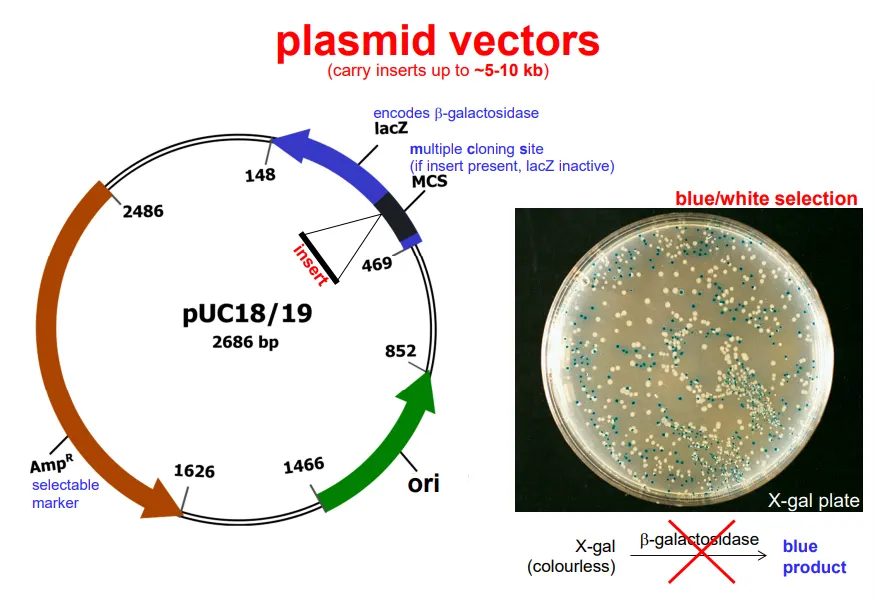
what are the key features of a phage vector?
dispensable ‘stuffer’ DNA that causes lysogenesis, so can be cut out and replaced with the DNA of interest
cos sites, which enable packaging and circularisation in the host
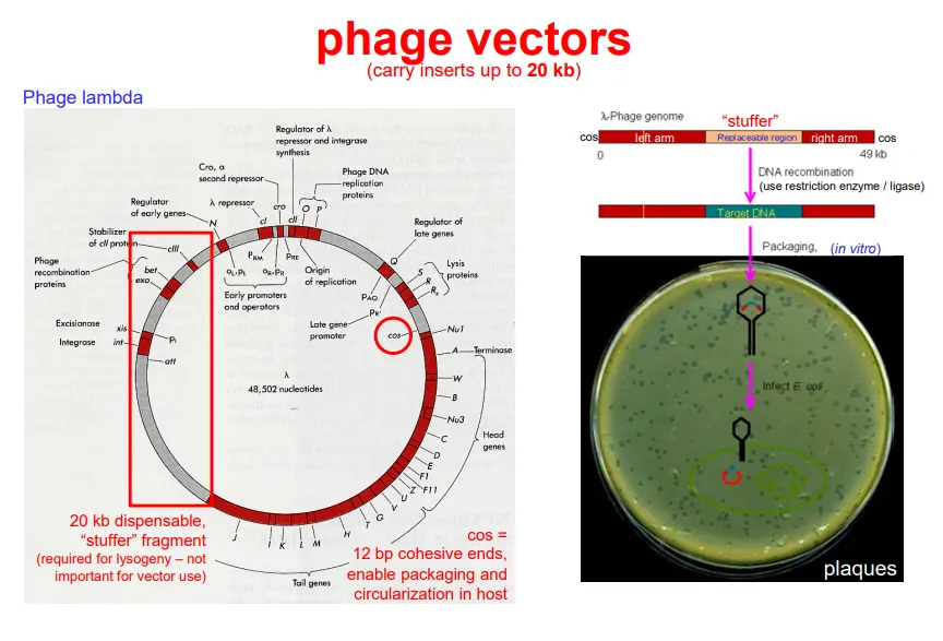
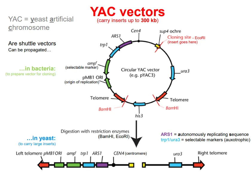
what are the key features of a YAC vector?
YAC- yeast artificial chromosome
telomeres at either end for stability, and a centromere
an origin of replication, so that it will be expressed in a yeasthost cell
selectable marker(s), which can be used to identify successful plasmid uptake eg. ampicillin resistance
a multiple cloning site (MCS), where the fragment is inserted- the location of this site is important, as it can be used for insertional inactivation
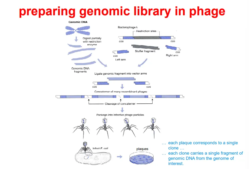
what is the process of preparing a genomic library using a phage vector?
break up the DNA into fragments by partial digestion using restriction endonucleases
use the same enzyme on the vector to produce compatible sticky ends, removing the ‘stuffer’ fragment
ligate the fragments into the phage to produce recombinant DNA, which can be inserted into a phage head
bacteria are infected with these bacteriophages and plated, so that each plaque contains a single foreign fragment (genomic library)
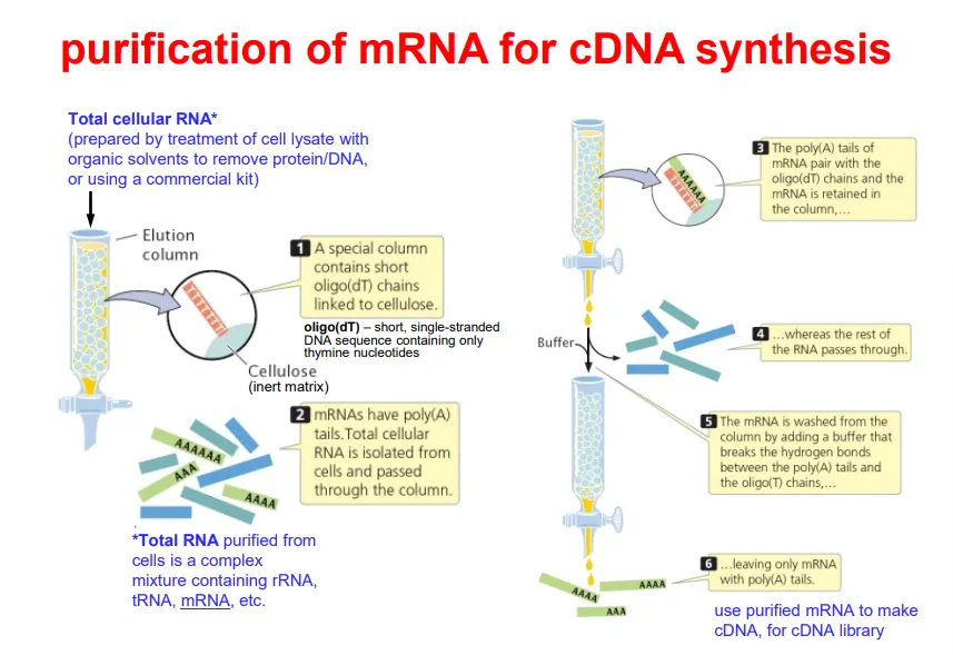
how can mRNA be purified for cDNA synthesis?
when mRNA is collected from a cell, a complex mix of coding and non-coding RNA will be collected
this has to be purified
all mRNA have a poly(A) tail, so we use a column containing oligo(dT) chains linked to cellulose, because this will bind all mRNA
once the non-coding RNA has been eluted/washed away, we are left with just mRNA (which gets separated from the oligo(dT) chains by a buffer that breaks the H bonds)
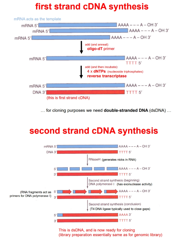
how is cDNA synthesised?
(following mRNA purification)
an oligo(dT) primer is used to prime synthesis, because the mRNA has a poly(A) tail
reverse transcriptase is used to add complementary dNTPs to the strand
this produces one cDNA strand bound to the mRNA strand
RNAase H produces nicks in the RNA strand, so that the RNA fragments can act as primers for DNA polymerase-I
this has exonuclease activity and can digest the RNA as it polymerises a complementary cDNA strand
DNA ligase must be used to close up the gaps
this is inserted into a vector and used to produce a cDNA library
how can genomic/cDNA libraries be screened?
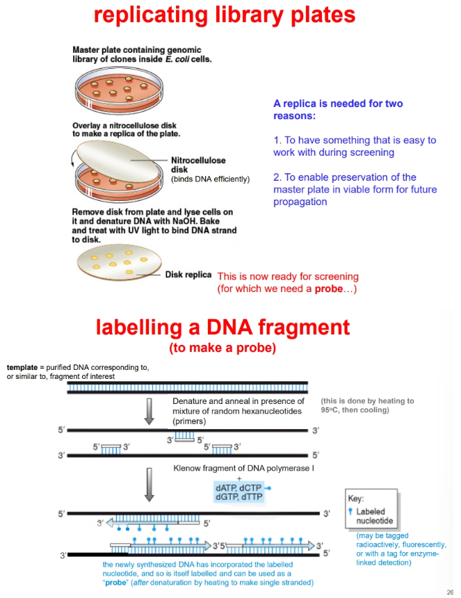
the plates must first be replicated, to make them easier to work with and preserve the master plate for future propagation
replica plating uses a nitrocellulose disc to stick to the cells
the cells are lysed, and the DNA is denatured (for hybridisation) using NaOH and stuck to the membrane by UV and heat
screening requires a double stranded probe, which is produced using a DNA template similar to our gene of interest
this fragment is denatured using heat and random hexanucleotide primers are annealed
the Klenow fragment of DNA polymerase-I produces a new strand using dNTPs, where one is tagged
denaturation by heating produces a single strand of this probe DNA, which will hybridise to our single-stranded DNA of interest when added to the replica plate in buffer
the correct clone plaque on the replica plate can then be detected radioactively/fluorescently/enzymatically, depending on the tag used
this is used to determine the correct colony on the master plate