Infections of kidneys and Fluid collections
1/103
There's no tags or description
Looks like no tags are added yet.
Name | Mastery | Learn | Test | Matching | Spaced | Call with Kai | Chat |
|---|
No analytics yet
Send a link to your students to track their progress
104 Terms
Most infections occur in the ___________ ascend into the kidneys
Bladder
Kidney infections are most commonly caused by …
Bacteria from the intestinal tract
Acute glomerulonephritis
Bilateral inflammation of glomeruli
Occurs as late complication of pharyngitis
Acute or chronic
Sudden onset of hematuria, azotemia, proteinuria
Acute glomerulonephritis is also caused by …
Other types of infection, autoimmune response, medications, toxins
Acute glomerulonephritis Labs
Increased serum BUN, and creatinine
Hematuria, proteinuria
Decreased GFR
Acute glomerulonephritis USA
Bilateral renal enlargement
May see increased cortical echogenicity with prominent pyramids
Increased RI
Ischemia or infarction causes hypoechoic foci in cortex
Acute glomerulonephritis
Chronic glomerulonephritis
Irreversible and progressive fibrosis
Usually progresses to end stage renal failure
Can lead to significant proteinuria, if chronic
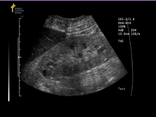
Chronic glomerulonephritis USA
Atrophic, smooth kidneys with increase echogenicity
Chronic glomerulonephritis
Acute Pyelonephritis
Inflammation of renal collecting system
Caused by bacteria from ascending UTI
Females 15-35yrs
Unilateral
Antibiotic treatment and imaging studies not necessary
Acute Pyelonephritis is most commonly caused by:
E. Coli
Acute Pyelonephritis labs
Diagnosis made with increased WBC, bacteria and pus in urine
Acute Pyelonephritis USA
Varied appearance, mostly normal kidneys
Loss of distinction between renal cortex and medulla
Diminished sinus echoes in affected kidney
Diffuse:
Unilateral or bilateral renal enlargement with diffuse swelling, loss of corticomedullary definition, calyceal clubbing
Focal:
Normal renal size with focal, indistinct hypoechoic wedge-shaped segment parenchyma, similar to renal infarct
Chronic pyelonephritis
Nephritis associated with urinary bladder reflux and stasis
Chronic untreated bacterial infection
Begins in childhood
Uni or bilateral
Chronic pyelonephritis is more common in…
Women
Chronic pyelonephritis causes:
Scarring of cortex if untreated renal failure will occur
Chronic pyelonephritis Labs
Diagnosis usually made clinically with increased WBC and pus in urine
Chronic pyelonephritis Symptoms
High fever, malaise, lethargy, anemia, N/V
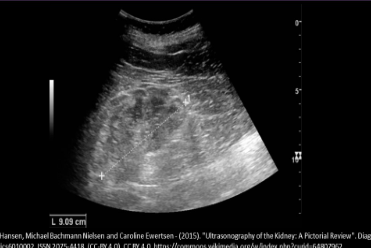
Chronic pyelonephritis USA
Small, shrunken, misshapen kidney with clubbing of calyces
Small kidney with clubbing of the calyces
Overlying cortical atrophy/scarring causes irregular kidney contour and cortical thinning
Increased corticomedullary echogenicity
Compensatory hypertrophy of contralateral kidney
Pyelonephritis
Emphysematous pyelonephritis
Bacterial infection associated with renal ischemia
Infection of parenchyma with gas formation
Commonly caused by E. Coli
Abscess formation with intrarenal air
Extend into perirenal space
LIFE THREATENING
Emphysematous pyelonephritis is most commonly seen in
Women, diabetics and IMMUNOSUPPRESSED pts
Emphysematous pyelonephritis Symptoms
Fever, leukocytosis, flank pain, dehydration, electrolyte imbalance
Emphysematous pyelonephritis Labs
Elevated WBC and glucose level, positive serum and urine bacterial cultures
Emphysematous pyelonephritis USA
UNILATERAL, enlarged, hypoechoic kidney
Multiple echogenic foci with reverberation/ring down artifact in the sinus or parenchyma
May demonstrate areas of echogenicity with dirty posterior shadowing
Emphysematous pyelonephritis
Malakoplakia
Chronic E. Coli infection
Unilateral enlarged kidney with multiple poorly defined cortical masses
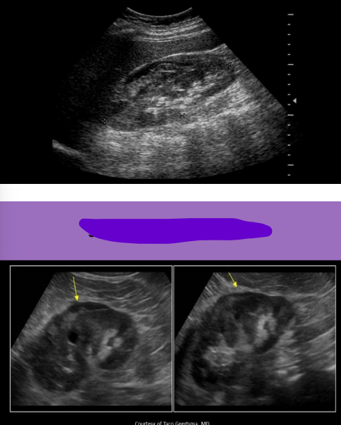
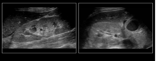
Xanthogranulomatous pyelonephritis
Chronic bacterial infections cause chronic obstruction which leads to destruction of parenchyma and replacement with lipid laden macrophages
Unilateral and diffuse
70% with stones
More common in women and diabetics
Requires nephrectomy
Xanthogranulomatous pyelonephritis is assocaited with
Phlegmon formation
Xanthogranulomatous pyelonephritis Labs
Bacteria in urine, anemia, leukocytosis
Xanthogranulomatous pyelonephritis symptoms
Dull persistent pain, weight loss, UTI
Xanthogranulomatous pyelonephritis USA
Unilateral renal enlargement
Loss of corticomedullary definition
Sinus very echogenic
Debris filled calyces
Multiple echogenic foci with shadowing
Staghorn calculus and hydronephrosis are common
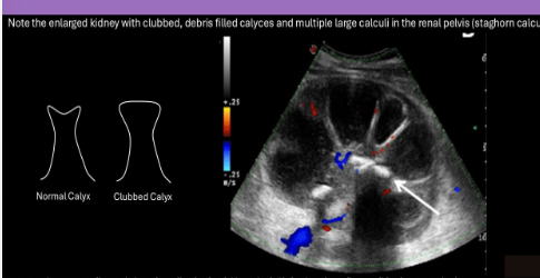
Xanthogranulomatous pyelonephritis
Mycetoma is also known as
Fungal ball
Mycetoma is associated with
Candidiasis infection that is caused by candida albicans
Most common fungal infection in the kidneys
Candidiasis
Mycetoma
Commonly seen in diabetics and immunosuppressed patients
Seen in pts that require long term use of indwelling urinary catheters, those with Hx of IV drug use, or long term steroid or antibiotic therapy
Treated with antifungal medication
Mycetoma Labs
Hematuria, bacteriuria, pyuria
Mycetoma Symptoms
Flank pain, chills and fever
Mycetoma USA
Hyperechoic
Non-shadowing mass within collecting system
Can be mobile, eval with different pt positions

Mycetoma
HIV nephropathy
Immunocompromised pts are at risk of numerous infections that damage the kidneys
INCREASED ECHOGENICITY OF KIDNEYS
Globular-shaped kidneys, decreased corticomedullary definition, decreased renal sinus fat
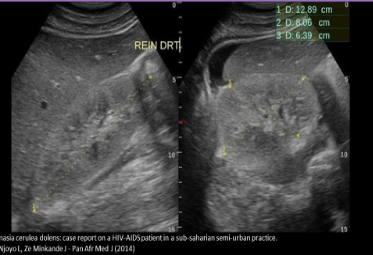
HIV Nephropathy
Corticomedullary renal abscess
Caused by ascending spread of bacteria in the urinary tract
End result of acute bacterial nephritis
Fever, chills, N/V, leukocytosis
Associated with diabetes, urinary tract obstruction, infected renal calculi, IV drug use
Renal carbuncle (abscess)
Refers to a renal abscess that forms in the parenchyma
Caused by hematogenous spread of staphylococcus aureus
Fever, chills, N/V, increased WBC
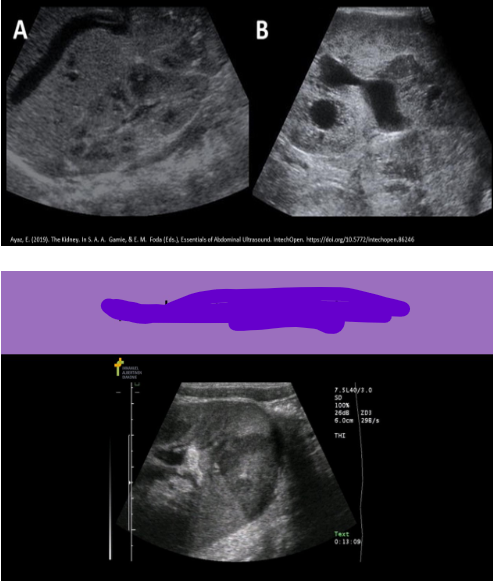
Renal abscess USA
Hypoechoic, complex mass with irregular borders
Fluid/debris levels
Thick walls
Shadowing from gas; may see ring down
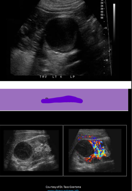
Renal abscess
Hematoma
Collection of blood outside the kidney
Trauma or surgery
Vary in size and shape with location
Blood usually fills space available, causing shape irregularity
Subcapsular/Intracapsular: Between the renal cortex and capsule
Extracapsular/Perirenal: Outside the capsule, between the capsule and the liver (Morison pouch) or spleen
Hematoma symptoms
Pain and hematuria
USA Hematoma
Varied appearance with age
Complex mass
Debris
Septations
Intracapsular will usually indent or deform renal parenchyma
Extracapsular does NOT indent parenchyma
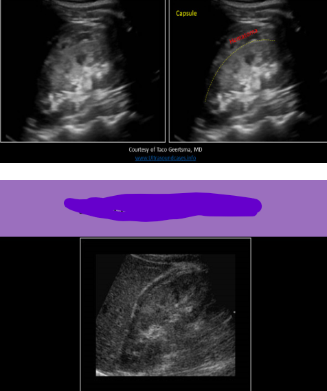
Extracapsular hematoma
Subcapsular hematoma
Urinoma
Leakage of urine from UPJ or UVJ
Post-op complication
Usually presents 1-2 weeks post transplant
May cause hydronephrosis due to ureter compression
USA Urinoma
Cystic collection adjacent to kidney, may see debris
Subcapsular fluid collections usually distort of indent renal parenchyma
Urinoma
Lymphocele
Encapsulated collection of lymph
Associated with trauma or surgery
Can have delayed presentation of up to 2-6 months after surgery
USA lymphocele
Cystic structure between kidney and bladder
The fluid commonly contains debris and septations
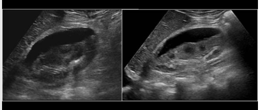
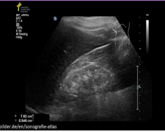
Hydronephrosis
Obstruction of urine outflow with dilated collecting system
Unilateral - UPJ stone, extrinsic compression from mass, malformed UVJ
Bilateral - Prostate enlargement, bladder mass, urethral obstruction, pregnancy (R>L)
Can lead to obstructive nephropathy
RI > 0.7 in arcuate arteries = associated nephropathy
Chronic hydronephrosis can cause
HTN, renal failure and sepsis
Most common site of obstruction by renal calculi
UVJ
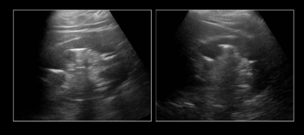
USA hydronephrosis
Dilation of normal collecting system
Minimal - Prominent pelvis, small amount of fluid
Mild - Dilated pelvis and calyces, sinus echoes visible
Moderate - Dilated renal pelvis and calyces, no sinus echoes visible “bear claw”
Severe - Extremely dilated collecting system, compression of cortical tissue, formation of parapelvic cysts
Follow ureter to junction with bladder to determine level of dilation
Ask pt to void bladder and see if hydronephrosis persists
= prox to urethra indicating possible urethrocele, UPJ obstruction or ureteral obstruction
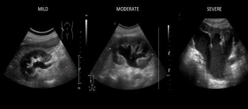
Hydronephrosis
Pyonephrosis
Pus in collecting system
Infection in an obstructed system, usually UPJ obstruction
Fever, flank pain, leukocytosis and other symptoms
Dysuria, pyuria, hematuria
Associated with perinephric abscess, bacteriemia and septic shock
USA Pyonephrosis
Enlarged echogenic kidney with significant dilation of the calyces
Mobile echogenic debris and pt position dependent debris levels
Stones possible
May also see dirty shadowing or ring down from gas produced by bacteria
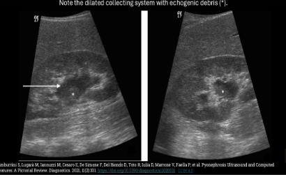
Pyonephrosis
Acute tubular necrosis (ATN)
Caused by prolonged ischemia or drug toxicity that damages renal parenchymal cells
Can be reversed if treated early enough
Most common cause of intrinsic acute renal failure
Acute tubular necrosis
USA ATN
Enlarged echogenic kidneys
Increased resistive index in parenchymal vessels
Pyramids appear more prominent
Acute renal failure
Acute renal failure
Acute inability to remove metabolites from the blood
CRITICAL but REVERSIBLE if caught early
Leads to abrupt decrease in renal function and acute reduction in urine output (oligouria)
Prerenal failure - Hypotension, volume depletion, heart failure, acute occlusion of the renal artery
Intrinsic failure - acute tubular necrosis, nephritic syndrome, interstitial nephritis, autoimmune diseases
Postrenal failure - Bilateral renal obstruction, oliguria, and empty bladder
Most common cause of acute renal failure
Tubular necrosis
Acute renal failure is caused by
Obstruction of urine flow, reduced renal perfusion or renal parenchymal disease
Acute renal failure labs
Increased potassium, BUN and creatinine
Acute renal failure symptoms
Hypovolemia, HTN, peripheral edema, hematuria, oliguria
Acute renal failure USA
Normal or increased echogenicity compared to liver/spleen
Kidneys are normal in size or slightly enlarged
Bilateral hydronephrosis indicates postrenal failure
Doppler evaluation should be performed on parenchymal arteries to measure the RI (>0.7 = intrinsic failure)
Chronic renal failure is also known as
Medical renal disease
Most common cause of Chronic renal failure
Diabetes mellitus
Other causes of Chronic renal failure are…
Renal vascular disease, chronic pyelonephritis, tuberculosis, lupus
Chronic renal failure Labs
Increased BUN and creatinine, hyperkalemia (elevated potassium)
Chronic renal failure
Considered end stage renal failure when kidney function is 10% or less
May require dialysis until transplant can be done
USA Chronic renal failure
Kidneys are small in size and very echogenic due to atrophy
Cortex is thinned (<10mm) with loss of corticomedullary border
Dialysis
Used to remove excess waste products and water from body when kidneys can no longer do it
Helps body control blood pressure and sodium potassium levels
Pts may experience HTN and uremia; N/V, swelling and fatigue
Hemodialysis
AVF created by connecting an artery and a vein, usually in the arm
Dialysis machine connected to the pt using two separate needles
Blood is removed from the body, sent through the machine and returned to body through AVF
Requires regular visits
Peritoneal dialysis
Used for pts with some residual kidney function
Eventually function will decline to a point that hemodialysis will be needed instead
Cleansing fluid is injected through an intraperitoneal catheter
Fluid filters waste products from the blood for a specified amount of time then drains out of abdomen
Can be done at home
These pts commonly demonstrate ascites
Chronic renal failure
What renal infection frequently occurs as a late complication of pharyngitis?
Acute glomerulonephritis
Which of the following is a symptom of acute glomerulonephritis?
Foggy urine
Hematuria
Proteinuria
All of the above
All of the above
Acute glomerulonephritis presents as ___________, while chronic glomerulonephritis presents as ___________
Bilateral renal enlargement; bilateral renal atrophy
What causes acute pyelonephritis
Ascending bacterial infection
Which of the following is a sign of acute pyelonephritis?
Loss of corticomedullary definition
Diminished sinus echoes in affected kidney
Calyceal clubbing
All of the above
All of the above
All the following are characteristics of Emphysematous pyelonephritis, except?
Commonly caused by E. Coli
Abscess formation with intrarenal air
Areas or ring down/dirty shadowing
Multiple renal calculi within parenchyma
Multiple renal calculi within parenchyma
What is a critical renal infection that requires an emergency nephrectomy
Emphysematous pyelonephritis
_____________________________ are more common in women and diabetics
Xanthogranulomatous pyelonephritis and emphysematous pyelonephritis
Mycetoma formation is associated with:
Candidiasis infection
Mycetoma formation is associated with which of the following?
Long term use of indwelling urinary catheters
Those with history of IV drug use
Long term steroid or antibiotic therapy
All of the above
All of the above
______________ refers to a renal abscess that forms in the parenchyma caused by hematogenous spread of staphylococcus aureus
Carbuncle
When evaluating a suspected renal hematoma, it is most important to document the location of the hematoma relative to:
Renal capsule
How is an intracapsular fluid collection differentiated from an extracapsular fluid collection of the kidney?
Presence or absence of parenchymal distortion
What sonographic characteristic indicates a lymphocele is present and not a urinoma
Lymphocele has septations in fluid