6.2: The Blood System
1/23
There's no tags or description
Looks like no tags are added yet.
Name | Mastery | Learn | Test | Matching | Spaced | Call with Kai |
|---|
No analytics yet
Send a link to your students to track their progress
24 Terms
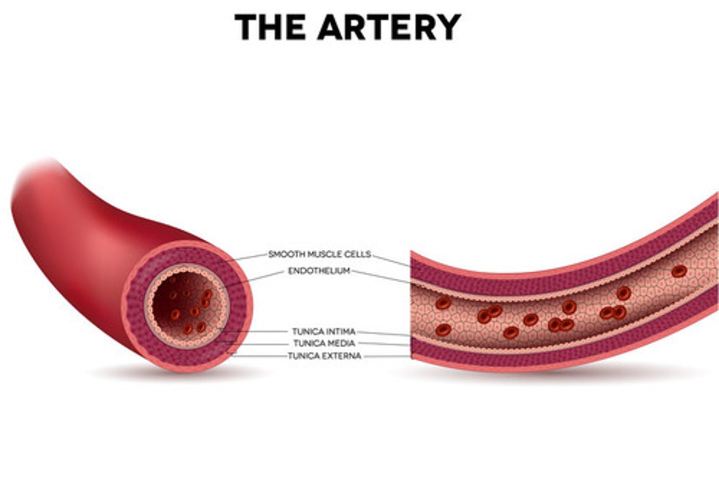
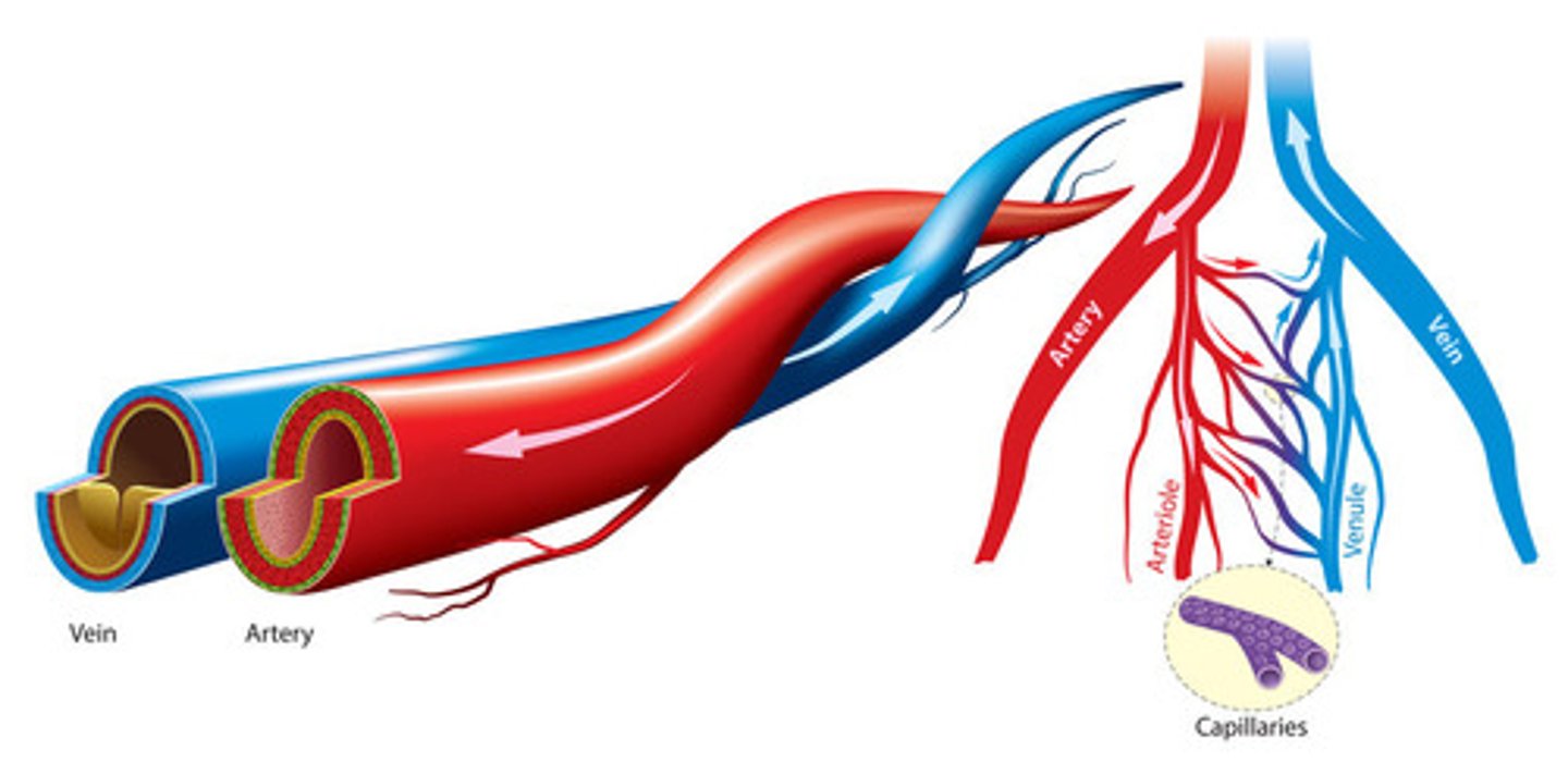
State the function of arteries
Arteries carry blood away from the heart
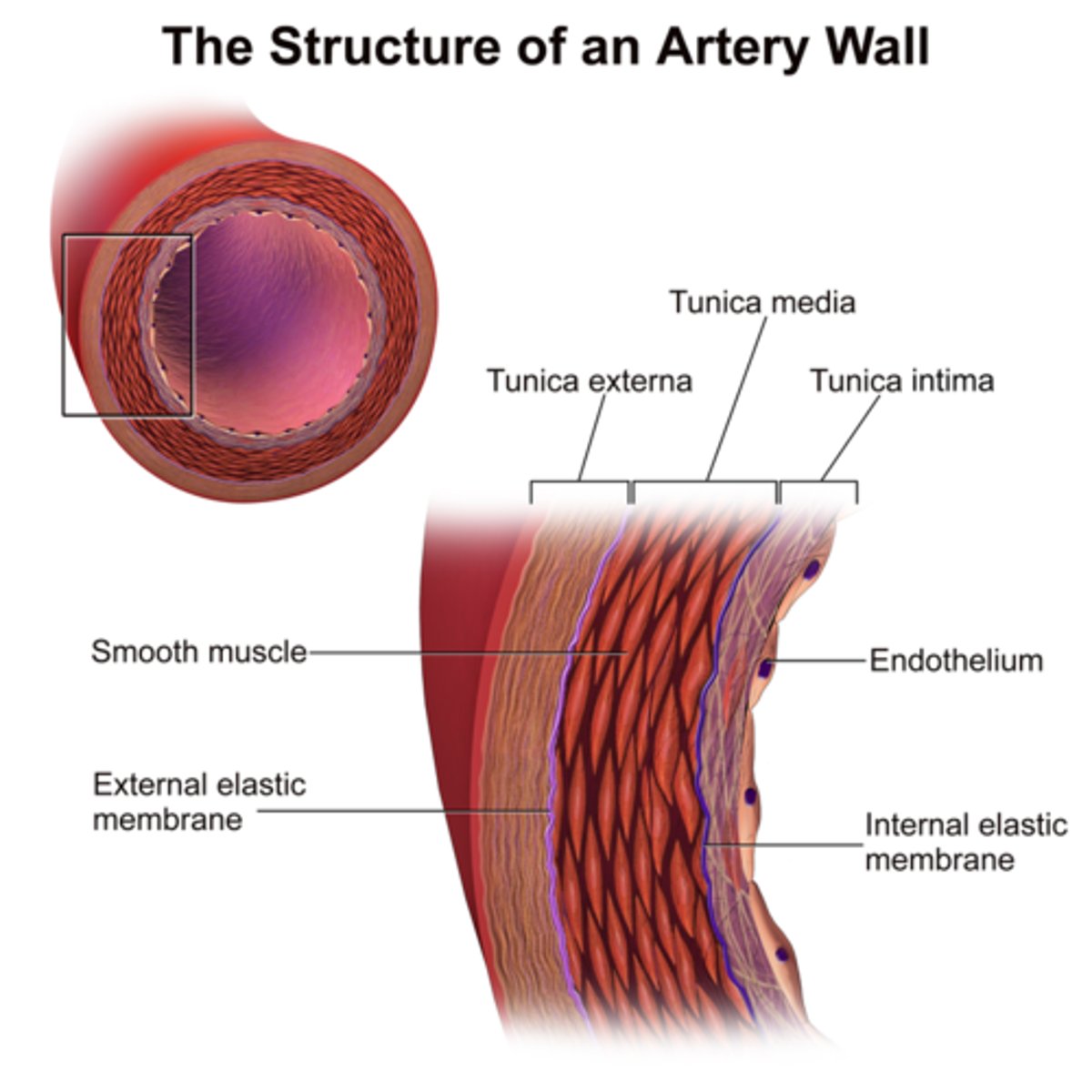
have a thick layer of muscle to help move blood through vessels
high internal pressure
have a layer of elastic collagen fibres to allow expanding and contracting
State the reason for toughness of artery walls
Arteries must be able to withstand the pressure of the blood as it moves with each heartbeat
Explain how arteries are able to transport the blood under high pressure from the heart to the rest of the body
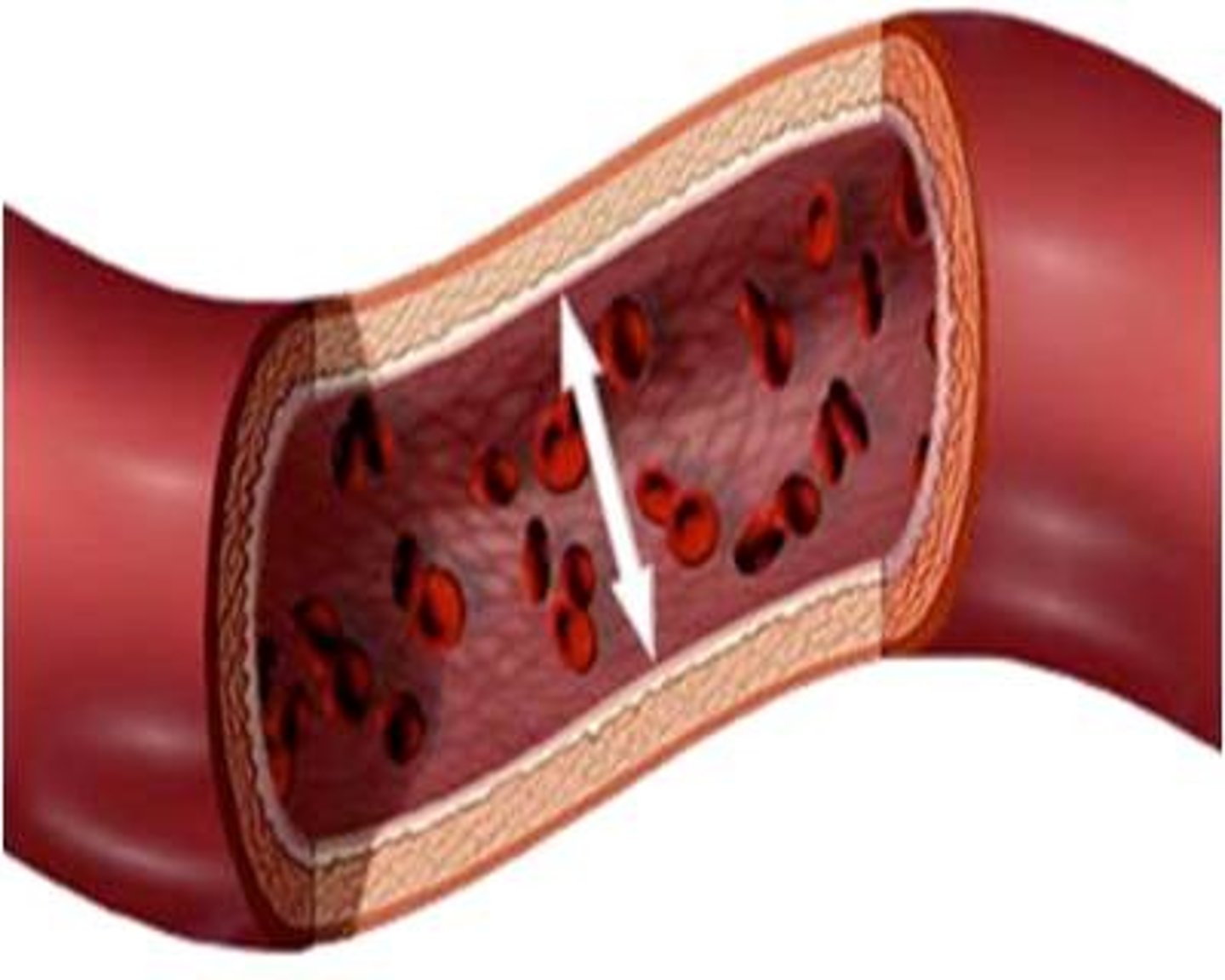
Blood leaves the heart through arteries under high pressure as a result of the contraction of the ventricles. The thick muscular walls and narrow lumen of arteries maintains the pressure as the blood moves to the rest of the body. Additionally, the elastic recoil of the arterial walls helps to push blood between contractions of the heart.
Define "blood pressure."
Blood pressure is the pressure exerted by blood on the walls of an artery.
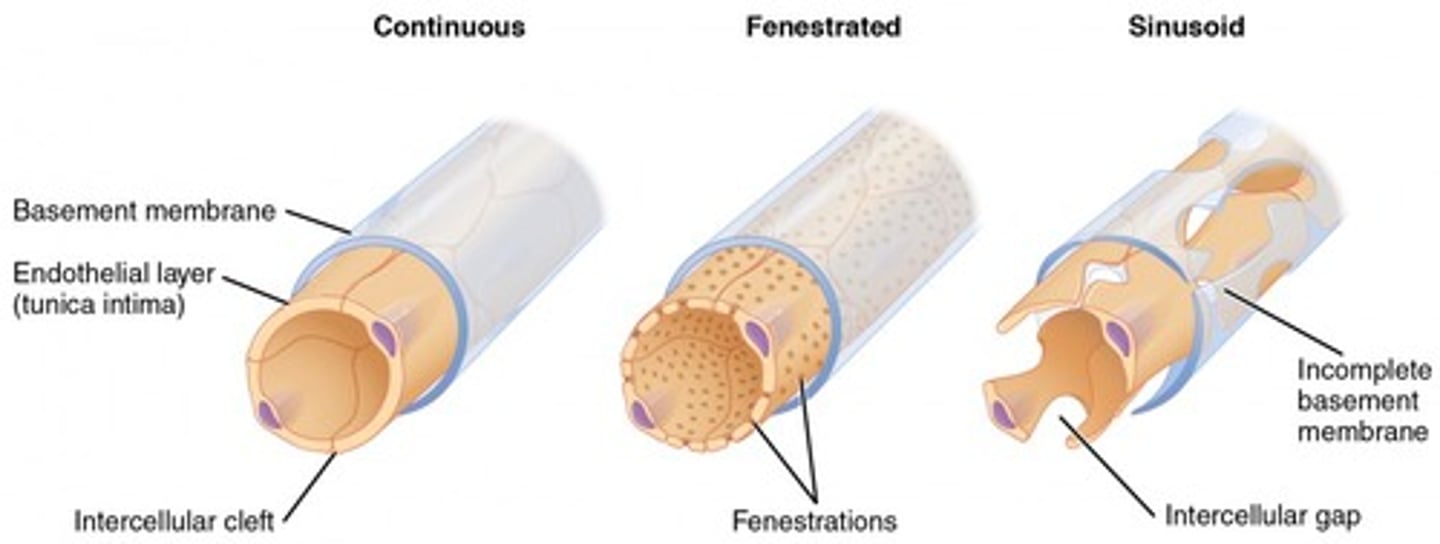
Describe the structure and function of capillaries
Capillaries are the smallest blood vessels in the body
The capillary lumen is so narrow
one cell thick to allow diffusion to occur from blood cells to the cells of other tissues
State the function of veins
Veins carry blood towards the heart
thin walls/thin muscle layer
low internal pressure
have valves inside to make sure blood only flows one way
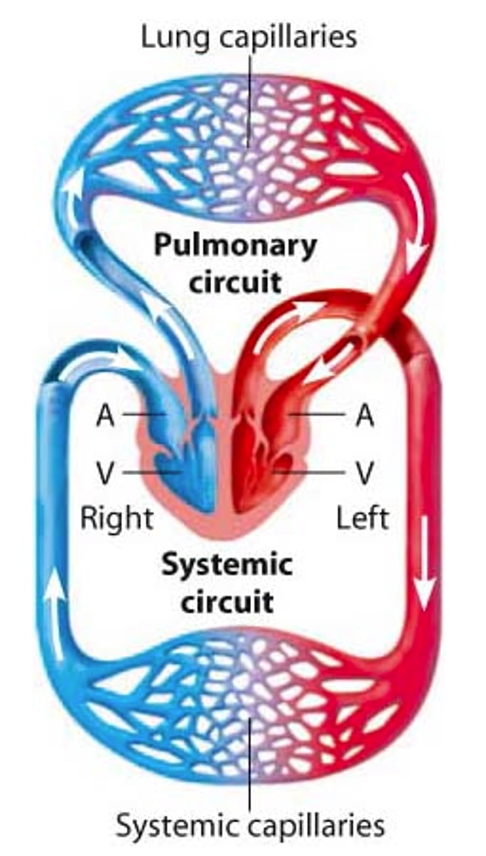
Draw a diagram to illustrate the double circulation system in mammals
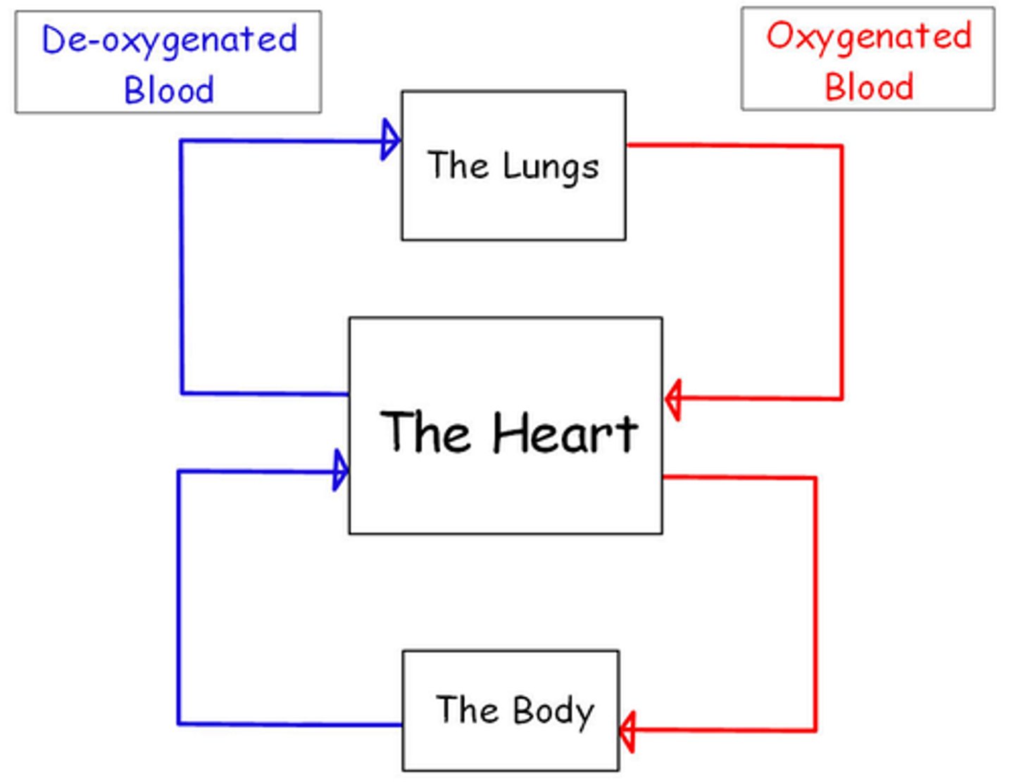
The majority of mammals (including humans) utilize a double circulatory system. It is called a double circulatory system because blood passes through the heart twice per full circuit.
The right pump sends deoxygenated blood to the lungs where it becomes oxygenated and returns back to the heart. The left pump sends the newly oxygenated blood around the body
Compare the circulation of blood in fish to that of mammals
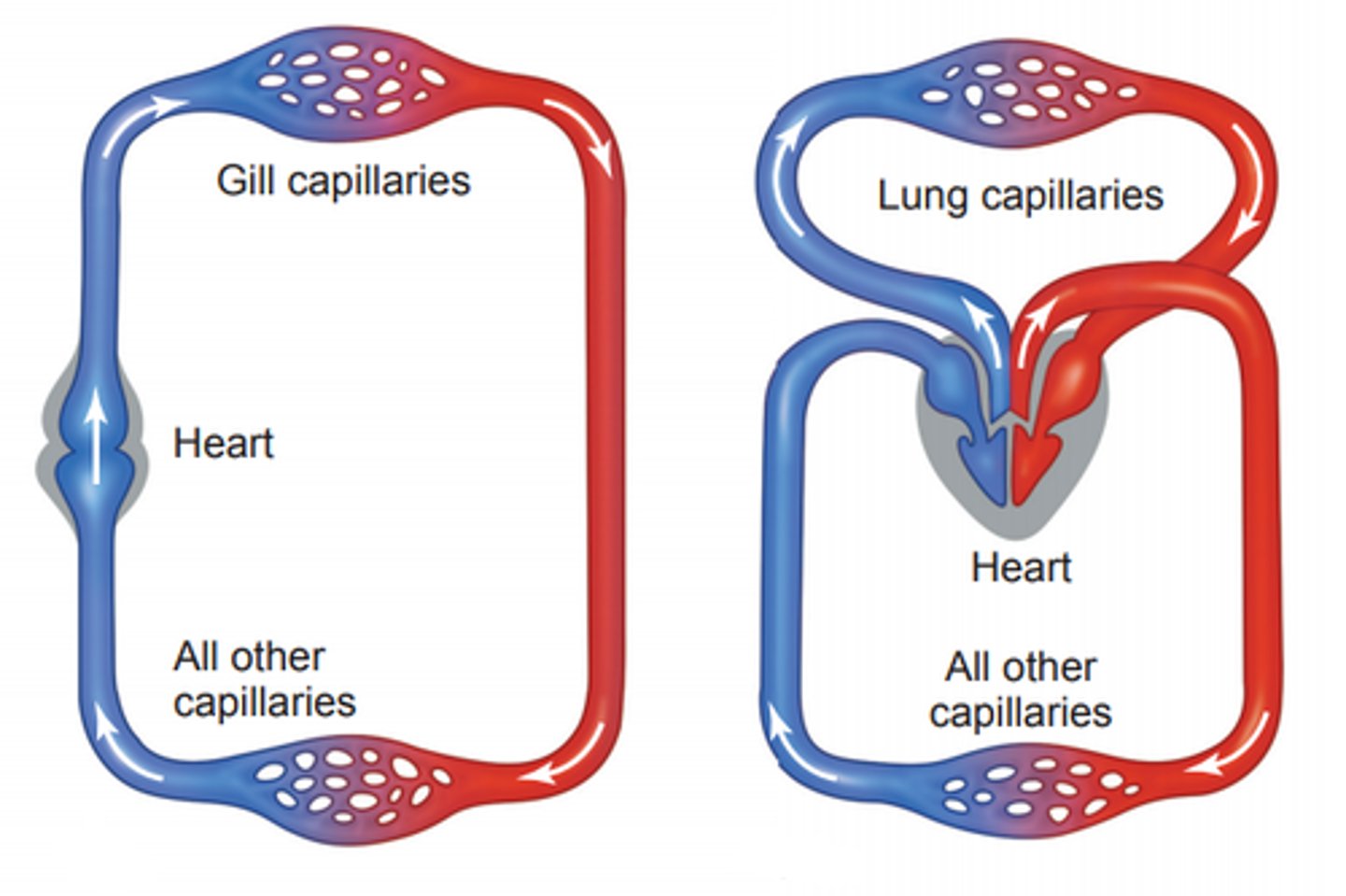
In fish, there is single pump circulation. The heart only has one atrium and one ventricle. The oxygen-depleted blood that returns from the body enters the atrium, and then the ventricle, and is then pumped out to the gills where the blood is oxygenated, and then it continues through the rest of the body
Explain why the mammalian heart must function as a double pump
There must be a double pump in order to create enough pressure to move the blood throughout the entire body. Pressure is needed to move blood through the resistance of a large network of blood vessels like arteries, capillaries, and veins
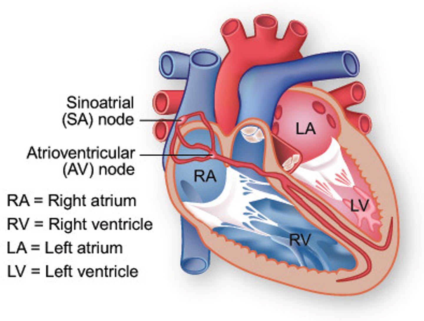
Define "myogenic contraction."
Myogenic contractions are contractions that are initiated in the heart muscle itself rather than by stimulation from nerve impulses.
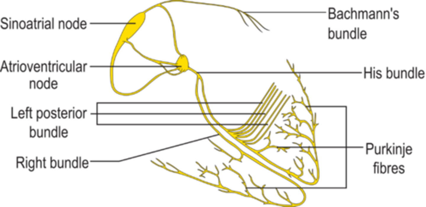
Outline the role of cells in the sinoatrial node
The sinoatrial node (SA node)
produce an electrical impulse (action potential) that travels through the heart causing it to contract.
The SA node is known as the heart's "pacemaker."
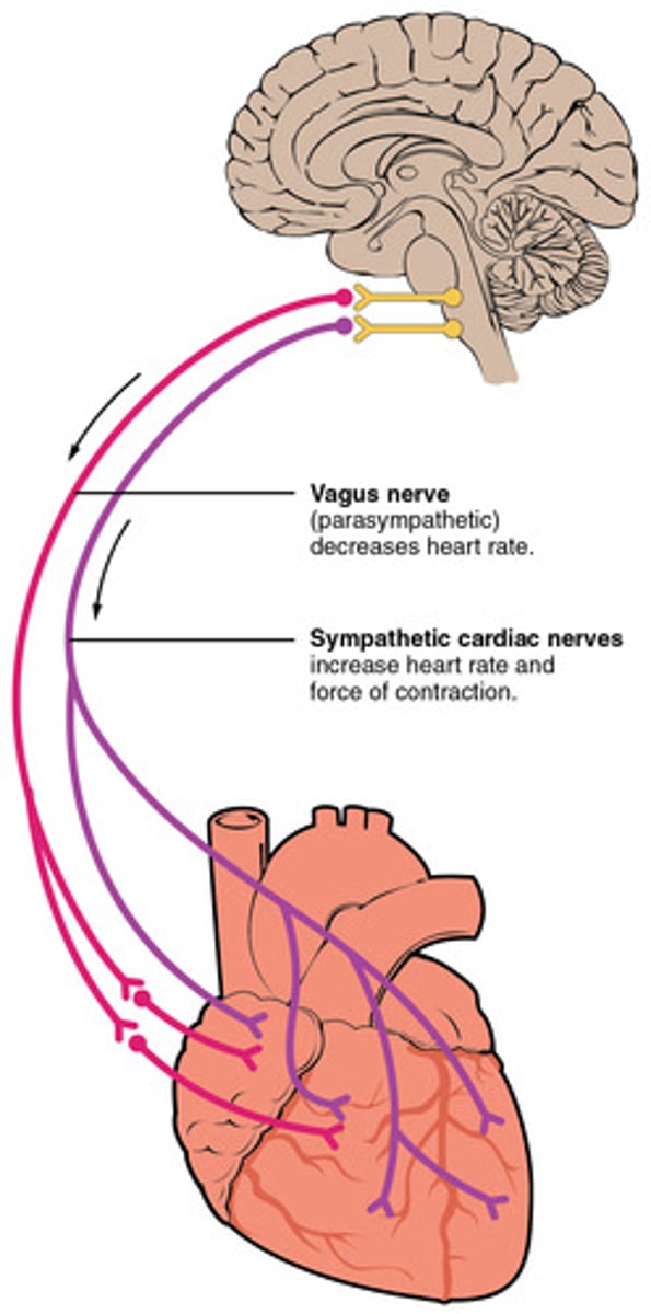
Outline the action of nervous tissue that can regulate heart rate
Heart rate is intrinsically determined by the pacemaker activity of the sinoatrial node (SA node) located in the wall of the right atrium. However, the pace of the SA node can be changed by impulses (action potentials) brought to the heart through nerves from the medulla of the brain. Neural input can influence heart rate, cardiac output, and contraction forces of the heart.
The vagus nerves (parasympathetic) can reduce the heart rate and the force of contraction of the heart.
Cardiac sympathetic nerves (sympathetic) can increase the heart rate and the force of contraction of the heart.
Outline William Harvey’s role in discovery of blood circulation
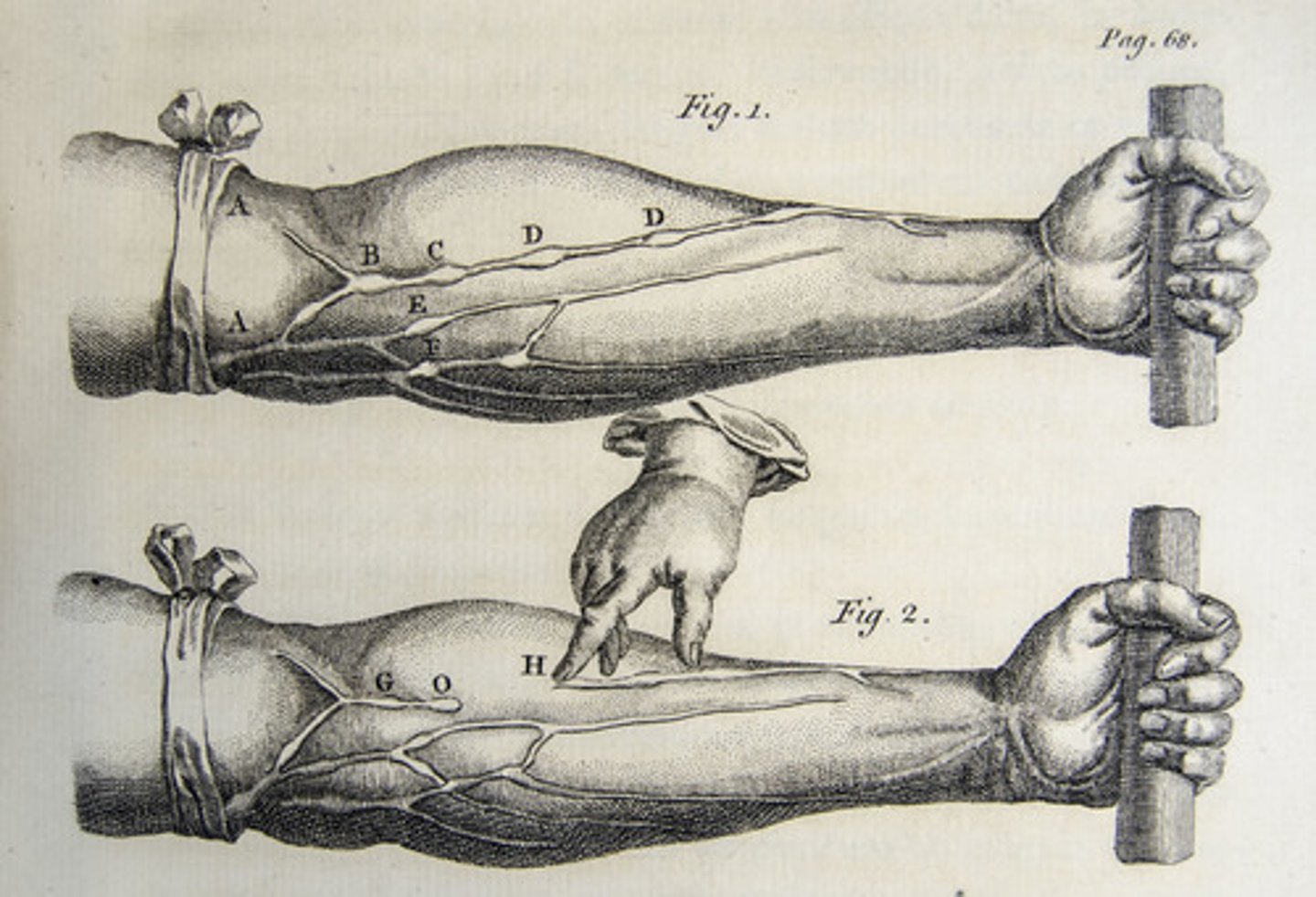
William Harvey (1578-1657), observing the heart in living animals, he was able to show that the valves in the veins permit the blood to flow only in the direction of the heart and to prove that the blood circulated around the body and returned to the heart
Describe the cause and consequence of atherosclerosis
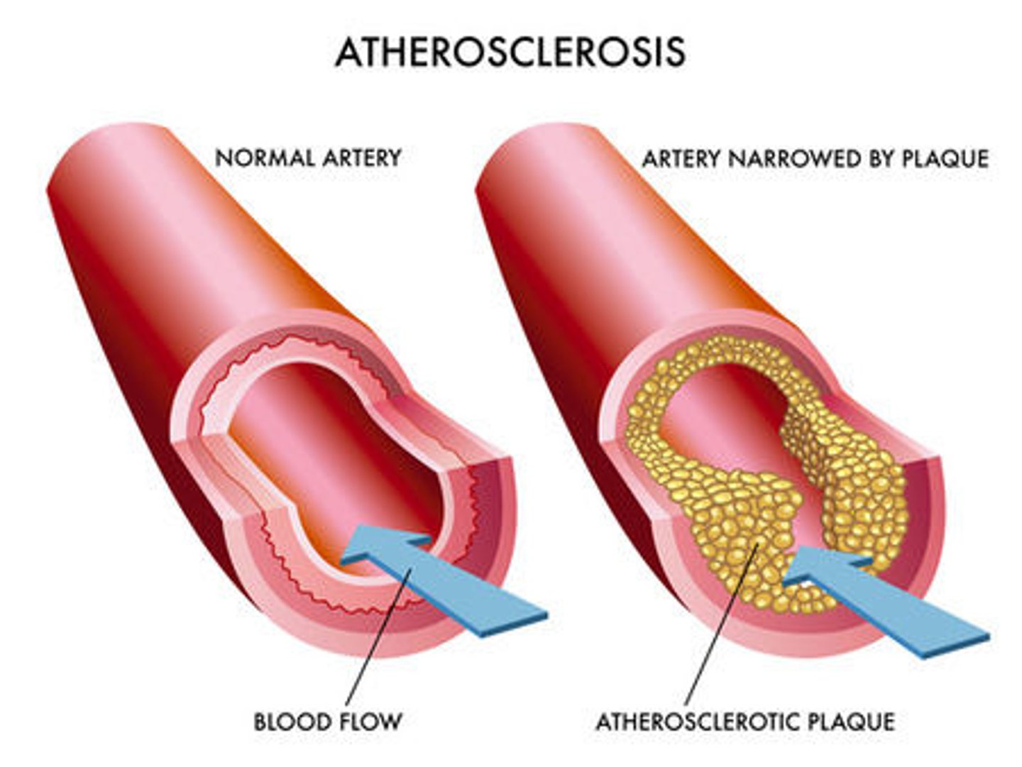
Atherosclerosis is the narrowing of arteries due to the buildup of fats, cholesterol and other substances in and on artery walls (plaque).
Plaques from atherosclerosis can:
- increase blood pressure
- cause the vessel to rupture allowing blood to clot inside the artery (potentially leading to stroke or heart attack).
Define "cardiac cycle”
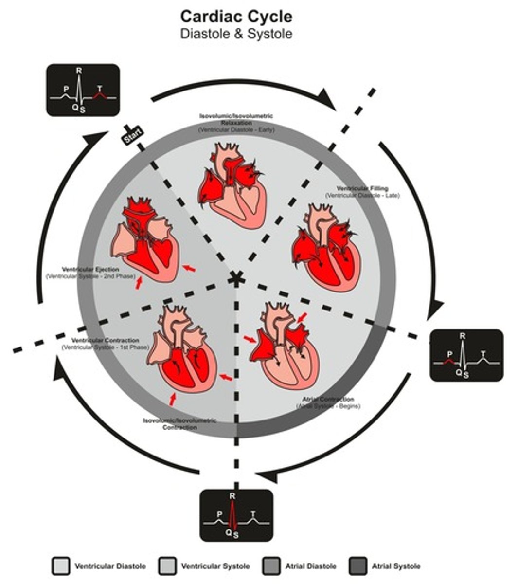
The cardiac cycle describes the series of events that take place in the heart over the duration of a single heart beat
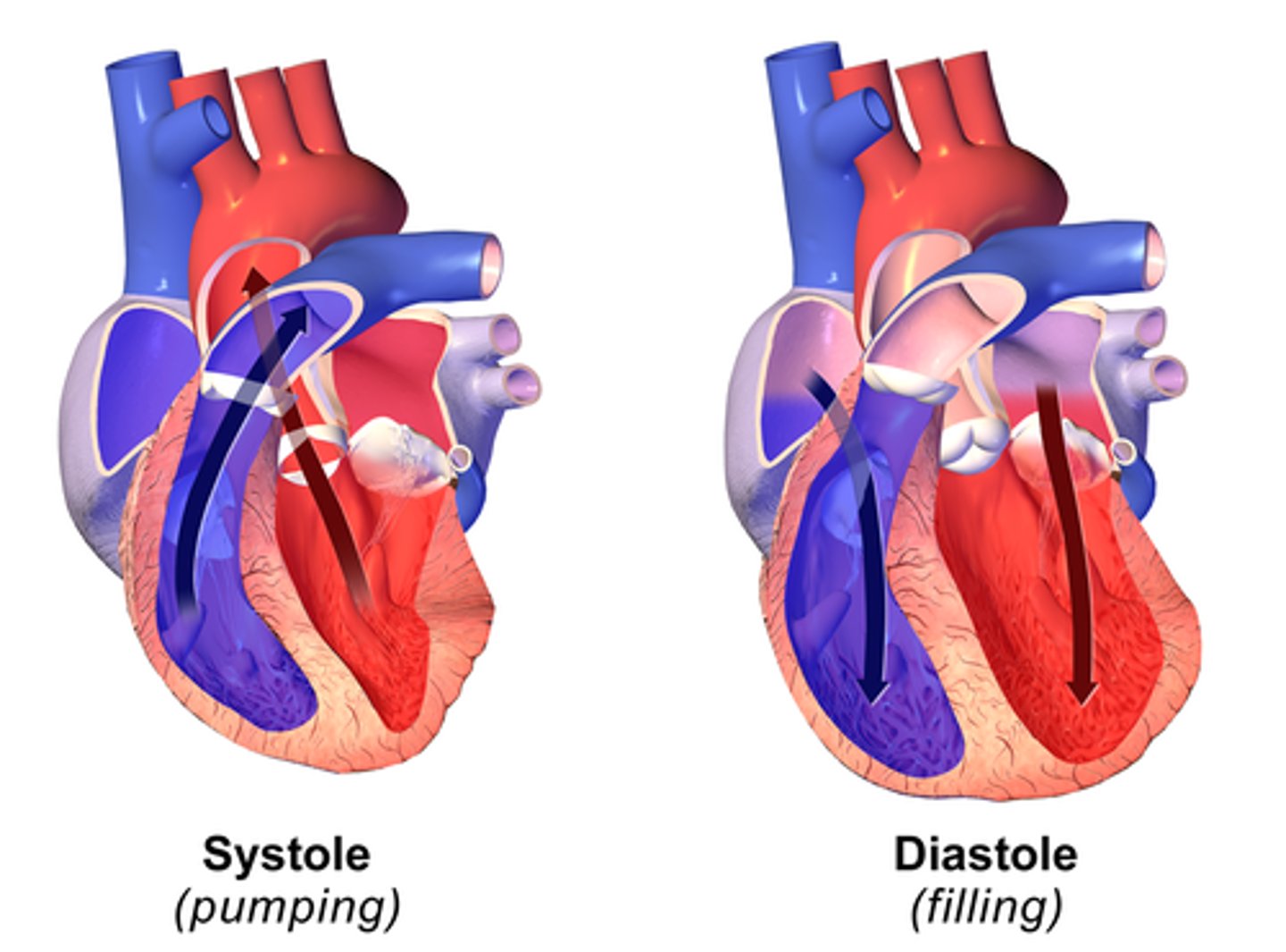
Define systole
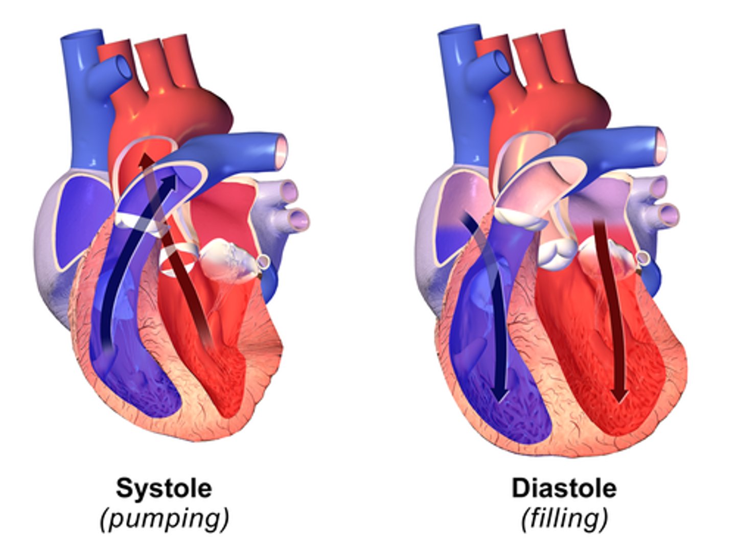
Systole is the part of the cardiac cycle during which the heart muscle contracts and moves blood out of the chambers. There is an atrial systole and a ventricular systole.
Define diastole
Diastole is the part of the cardiac cycle during which the the muscle muscle relaxes and allows the chambers to fill with blood There is an atrial diastole and a ventricular diastole.
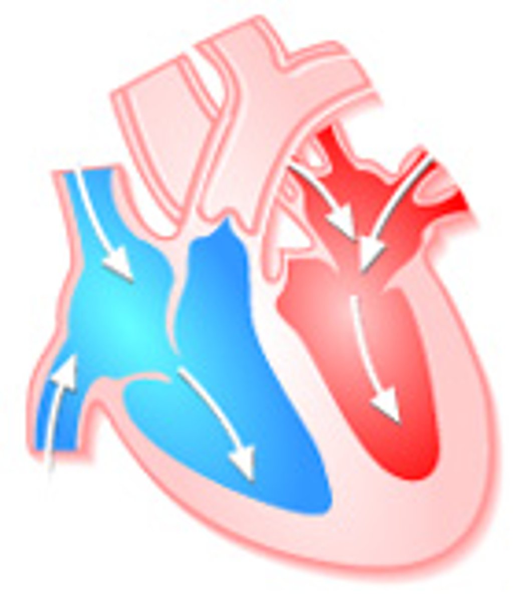
Outline events of atrial systole
Contraction (systole) of the atria is triggered by the firing of the sinoatrial node. As the atrial muscles contract, pressure rises within the atria and blood is pumped into the ventricles through the open AV.
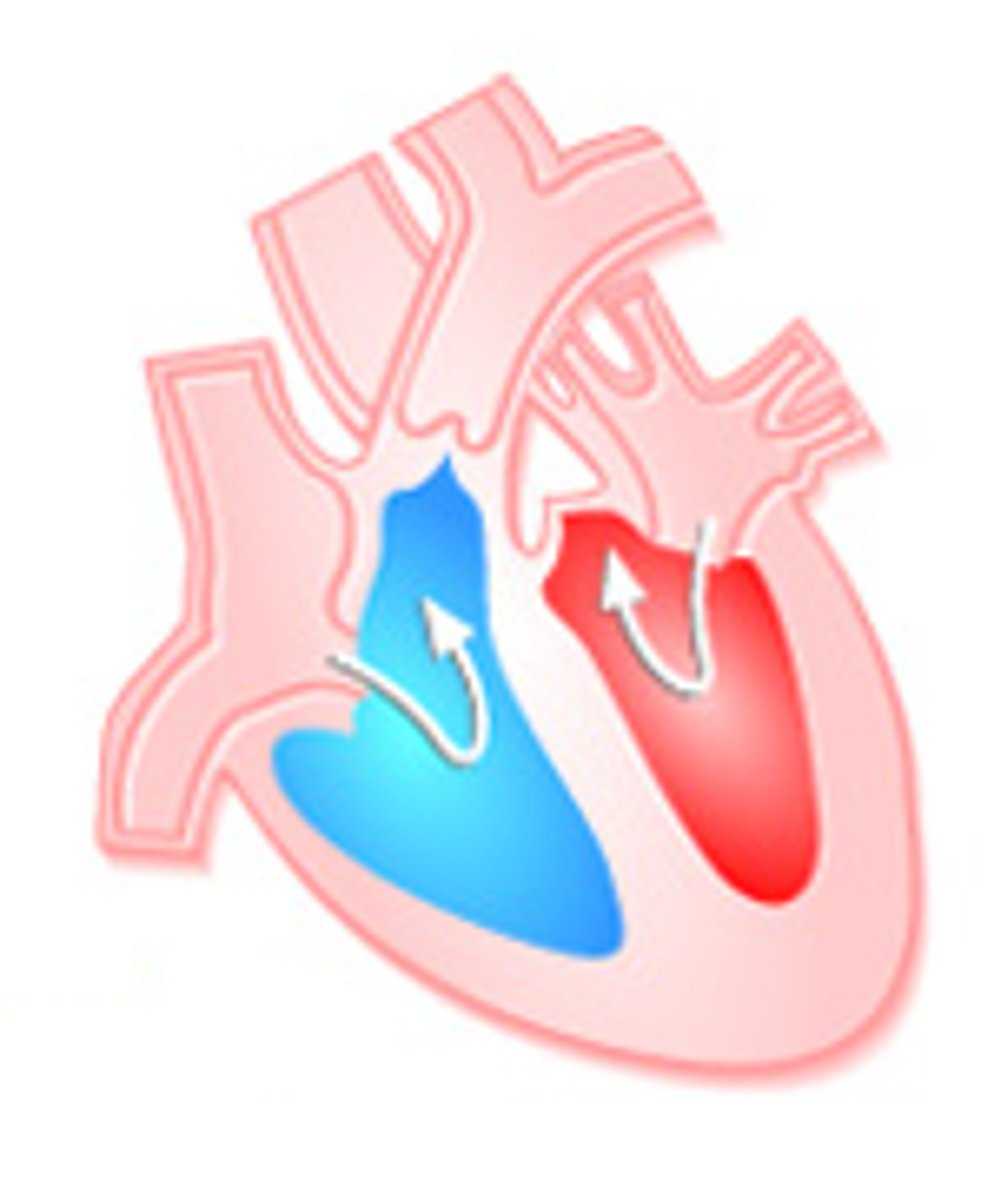
Outline events of ventricular diastole
During the early phase of ventricular diastole, as the ventricular muscle relaxes, pressure on the remaining blood within the ventricle begins to fall. The semilunar valves close to prevent backflow into the heart
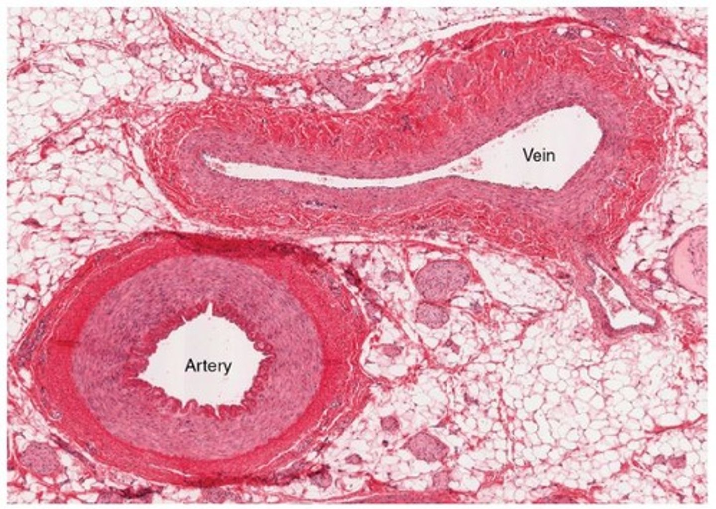
Given a micrograph, identify a blood vessel as an artery, capillary or vein
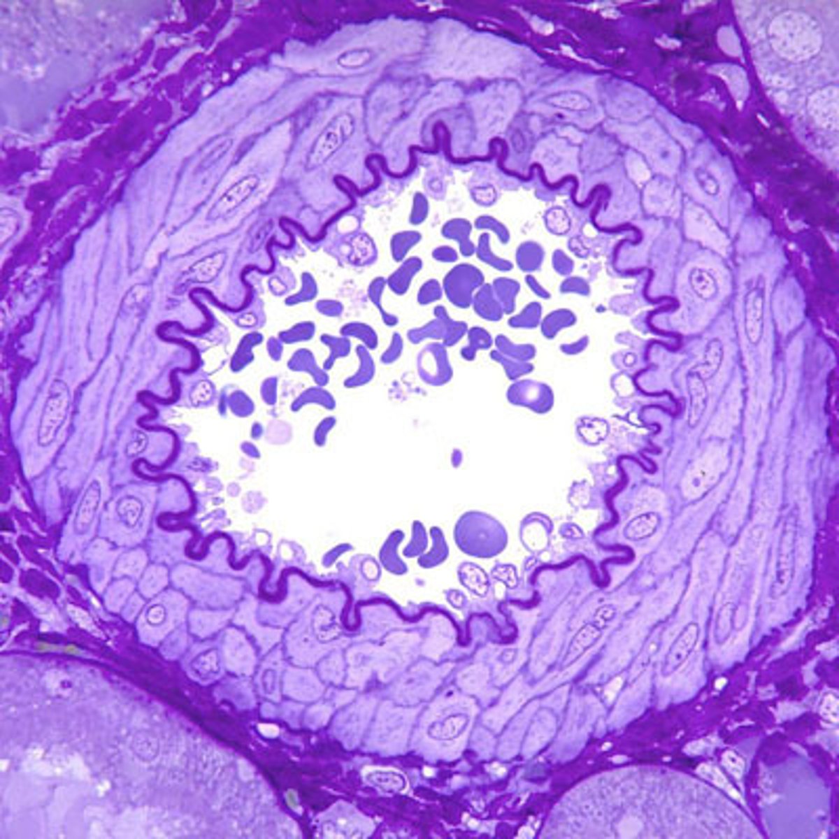
The walls of arteries are much thicker than those of veins because of the higher pressure of the blood that flows through them. The artery walls also tend to have a more distinct round shape, held in place by the fibers of the tunica media.
Capillaries can be distinguished by the very narrow lumen size (only 1 red blood cell wide).
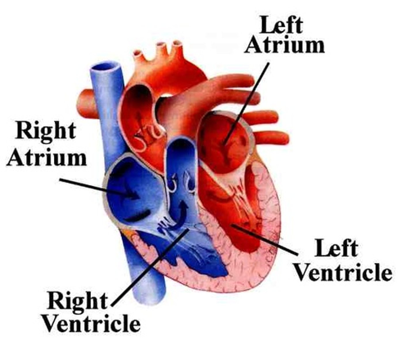
Label the chambers on a diagram of the mammalian heart
The mammalian heart has four chambers: two atria and two ventricles.
The right atrium receives oxygen-poor blood from the body via the vena cava and pumps it to the right ventricle.
The right ventricle pumps the oxygen-poor blood to the lungs via the pulmonary artery.
The left atrium receives oxygen-rich blood from the lungs via the pulmonary veins and pumps it to the left ventricle.
The left ventricle pumps the oxygen-rich blood to the body via the aorta.
Label the vessels on a diagram of the mammalian heart
Veins are vessels that bring blood to the heart.
The superior and inferior vena cava bring oxygen-poor blood from the body to the right atrium.
The four pulmonary veins bring oxygen-rich blood from the lungs to the left atrium.
Arteries are vessels that move blood away from the heart.
The pulmonary artery takes oxygen-poor blood from right ventricle to the lungs.
The aorta takes oxygen-rich blood from the left ventricle to the rest of the body.
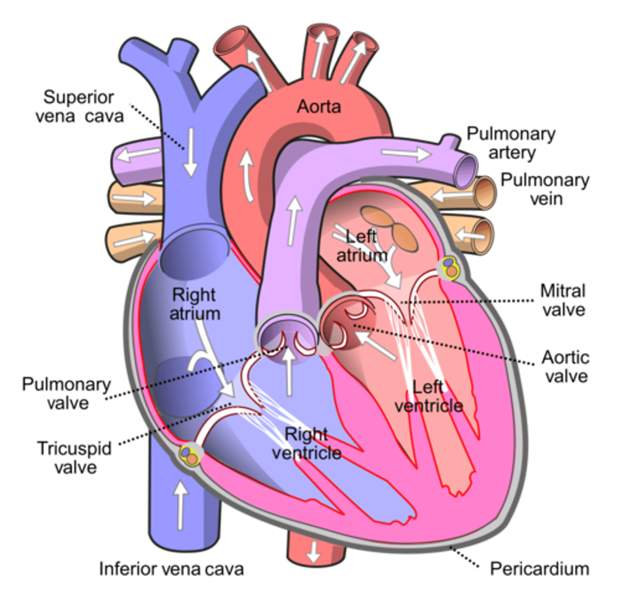
Label the valves on a diagram of the mammalian heart
The semilunar (SL) valves are located between the ventricles and the arteries leaving the heart. The pulmonary SL valve is located between the right ventricle and the pulmonary artery. The aortic SL valve is located between the left ventricle and the aorta.

Describe how Harvey was able to disprove Galen’s theory
Prior to Harvey’s findings, scientists held to the antiquated views of the Greek philosopher Galen, who believed that:
Arteries and veins were separate blood networks (except where they connected via invisible pores)
Veins were thought to pump natural blood (which was believed to be produced by the liver)
Arteries were thought to pump heat (produced by the heart) via the lungs (for cooling – like bellows)
Based on some simple experiments and observations, Harvey instead proposed that:
Arteries and veins were part of a single connected blood network (he did not predict the existence of capillaries however)
Arteries pumped blood from the heart (to the lungs and body tissues)
Veins returned blood to the heart (from the lungs and body tissues)