Exam 1 Study Guide
 Call Kai
 Learn
Learn Practice Test
 Spaced Repetition
 Match
Match Flashcards
 Knowt Play
1/237

There's no tags or description
Looks like no tags are added yet.
Name | Mastery | Learn | Test | Matching | Spaced | Call with Kai | Chat |
|---|
No analytics yet
Send a link to your students to track their progress
238 Terms
PIONEERS + IMAGE RECEPTORS
LECTURE 1
X-rays
Energetic form of electromagnetic radiation capable of imaging the structure of an object on film
What are some examples of electromagnetic radiation?
A range of energies from radio/TV waves to x rays

radiograph
Any picture of the teeth and oral structures produced by x-ray exposure and chemical or digital processing of the film.
radiology
The branch of medical and dental sciences that uses x-ray energy in the diagnosis and treatment of disease.
radiography
The making of film records (the actual procedure) of the internal structures of the teeth and surrounding bone by exposure of film specially sensitized to x rays.
Dr. Roentgen
- Discovered x-rays
- unknown "x" ray emitted
- First radiograph
- Invisible ray "X" caused a fluorescent glow of a photographic screen during his experiment with Crookes tube. -x-ray of wifes hand
Dr. Walkhoff
- First dental radiograph; on January 14, 1896, which required 25 minutes of exposure
- extraoral radiographs (glass plate wrapped in dental dam)
- Established 1st dental x-ray laboratory
-Observed hair loss on exposed patients
Dr. Kells
- 1st dental radiograph in America April 1896
- Invented film holder
- Advocated paralleling technique
-Skiagraphs - "Shadow Pictures"
- Gave 1st clinic on dental radiology at a dental meeting
-1st US radiographic clinic and laboratory
Accident
Radiation damage from"tuning the tube" to test machine x-ray output led to him losing his left hand and arm due to over exposure to x-rays
-Had no regrets, though, because his work brought so much information to the realm of radiology. Later killed himself in 1928 as to not be a burden to his family.
Dr. Rollins
"Forgotten Man of Dentistry"
- Invented safety procedures
- Invented x-ray machine arm and bracket
- Developed beam filters and devices to restrict the beam size
Accident
In 1898, received a severe burn while exposing his hand to the X-ray tube and high-voltage wire.
-Injury prompted research followed by these recommendations: Wear leaded glasses, Enclose x-ray tube inside leaded or non-radiable housing, Irradiate only the area of interest, and cover the rest with protective material.
Dr. Raper
Graduated IUSD in 1906
-Introduced dental radiology into curriculum in 1909
- Wrote 1st radiology textbook in 1913, Elementary Dental Radiology
- invented the tab bitewing technique 1925
- Coined the term radiodontia
-Opened one of the first oral radiology offices rather than lab
- 1st specialist
Dr. Paatero
- panoramic machine developed 1948
- Coined term pantomography
- 1st practical application of slit beam tomography
What is the difference between the term x-ray and radiograph?
X-rays are energetic forms of electromagnetic radiation capable of imaging the structure of an object on film. Radiographs are the actual pictures of the teeth and oral structures produced by X-ray exposure and chemical or digital processing of the film.
How do the terms radiography and radiology differ?
Radiology is the branch of medical & dental sciences that uses x-ray energy in the diagnosis and treatment of disease. Radiography is the making of the film records (the actual procedure)
How and when were x-rays discovered? Who discovered x-rays?
On November 8, 1895, Roentgen discovered the x-ray. He was performing an experiment with Crookes tubes that resulted in an unknown, "X" Ray emitting a fluorescent glow on a photographic screen
*Had his wife, Bertha, place her hand down and it showed the bones in her hand.
Who are the individuals who are credited with the 1st dental radiograph in the world and 1st dental radiograph in America?
World: Walkhoff
America: Kells
What did Dr. Rollins contribute to dental radiology and what was the signifance of his research?
Invented the x-ray machine and bracket, and later invented safety procedures
How did Dr. Howard Raper's inventions change dental radiography?
Invented the tab bitewing technique
When and by whom was panoramic imaging invented?
Dr. Paatero, 1948
Image receptor
Any device that transforms x-rays into a latent image
2 types:
-Traditional film-based
-Digital
Latent image
An invisible image
Visible image
A latent image that has been processed by chemicals (traditional film based) or computer (digital)
Radiopaque
Shows up as white on the film
Image is stopped and does not pass through (crystals are not energized and are removed in the fixer)

Radiolucent
Shows up as black or dark areas on film
(Crystals are energized)

Attenuation
The ability of the crystals to absorb different amounts of radiation based on the thickness of the tissue.
Surveys or Series is..
A combination of several periapical films and/or bitewings taken on one patient at one appointment
Basic components of x-ray film
- Film base
Supports and attaches the emulsion layer
- Adhesive layer
Attaches the emulsion to the film base
- Film emulsion
cont.
- protective coating layer
Transparent coating that protects the emulsion

Emulsion Layer
- Stores the latent image after the x-ray interacts with the emulsion crystals
- Gelatin that contains silver halide crystals (Halide - chemical compound sensitive to radiation or light)
- On both sides of film base, requiring less radiation to produce the image.
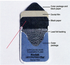
Intraoral film packet makeup
• Film wrapper
• Saliva proof
• White side toward the x-ray source
• May be a single or double film pack
• The Dot
Determines where the film is oriented in the mouth
• Black paper wrapper
Protects the film from light that may leak in
• Lead foil
• Placed away from the radiation source
• Prevents film fogging from radiation scatter back from the patient.
How should x-ray film be stored?
Away from:
• Heat (optimum temperature 50 - 70 degrees F)
--Refrigeration extends the film's life
• Intense light
• X-ray source
• Moisture
• Chemicals
-Keep inoriginal packaging
-Nothing placed on top
-Check expiration dates
QUALITY ASSURANCE
Define film fog and explain how it occurs.
*Darkening of radiograph by sources other than the radiation of the primary beam to which the film was exposed
-Occurs when film is exposed to temp over 80 degrees F
Explain the relationship between film speed and radiation exposure.
**The faster the film speed, the less radiation
A (slowest film speed) - F (Fastest film speed)
(Currently available: D, E, F)

Traditional film-based image receptors
• Radiographic film (Intraoral)
• Intensifying screens (Extraoral)
Digital Receptors
-Direct
-Indirect
**Both can be used intra- or extra- orally
Intraoral Radiographs (Digital or Film)
**The image receptor is placed inside the mouth behind the teeth with the x-ray source outside of the mouth
--The film reacts to direct exposure to the x-ray (No screens - traditional film)
1. Periapical Radiograph
2. Bitewing Radiograph
3. Occlusal Radiograph
Periapical Radiograph
** Shows the entire tooth or a group of teeth (this determines the amount of pics taken) from the crown to the apex including supporting bone and other structures around the teeth.
• Used to determine caries, periodontal disease, & pathology
Sizes
0 - Used for small children
1 -Used for older children & anterior films on adults
2 - adult size - Universal film size
4 - occlusal radiograph

Bitewing Radiograph
**Interproximal view of the posterior teeth
• Important to clearly see the area between teeth in occlusion
to detect interproximal caries and alveolar bone height. Patient bites on a tab or film holder to position the image receptor.
**Adult surveys usually consist of four films and child surveys usually consist of two films.
Sizes
0, 1, 2, and 3
• Size 3 is a longer film used for bitewings

Occlusal Radiograph
**Usually anterior and shows a large portion of the maxilla or mandible
** 2 types (Either type may be Maxillary or Mandibular views):
- Topographical
- Cross-sectionalize
Adult: Size 4 film
Children: Size 2 film
Topographical Occlusal Radiograph
Views a broader area of the maxilla & mandible, impacted teeth, bony lesions, palate, the floor of the mouth
Cross-Sectionalize Occlusal Radiograph
Used to localize objects such as impacted or supernumerary teeth, foreign objects, salivary stones
Extraoral Radiographs (Digital or Film)
•Image receptor outside the mouth and the x-ray source outside of the mouth
--The film does not respond well to direct x-ray exposure. The film reacts to light emitted by the screens inside the cassettes
1. Panoramic Imaging
2. Lateral jaw Radiograph
3. Cephalometric Radiograph
4. Cone-Beam Computed Tomography
Panoramic Radiograph
• Probably the most common
• Views entire dentition and related structures on one film
• Image not as well defined
• Object to film distance is great
• Numerous uses
Lateral Jaw Radiograph
• Used to view the posterior region of the mandible• Can be used to evaluate impacted teeth, fractures and lesions• Useful with children and patients with limited jaw opening
Cephalometric Radiograph
-There are many different types of projections but the most common view is the lateral head plate.
-Profile view of the skull and bones in the face, teeth, and tissue
-View of the relationship between the maxilla and t he mandible
-Used in Orthodontics and Pedodontics to evaluate growth assessment
Cone-Beam Computed Tomography
**DIGITAL ONLY
• Also known as 3D imaging
• Provides 3D views of the mouth, face, and jaw from any direction
• Specialized software program is necessary
• Specialized operator training needed
• Digital images can be easily manipulated, adjusted and colorized on the computer
• Used for the positioning of implants, position of impacted teeth, localizing the mandibular nerve for surgery, sinus and TMJ evaluation, and many more uses.
What is a cassette and what are its component parts?
**Film holder
- Can be plastic or metal (depending on machine)
- Protects from light exposure
- L or R on the outside or embedded into the screen
- Front side faces the patient during exposure
**NOT the actual image receptor
2 types:
• Rigid-type film cassette w/ intensifying screen
• Flexible film cassette
Digital Imaging
**Can be used intra- or extra- orally
• Computer-based technology
• Sensor receives the exposure -it is the image receptor
• Data is communicated to the computer
• Computer processes the latent image
• The sensor coverts the x-ray to light and then uses a chip to create a digital image
• Image is displayed on the monitor or may be printed out or transmitted electronically
Direct digital sensor
*Rigid, corded
Transfers information to the computer without intermediary processing, and connects directly to the computer.
2 Types
* (CCD) Charge-coupled device
* (CMOS)Complementary Meatal Oxide Silicon
CCD & CMOS
* Transfers information to the computer without any intermediary processing
• Image is obtained directly from the sensor via connection with the computer (wire)
***The incoming light or X-ray interacts with the silicone and creates an electrical charge in the pixel. The total charge in each individual pixel is proportional to the energy striking the detector, allowing the image is to be displayed on the monitor
Pixels
**Schematic representation of a digital image
**The bigger the pixels, the less detail in the image
Indirect digital sensor
**PSP (PhotoStimuable phosphor plate) Plates
Gathers information that must be "interpreted" in an intermediary step and then transferred to the computer. Requires an additional piece of technology, a laser scanner: that "reads", or digitizes the latent image
**• Phosphor plates store the phosphors, Laser scanner digitizes the latent image, Image is displayed on a monitor
• Phosphor plates are flexible and thinner than the direct digital sensor
Advantages and Disadvantages of digital imaging
Advantages:
- No film or processing used
- Less radiation to the patient (70-80% reduction compared to F speed film)
- Large database
- Computer processing & and storage, rather than storing darkroom chemicals
-Can be stored permanently in the patient's electronic record or transmitted electronically
-Instant image capture, display, enhancement, and printing
- On-screen patient education
Disadvantages:
- Equipment is costly (10k-14k)
- Can be thicker and more rigid than conventional
-Can be tampered w/ (court)
-Wire makes mobility more difficult
- Printed images- unsharp, low density
INTRAORAL RADIOGRAPHIC THEORIES + TECHNIQUES
LECTURE 2
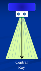
Central Ray (CR)
**Center of the diameter (bullseye) of the x-ray beam and PID (Position Indicating Device)
- Directed toward the center of the receptor
- External landmarks used to guide the CR entry for each view
long axis (LA)
**An imaginary line that extends from the apex of the tooth to the crown
- vertical orientation of the tooth in the mouth
- The teeth roots (apices) tend to be angled more lingually than the crowns (don't side straight up and down)

Vertical angulation (VA)
- Upward or downward movement of the x-ray beam
- Controls long axis dimension or length of the object
Positive angle: x-ray head above horizon, PID directed ↓
Negative angle: x-ray head below horizon, PID directed ↑
How do you get Elongation or Foreshortening?
Incorrect vertical angulation
Elongation: low vertical angle!!! (sun shadow analogy)

Horizontal angulation (HA)
**Forward or backward movement of the PID along the horizon
- Controls width dimension and interproximal or contact entry of the x-ray beam
How do you get overlapping or widening?
Incorrect horizontal angulation
Rules of Accurate Image Formation
1.) Small focal spot size
2.) Short object-to-receptor distance
3.) Long source-to-object distance
4.) Object to receptor parallelism
5.) Right angle central ray entry
1.) Small Focal Spot Size
The target area or spot inside the X-ray head where X-rays are produced is directed to a confined, specific area of wanted imaging
-This reduces penumbra & magnification
Unsharpness (Penumbra)
A secondary shadow or fuzziness that surrounds the periphery of the actual image (umbra)
-Edge Gradient

Magnification
Equal or uniform enlargement of the object recorded
2.) Short object-to-receptor distance
Distance between the tooth or structures and the image are as close as practical to the receptor
-This reduces unsharpness/penumbra & magnification

3.) Long source-to-object distance
**Distance between where x-rays are produced inside the x-rayed and the tooth or other structures
•Use the longest source-to-object distance as practical•Long PID (position indicatingdevice/x-ray cone)•16" Source-Object Distancelongest PID length available•Other distances are 12" or 8"
-This reduces penumbra & magnification

4.) Object to receptor parallelism
-Place the receptor parallel to the object
-Establish vertical and horizontal parallelism
**Reduces...
-shape distortion
-Vertical Foreshortening & Elongation
-Horizontal Overlapping & Widening
-Improves anatomic accuracy

Shape Distortion
• Vertical - Length
• ← Elongation (due to underangulation)
• ↙Foreshortening (overangulation)
• Horizontal - Width
• ↓Overlapping & Widening

Anatomic Accuracy
•Surrounding structures should be recorded in their true spatial relationship to the teeth
•Distortion can obscure structure sand mask dental disease like caries and periodontal bone loss
Picture: these two images are the same teeth at different angulation

5.) Right angle central ray entry
Direct the central ray perpendicular or at a right angle (90°) to the object and receptor
-Reduces shape distortion & Improves anatomic accuracy

Intraoral techniques
Preferred:
- Paralleling periapical technique
- Bitewing technique
Secondary:
- Bisecting angle periapical technique - "BAT" Technique
Paralleling Periapical Technique
- Applies most of the rules of accurate image formation
- Produces the most accurate and representative image of the teeth and surrounding bone
- Preferred technique for periapical imaging

Bitewing Technique
- Applies principles of paralleling technique to imaging the teeth crowns
- Used to record interproximal surfaces of the teeth
- Most accurate view of the crowns and bony margins
-Can be taken vertically or horizontally

Bisecting Angle Periapical Technique "BAT" Technique
- Receptor is placed close to the object to be imaged
- Reduced source to receptor distance (8" PID used)
- Requires increased vertical angulation compared to paralleling technique
- Round PID's are recommended to reduce cone cutting
- Images will be distorted because the central ray is not perpendicular to the teeth and the image receptor
- An increased vertical angulation is used
**Used when patient has an anatomical anomaly or physical/mental disbility

Preparing for taking Radiographs
1. Prepare operatory using effective infection control and covering methods
2. Gather necessary materials
3. Set x-ray machine exposure facctors
4. Assemble XCP (RNN) instruments
5. Seat ad adjust patient's head position
6. Place lead apron and thyroid collar
7. Ask patient to remove eyeglasses and intraoral prosthesis (dentures, partials, retainers)
8. Explain procedure + get consent before beginning
XCP Assembly
Anterior PA's: Vertical blue biteblock with anterior ring (flip up and down to take maxillary and mandibulaar)
-never have to take apart
Bitewings: red tab biteblock with anterior ring (look like airplane) (flip side to side to take right and left bitewings)
Posterior PA's: Horizontal yellow biteblock with posterior ring
-upper right and lower left (tab faces you)
-upper left and lower right (tab away from you)

One arm XCP
*

Recommended patient head position for maxillary periapicals & Bitewings
**Sitting straight up with midsagittal plane perpendicular to the floor
- Ala (corner of nose), tragus (tab in ear) plane parallel to the floor

Recommended patient head position for mandibular periapicals
**Head leaned back a little with midsagittal plane perpendicular to the floor
- Commissure (corner of mouth) - tragus (tab in ear) plane parallel to the floor (line from corner of the mouth to the tab in the ear)

What are the central entry points for each maxillary periapical?
- Incisor - tip of nose
- Lateral - nares (nostrils)
- Canine - ala (corner of the nose)
- Premolars - pupil to ala-tragus plane
- Molars - outer canthus (outer corner of the eye) to ala-tragus plane

What are the central ray entry points for each Bitewing?
-premolar: pupil point to occlusal plane
-molar: outer canthus point to occlusal plane

What are the central ray entry points for each mandibular periapical?
- Incisor - tip of nose to center of chin
- Canine - ala to center of the chin
- Premolars - pupil to center of mandible
- Molars - outer canthus to center of mandible

Characteristics of Quality Periapical Images
• Correct anatomical area recorded
• Teeth recorded including at least 3-4 mm of bone beyond apices
• Tooth is of correct length
• Interproximal contacts open
• Acceptable density and contrast
• Free of technical, handling and processing errors
Characteristics of Quality Bitewing Images
-Dot convexity toward clinician and mandible
-Correct anatomical area recorded
-Interproximal contacts open
- Equal display of maxillary and mandibular crowns
- Alveolar bone margins displayed
-Horizontal or slight smile of occlusal plane
- Acceptable density and contrast
- Free of technical, handling and processing errors
LECTURE 3:
Xray Production and Behavvior
Radiation
The emission or transfer of energy through space in the form of waves or particles
higher frequency=higher penetrating power

Electromagnetic radiation
PURE ENERGY:
- no mass
- no electrical charge
- Travels in a straight path but in an oscillating pattern
- Broad range of energies that vary in wavelength and frequency

wavelength
distance from one crest or trough to another

Shorter wavelengths
high energy and high frequency thus high penetrating ability

long wavelengths
low energy, low frequency thus poor penetrating ability

Short wavelengths deliver enough energy to...
ionize atoms
because of their high penetrating ability
Frequency
# of wavelengths per unit time
Measured in Hertz (Hz)-repitions per second

Determine Energy level
Wavelengths & Frequency together
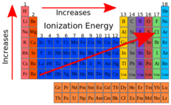
Ionization
When an atom or molecule gains or loses an electrical charge
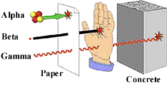
Particulate radiation
**X-rays are "packets" of energy that travel with properties of:
-Waves (Electromagnetic Radiation)
-Particles (X-ray photon or bundle of energy)
* "Mass in Motion"
* Possesses an electrical charge
* Travels in a straight line that creates tiny fast moving subatomic particles that have energy and mass
What unique characteristics do x-rays possess that make them useful in dental imaging?
- Able to penetrate matter
- Absorbed differentially by matter *(The composition of the object determines if the x-ray penetrates or passes through it)
Properties of X-Rays: (electromagnetic and particulate combined)
-not detectable by senses
-travel at speed of light in straight path (oscillating)
-able to penetrate matter
absorbed differently by matter
-causes certain substances to fluoresce and emit light
-capable of producing biologic change via iionization
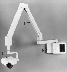
What are the components of the x-ray machine
- X-ray tube head
- Support arm
- Power supply
- Control panel (outside room)
- X-ray tube
Function of the Tube head and supporting arm
**Metal encasement enclosing x-ray tube, transformers, and oil
- Electrical cables inside supporting arm
- Supporting arm and yoke enable the tube head to be positioned around the patient's head
What percent of the x-ray gets absorbed and what percent goes out of the tube?
99% gets absorbed and 1% goes out the tube
Components of x-ray tube
located inside the x-ray head!
*leaded glass envelope with portal, cathode, anode, and electrical power supply components
