N222 - Exam #1
 Call Kai
 Learn
Learn Practice Test
 Spaced Repetition
 Match
Match Flashcards
 Knowt Play
1/11

There's no tags or description
Looks like no tags are added yet.
Name | Mastery | Learn | Test | Matching | Spaced | Call with Kai | Chat |
|---|
No analytics yet
Send a link to your students to track their progress
12 Terms
Cardiac Rhythms & Heart Failure
Basic ECG rhythm interpretation


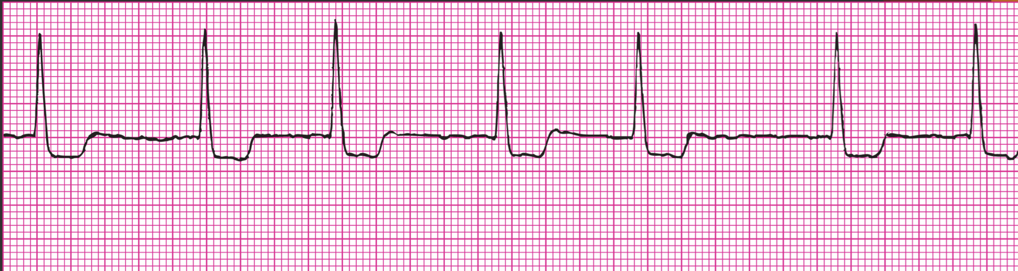
Atrial fibrillation (AF) is the most common dysrhythmia seen in clinical practice. AF can be encountered and treated in the ambulatory and acute care settings. It can impair quality of life and cause considerable morbidity and mortality, largely related to clotting concerns such as embolic stroke, deep vein thrombosis (DVT), or pulmonary embolism (PE).


Ventricular fibrillation (VF), sometimes called V fib, is the result of electrical chaos in the ventricles and is life-threatening! Impulses from many irritable foci fire in a totally disorganized manner, so ventricular contraction cannot occur. There are no recognizable ECG deflections. The ventricles merely quiver, consuming a tremendous amount of oxygen. There is no cardiac output or pulse and therefore no cerebral, myocardial, or systemic perfusion. This rhythm is rapidly fatal if not successfully ended within 3 to 5 minutes.
VF may be the first manifestation of coronary artery disease (CAD). Patients with myocardial infarction (MI) are at great risk for VF. It may also occur in those with hypokalemia, hypomagnesemia, hemorrhage, drug therapy, rapid supraventricular tachycardia (SVT), or shock. Surgery or trauma may also cause VF.


Normal sinus rhythm. Both atrial and ventricular rhythms are essentially regular (a slight variation in rhythm is normal). Atrial and ventricular rates are both 87 beats/min. There is one P wave before each QRS complex, and all the P waves are of a consistent morphology, or shape. The PR interval measures 0.18 second and is c

Sinus rhythms. (A) Sinus tachycardia (heart rate, 115 beats/min; PR interval, 0.12 second; QRS complex, 0.08 second). (B) Sinus bradycardia (heart rate, 52 beats/min; PR interval, 0.18 second; QRS complex, 0.08 second).

Telemetry electrode placement
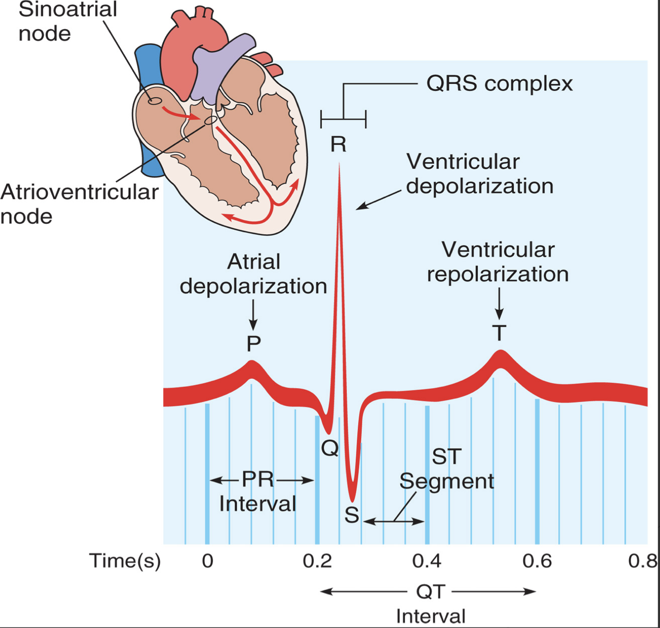
The electrocardiogram (ECG) provides a graphic representation, or picture, of cardiac electrical activity. The cardiac electrical currents are transmitted to the body surface. Electrodes, consisting of a conductive gel on an adhesive pad, are placed on specific sites on the body and attached to cables connected to an ECG machine or a monitor. The cardiac electrical current is transmitted via the electrodes and through the lead wires to the machine or monitor, which displays the cardiac electrical activity.