Looks like no one added any tags here yet for you.
Identify the basic symptoms associated with influenza
headache, chills, cough, high fever, myalgia, malaise and anorexia
Define important influenza characteristics used for classification
Type:
Influenza A and B are the most common types that cause illness in humans. Influenza A can also infect animals like birds and pigs and has more potential to cause pandemics. Influenza B mainly infects humans.
Surface Proteins (HA and NA):
Influenza A is divided into subtypes based on two proteins on the virus surface:
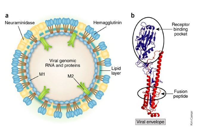
Hemagglutinin (HA): Helps the virus enter cells (there are 18 types, like H1, H3).
Neuraminidase (NA): Helps the virus leave the cell after it multiplies (there are 11 types, like N1, N2).
These proteins create subtypes like H1N1 or H3N2.
Identify major events in influenza history
1918: H1N1 (Spanish flu),
1957: H2N2 (Asian flu)
1968: H3N2 (hong Kong flu)
1977: H1N1 (Russian flu)
2009: H1N1
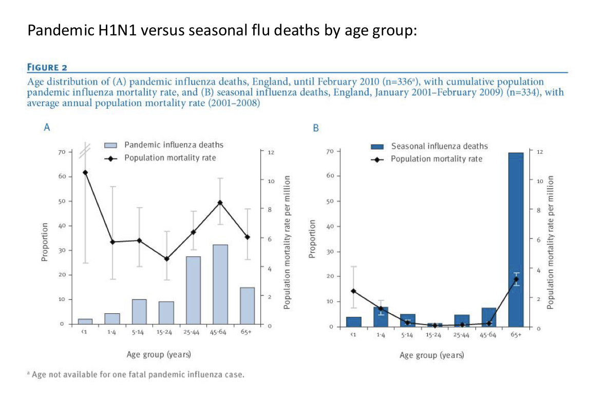
Explain the characteristics and trends that differ in pandemic vs seasonal influenza
For a virus to have pandemic potential it must:
Be antigenically distinct from previously circulating strands, transmit efficient from person to person, MAY posses increased pathogenicity
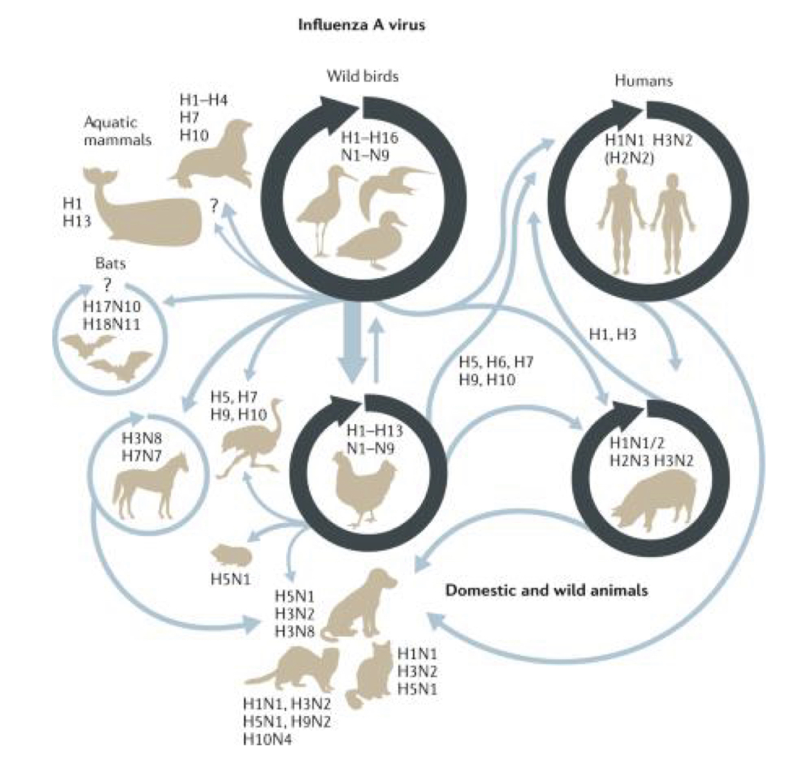
Explain influenza host range and adaptation
Cross species transmission exists, but not between ALL animals
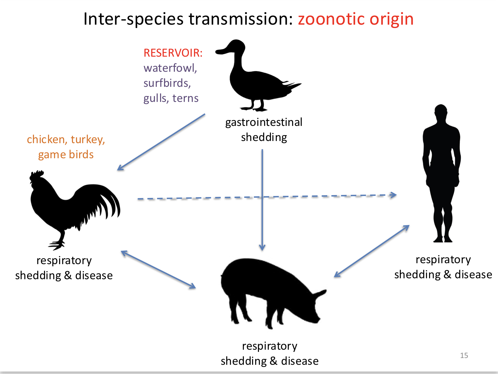
Explain inter-species transmission between avian, swine, and humans
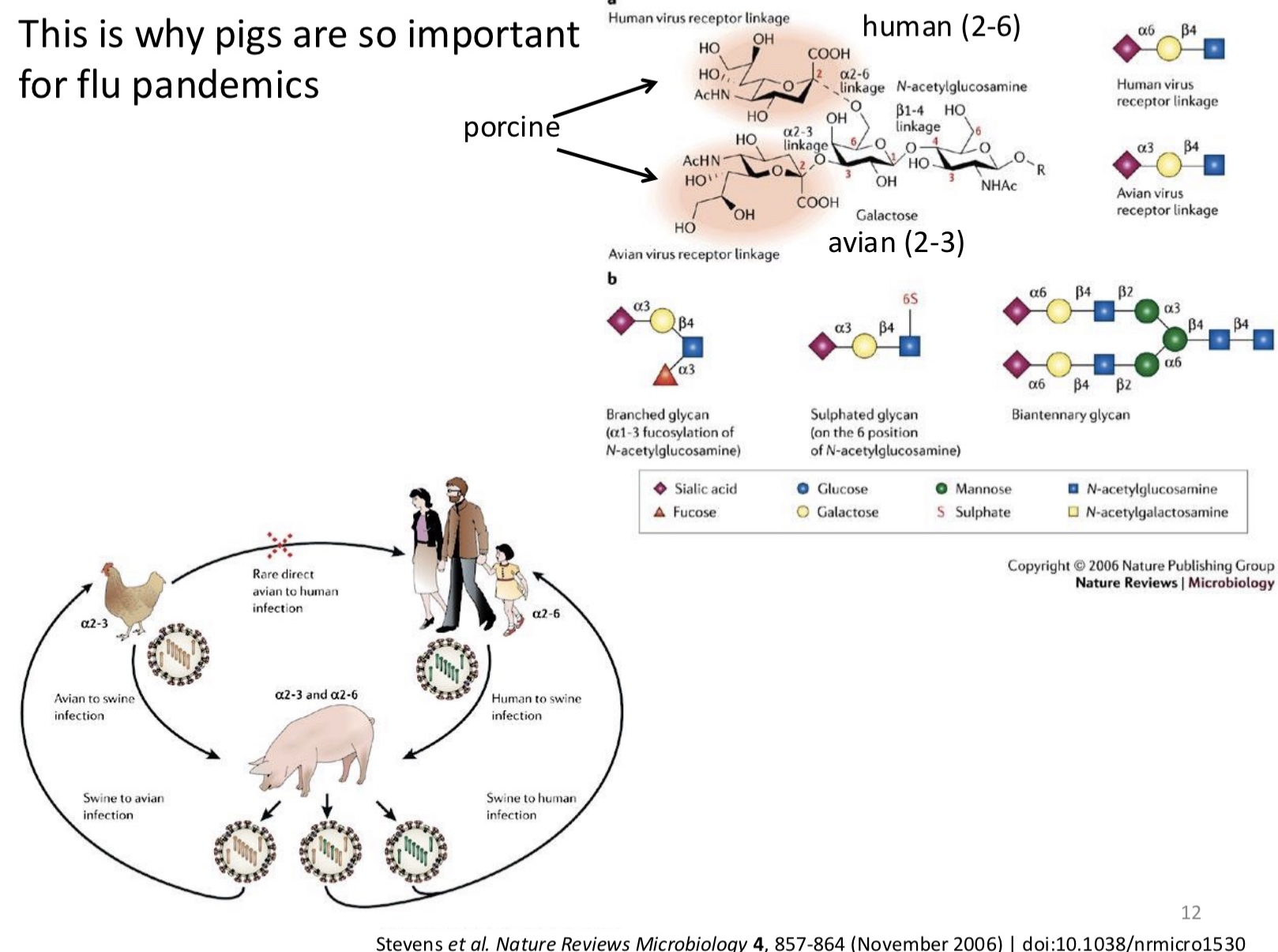
Inter-species transmission of influenza occurs when a virus jumps from one species to another, such as from birds (avian flu) or pigs (swine flu) to humans. Influenza A viruses can infect multiple species due to their ability to mutate and adapt. For example, a pig can be infected with both avian and human flu strains, allowing the viruses to swap genetic material (reassortment) and create a new virus. This new strain can then infect humans, who may have little immunity, potentially leading to outbreaks or pandemics.
Identify current influenza strains of major concern
H1N1 (seasonal), H3N2 (seasonal), influenza B, H5N1 and H7N9 (avian)
Explain the molecular interactions involved in each of the following steps of the influenza single-cell replication cycle: attachment
Attachment in the influenza replication cycle is the first step where the virus binds to a host cell. involves specific molecular interactions between the viral surface protein hemagglutinin (HA) and the host cell receptors.
The HA protein consists of: HA1 (which is responsible for receptor binding) and HA2 (which helps with membrane fusion later in the cycle).
Cellular receptor molecule – sialic acid, the terminal addition to amino-linked glycan moieties on cellular glycoproteins.
In humans, sialic acid is predominantly linked to galactose by an α-2,6 linkage. In birds, sialic acid is linked by an α-2,3 linkage.
Once HA binds to the sialic acid receptors on the host cell, the virus is anchored to the cell surface. This attachment is the first essential interaction that allows the virus to proceed with entry into the host cell for further replication.
Explain the molecular interactions involved in each of the following steps of the influenza single-cell replication cycle: transcription
Active template for transcription is the helical NP coated vRNA. 5’ and 3’ ends form a “panhandle”
Need polymerase basic protein 1 (PB1), polymerase basic protein 2 (PB2), and polymerase acidic protein (PA) in complex.
PB1 has RNA dependent RNA polymerase activity PB2 binds to cap on the 5’ end of cellular mRNAs
PA has endonuclease activity
cap snatching primes transcription: PB2 binds cellular mRNA cap, PA cuts it off and PB1 uses the oligonucleotide to prime synthesis.
Polymerase slips to add poly(A) and terminate
mRNA is not a full-length copy of the vRNA. Missing sequence from 5’ end of vRNA, extra sequence from cellular mRNA
Explain the molecular interactions involved in each of the following steps of the influenza single-cell replication cycle: translation
Outsourced to host
Explain the molecular interactions involved in each of the following steps of the influenza single-cell replication cycle: replication
Full-length copy of vRNA (no termination at polyA signal)
NP, PA, PB1, and PB2 are imported into the nucleus after it their translation and bind to vRNA template
Switch to from transcription to replication mediated by free NP levels
The viral polymerase synthesizes complementary RNA (cRNA), which is a positive-sense RNA strand.
Different form of the polymerase replicates than transcribes (conformation different, same viral proteins)
Once the cRNA is formed, it is used as a template to synthesize more negative-sense viral RNA (vRNA)
The newly synthesized vRNA is again bound by NP, forming new viral ribonucleoproteins (vRNPs).
These vRNPs will be exported out of the nucleus and packaged into new virions.
Replication is error prone (approx error rate of 1 in 10,000 nt incorporated)
+ strand antigenome is then copied in a similar fashion to make the – strand genome
Understand how each step in the cycle flows to the next and describe how an antiviral at each step might impact the cycle.
1. Attachment:
- Flow: The influenza virus binds to the host cell by using its hemagglutinin (HA) protein, which attaches to sialic acid receptors on the host cell surface.
- Antiviral Impact: Drugs that block the HA-sialic acid interaction would prevent the virus from attaching to the host cell, stopping the infection before it even begins.
2. Entry:
- Flow: After attachment, the virus is engulfed by the host cell through endocytosis. Inside the endosome, the virus's hemagglutinin undergoes a change triggered by the acidic environment, allowing the viral membrane to fuse with the endosomal membrane and release the viral RNA (vRNA) into the cytoplasm.
- Antiviral Impact: Drugs that inhibit the fusion of viral and host membranes could block the release of vRNA into the cell.
3. Transcription and Replication:
- Flow: Once inside the nucleus, the viral RNA-dependent RNA polymerase complex (PB1, PB2, PA) transcribes the vRNA into mRNA for protein production and replicates the viral genome for new virions. The viral RNA must be replicated through an intermediate complementary RNA (cRNA) template.
- Antiviral Impact: Drugs that inhibit viral RNA polymerase would block both viral mRNA production (needed for protein synthesis) and vRNA replication.
5. Translation and Protein Processing:
- Flow: Viral mRNAs are exported from the nucleus into the cytoplasm, where the host ribosomes translate them into viral proteins like hemagglutinin (HA), neuraminidase (NA), matrix proteins (M1), and nucleoproteins (NP). These proteins are transported to their respective locations for virion assembly.
- Antiviral Impact: Drugs that disrupt viral protein synthesis or processing could block the production of essential viral components.
6. Assembly of Viral Components:
- Flow: Newly synthesized viral RNA segments are packaged with nucleoproteins (NPs) to form viral ribonucleoprotein complexes (vRNPs). The viral membrane proteins HA, NA, and M2 are inserted into the host’s plasma membrane, where new virus particles will bud.
- Antiviral Impact: Drugs that block viral assembly could prevent the virus from putting together functional viral particles.
7. Release:
- Flow: The new virus particles bud from the host cell membrane, but they remain attached to the cell surface via the interaction between hemagglutinin (HA) and sialic acid receptors. Neuraminidase (NA) cleaves this interaction, allowing the new virions to be released and infect other cells.
- Antiviral Impact: Drugs that inhibit NA would prevent the virus from releasing new particles, trapping them on the cell surface and halting further spread.
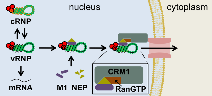
Explain how viral components are exported out of the nucleus
the M1 (matrix protein) and NEP (nuclear export protein) bind to the nucleoprotein (NP) and cover its NLS, which normally allows the nucleoprotein to enter the nucleus. By doing this, they expose an export signal, leading to the export of the vRNP complex
Explain how HA and NA end up in lipid rafts
Hemagglutinin (HA) and neuraminidase (NA) localize to lipid rafts due to specific sequences in their transmembrane and tail regions that have an affinity for these areas. After synthesis, HA and NA are trafficked to the lipid rafts. These rafts serve as platforms for viral assembly and budding, ensuring the proper incorporation of HA and NA into new virions.
Explain the importance of HA, NA cytoplasmic tails in viral assembly
1. HA Cytoplasmic Tail:
- The HA cytoplasmic tail is crucial for proper trafficking of HA to the plasma membrane, where viral assembly occurs. It helps HA localize to lipid raft regions, which are essential for viral particle formation.
- It also facilitates interactions with the viral matrix protein (M1), coordinating the incorporation of HA into the budding virion.
2. NA Cytoplasmic Tail:
- The NA cytoplasmic tail is involved in packaging NA into the budding virus and ensuring it is correctly positioned at the viral envelope.
- It also helps maintain the balance between HA and NA at the viral surface, which is necessary for efficient viral release after budding.
Explain the role of NA in viral release
Tetrameric
Cleaves sialic acid from glycans
Inhibition leads to particles failing to disperse from cells
Blocks the active site of neuraminidase
Define viral reassortment and explain its role in pandemics
Two antigenically distinct forms of flu infect a single cell. Gene segments mix during packaging creating new recombinant viruses.
Generates a completely new subtype of HA or NA for which there is no pre-existing immunity (antigenic shift)
Describe the different host sialic acid linkages and HA specificity
α-2,6-linked sialic acids: Found mainly in the human upper respiratory tract; preferred by human-adapted influenza strains.
α-2,3-linked sialic acids: Found mainly in the avian respiratory and intestinal tracts, as well as in the human lower respiratory tract; preferred by avian influenza strains.
The specific binding between the viral hemagglutinin (HA) and the type of sialic acid linkage on the host cells determines both the virus’s host range and the site of infection within the respiratory system.
Explain the role of swine in interspecies transmission
they can be infected by both avian and human influenza strains because they have both types of sialic acid receptors in their respiratory tract:
α-2,6-linked sialic acids (human-like receptors)
α-2,3-linked sialic acids (avian-like receptors)
Because pigs can host both types of influenza viruses, they act as "mixing vessels," where genetic reassortment can occur. When a pig is co-infected with human, avian, and/or swine influenza viruses, the different viral RNA segments can mix, potentially producing a new hybrid strain that can infect humans and animals. can lead to the emergence of novel viruses capable of causing pandemics, such as the H1N1 pandemic in 2009.
Compare and contrast antigenic shift and drift and the roles they play in pandemics and seasonal flu
Antigenic Shift – big antigenic changes caused by replacement of surface antigens (HA, NA)
Antigenic Drift – amino acid changes in surface antigens that allow escape from antibody
Antigenic drift is primarily responsible for the need for vaccine changes
There is approximately 1% drift/year. In the HA1 subunit that equates to about 3 amino acids. This means the vaccine has to be updated each year
Describe the vaccine design for the inactivated influenza vaccine
Vaccine composition is determined by constant monitoring of circulating influenza viruses. HA and NA from circulating viruses are introduced into the background of a vaccine strain to produce the vaccine.
Vaccine strains are grown in embryonated eggs, purified by chromatography or centrifugation.
Inactivated using either formalin (formaldehyde) or beta-propiolactone. These chemicals cross-link the proteins in the virus and prevent it from functioning.
The virus particles can be recognized and engulfed by antigen presenting cells and ultimately stimulate an antibody response to the HA and NA proteins.
Describe the vaccine design for live attenuated vaccine
Administered in nasal spray (FluMist®)
Virus is live but weakened
HA and NA from circulating strains are introduced into an attenuated background
Describe the differences between pathogenesis of uncomplicated flu and viral pneumonia
Uncomplicated Influenza:
- Site of infection: Primarily affects the upper respiratory tract
- Mechanism: The virus infects epithelial cells of the respiratory tract, leading to cell death, inflammation, and the release of pro-inflammatory cytokines.
- Immune response: A strong innate immune response clears the virus, and adaptive immunity helps in recovery within 1-2 weeks.
- Tissue damage: Limited to the upper respiratory tract, with mild to moderate damage and inflammation.
Viral Pneumonia:
- Site of infection: Extends to the lower respiratory tract, particularly the alveoli and lung parenchyma.
- Mechanism: The virus invades the deeper parts of the lungs, causing direct damage to alveolar epithelial cells. The immune response can lead to severe inflammation, edema with APC, and fluid accumulation in the alveoli, impairing gas exchange.
- Immune response: Overactive or dysregulated immune response can lead to widespread tissue damage and secondary bacterial infections.
- Tissue damage: Severe damage to the lung tissue, with alveolar inflammation, necrosis, and potential acute respiratory distress syndrome (ARDS) in severe cases.
- Outcome: Viral pneumonia can be life-threatening, especially in immunocompromised or high-risk individuals, and may require hospitalization.
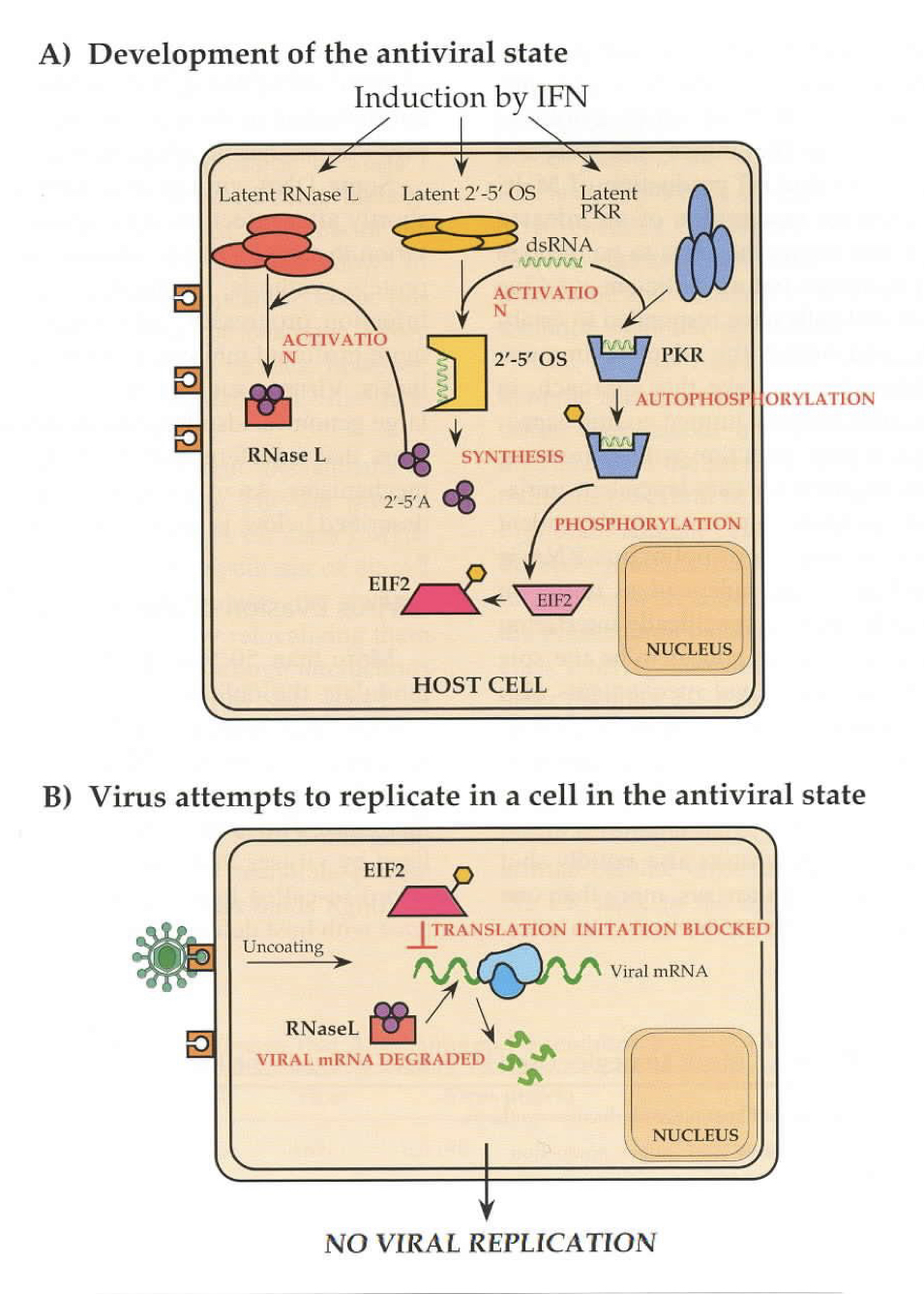
Describe the role of the innate response in influenza infection
TLR3 and TLR7 stimulation leads to production of pro-inflammatory cytokines such as IFN-alpha which leads to influx of neutrophils and molecular antiviral responses,
Protein Kinase R (PKR) and 2’-5’ oligoadenylate synthase (2’-5’OS) activated by dsRNA Products of 2’-5’OS activate RNaseL
PKR phosphorylates eIF2 inhibiting translation
RNaseL digests viral RNA.
Describe the role of NS1 in influenza infection
Inhibits transcription of interferon responsive genes
Describe the role of the adaptive immune response in influenza infection
APC clean up dying cells and present antigen to T-helper cells
CTLs recognize infected cells through T-cell receptor/CD8 recognition of MHC1 plus antigen
B-cells recognize antigen through surface antibody and phagocytose it.
Present it to T-helper cells that become activated then activate the B-cell to divide and differentiate to plasma cells
Main antibodies are to HA and NA. HA antibodies can be neutralizing (prevent infection) NA antibodies are non-neutralizing but inhibit NA function.
CTLs to HA, NA or M1, NP, PB2
Identify the important influenza antigens in the adaptive response
APC – antigen presenting cells Dendritic cells, macrophage, B-cells
T-helper cells – signal to expand immune response. Cause expansion of B-cells and CTLs
CTLs –cytotoxic T lymphocytes. Directly recognize and kill infected cells
B-cells – produce an antibody species. Differentiate into memory and plasma cells
Explain how differences in host receptors lead to different tropism and disease in influenza
Distribution of virus receptor alters tropism
Tropism alters disease
Alpha 2,6 linked sialic acid found in upper respiratory tract
Alpha 2,3 linked sialic acid found in lower respiratory tract
Seasonal flus bind alpha 2,6, H5N1 binds alpha 2,3 therefore if/when it infects it cause severe lower respiratory tract disease
Explain the mechanism of HA cleavage and its relationship to pathogenesis and tropism
For the virus to be infectious, HA must be cleaved into two subunits, HA1 and HA2, by host proteases. This cleavage exposes a fusion peptide that allows the viral envelope to fuse with the host cell membrane, enabling the virus to enter the cell.
Relationship to pathogenesis and tropism:
- Pathogenesis: Efficient HA cleavage is crucial for viral infectivity. Strains with HA that is easily cleaved by widely distributed proteases can cause more severe infections and systemic disease.
- Tropism: The availability of specific proteases in different tissues affects where the virus can replicate. For example, viruses with HA that require proteases found mainly in the respiratory tract are limited to infecting those tissues, whereas strains that can be cleaved by more ubiquitous proteases may have broader tissue tropism.
For HPAIs like H5N1 a polybasic run of amino acids (lysines and arginines, yellow box above) allows cleavage by ubiquitously expressed proteases (furins and PC6). These are found in all cells late in the secretory pathway hence cleavage can occur in any tissue and tropism is Dramatically expanded
If H5N1 escapes the respiratory tract (due to severe tissue damage) it can infect and replicate in other tissues causing other organs to fail (particularly kidneys)
Identify basic characteristics of S. pneumoniae
Gram positive, spheroidal, diplococcus bacterium.
Aerotolerant (doesn’t need oxygen but can be around it)
Leading cause of community-acquired pneumonia
Possesses a polysaccharide capsule which is the major antigen and a virulence determinant (hard for human immune system to deal with)
There are >100 capsule types
Pneumococcus is also naturally transformable (takes in external DNA)
Explain characteristics of non-invasive pneumococcal diseases
otitis media (ear infection), sinusitis (sinus infection), bronchitis. CM progress to invasive versions, but are very treatable.
Occur in non sterile environments in the upper respiratory tract
Explain characteristics of invasive pneumococcal diseases
Invasive disease occurs when the bacteria spread to previously sterile body compartments such as the lower respiratory tract (LRT), the blood stream and the CNS
Spread to the LRT is a result of uncontrolled spread from URT, further tissue damage allows bacteria access to the blood stream.
Meningitis usually occurs either as a consequence of skull trauma allowing continuity between the nasopharyngeal space and the CNS, or as a consequence of bacteria crossing the blood-brain barrier during sepsis (a secondary consequence of severe LRT infection)
Identify the basic epidemiological trends of pneumococcal infections
900,000 cases/year of pneumococcal pneumonia in U.S.
Incidence highest in the young (<2y.o.) and the elderly (>65 y.o.)
Significant problem in immunosuppressed (AIDS patients)
Pre-2000 approx 14.5 million cases in children annually (global), resulting in approx. 800,000 deaths
Vaccination since 2000 has reduced severe disease burden somewhat, but it is not clear that there has been a significant benefit to overall disease occurrence
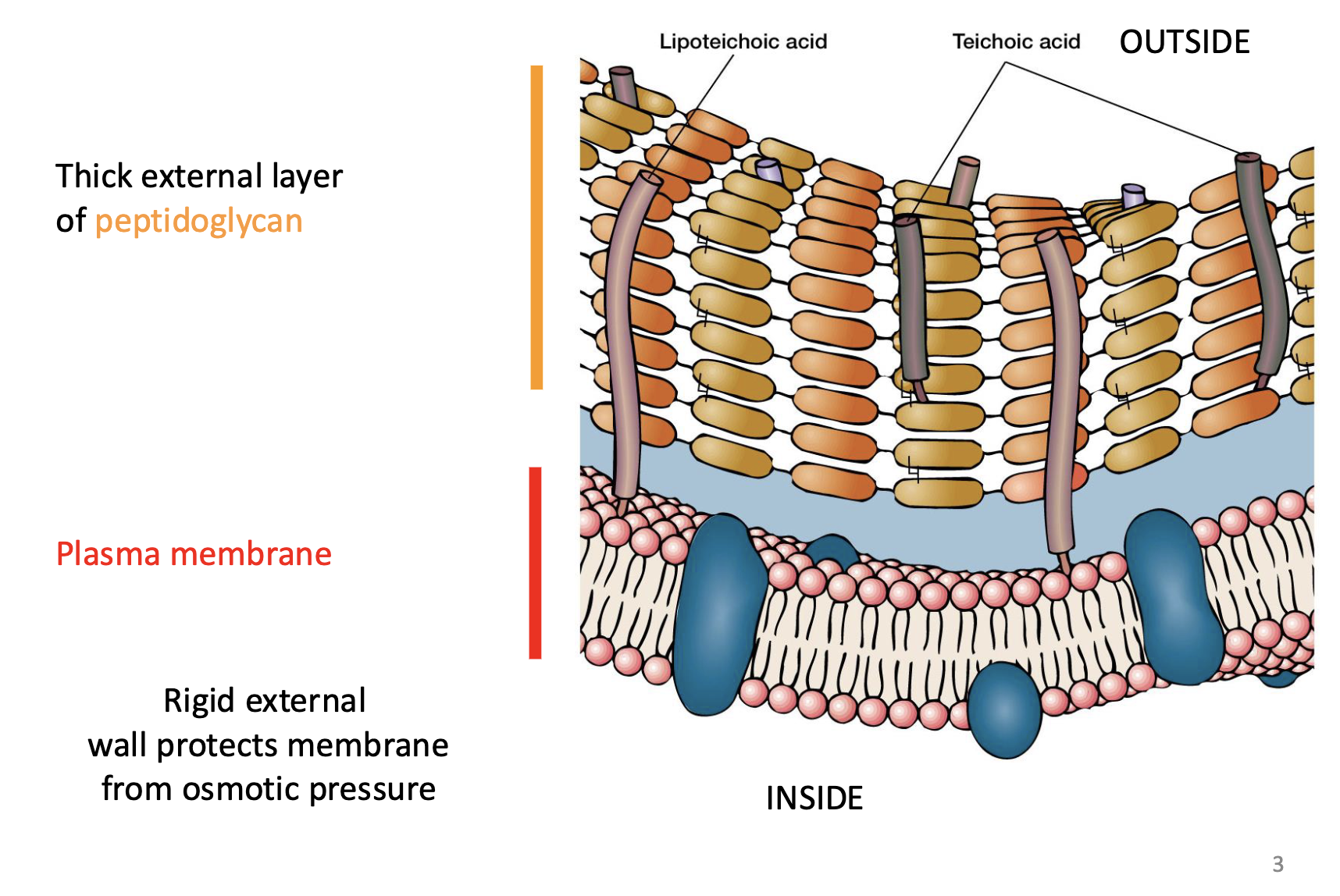
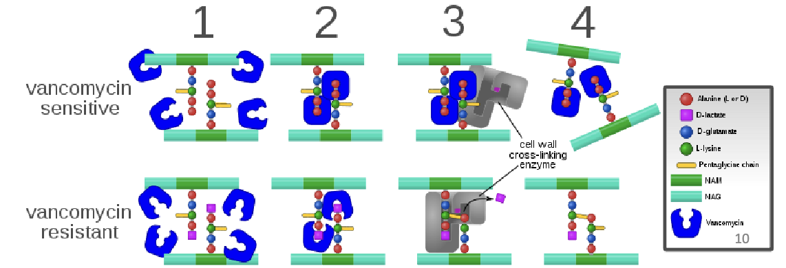
Describe the gram positive cell wall structure, focusing on components of peptidoglycan
1. Peptidoglycan Layer (Murein)
Thick and Rigid Layer: The peptidoglycan layer in Gram-positive bacteria is much thicker than in Gram-negative bacteria
Structure: Peptidoglycan is made up of long chains of alternating sugar molecules:
N-acetylglucosamine (NAG)
N-acetylmuramic acid (NAM) These sugars are linked by β glycosidic bonds to form linear chains.
Peptide Cross-links (protein component): Attached to the NAM molecules are short peptide chains that cross-link adjacent sugar chains, providing the peptidoglycan its strength and rigidity.
Unusual amino acids: increases peptidoglycan resistance to proteases, but also allows recognition by PRR (TLR2) of the innate immune system. use of D- form amino acids
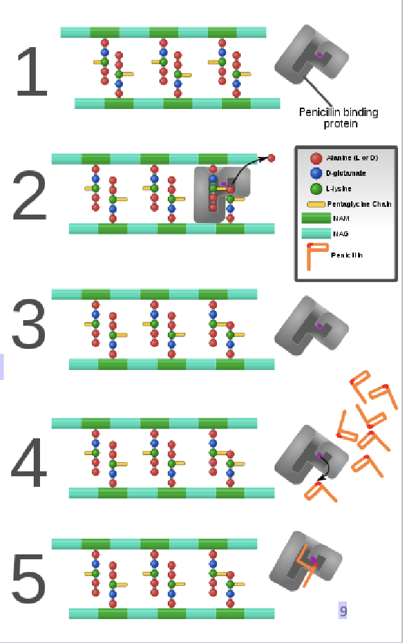
Explain the mechanisms of action of penicillin
Penicillin inhibits peptidoglycan synthesis
Involved in final stages of peptidoglycan synthesis
1. PBP binds to the sugar linked peptides
2. Catalyzes crosslink with interpeptide bridge releasing an alanine
3. PBP then dissociates from the cell wall
4. Penicillin binds in active site of PBP
5. the beta-lactam ring reacts with the serine group of the PBP active site becoming covalently linked
Different PBPs bind different versions of beta-lactams
Mutations in the PBPs can inhibit access of beta-lactams to the binding site preventing inhibition
Explain the defining features and role of the pneumococcal capsule
This capsule is an external, polysaccharide layer that surrounds the cell wall
Polysaccharide Composition:
- primarily composed of polysaccharides, which vary in structure between different strains. >100 serotypes of S. pneumoniae, major challenge in vaccine development
Capsule locus
1. Modular Structure: The capsule production system has different gene modules that each play specific roles.
2. Regulation (cpsABCD): These genes control capsule levels. Deleting B and D stops capsule production and reduces virulence.
3. Glycosyltransferases: These enzymes select which sugars to add to the capsule and how they’re linked.
4. NDP-sugar Biosynthesis: These genes make the sugars needed for capsule construction.
5. Capsule Variety: Parts of the capsule genes can be swapped through natural transformation, creating new capsule types and strains.
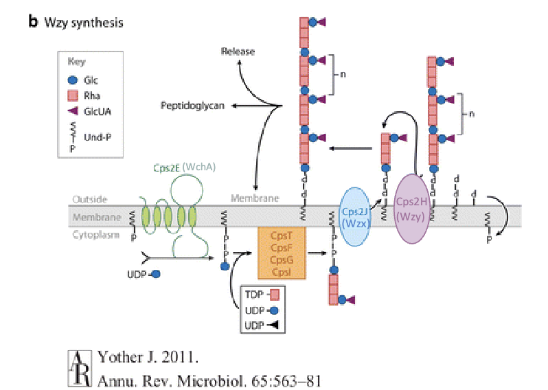
Explain how the pneumococcal capsule is synthesized
1. Primed Sugar Addition: A primed sugar (from NDP-sugar biosynthesis) is added to a lipid carrier (Und-P) by the initiating glycosyltransferase.
2. Sugar Chain Growth: More sugars are added by other glycosyltransferases to form an oligosaccharide.
3. Membrane Flip: The lipid-linked oligosaccharide is flipped to the opposite side of the membrane.
4. Polymerization: The polysaccharide polymerase adds a growing chain to the flipped oligosaccharide.
5. Anchoring: The finished polysaccharide is usually anchored to the peptidoglycan layer.
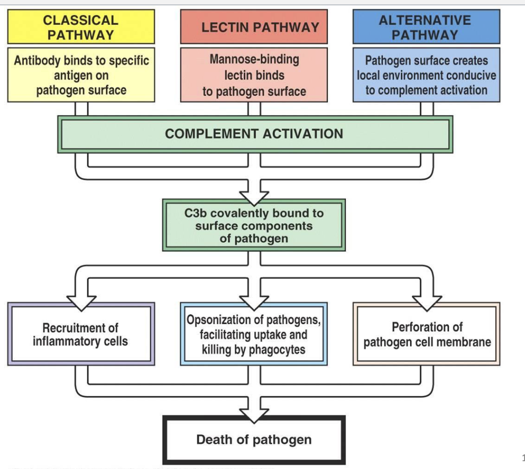
Explain complement pathway and how it relates to pneumococcus
The complement pathway is a part of the innate immune system that helps clear pathogens through opsonization, inflammation, and cell lysis. It involves three activation pathways:
1. Classical pathway (triggered by antibodies bound to pathogens),
2. Lectin pathway (activated by mannose-binding lectin binding to pathogen surfaces),
3. Alternative pathway (spontaneous activation on pathogen surfaces).
Relation to pneumococcus (Streptococcus pneumoniae):
Pneumococcus can activate the complement system, enhancing opsonization (marking for phagocytosis) and promoting its clearance by immune cells. However, pneumococcus has evolved mechanisms to evade the complement system, such as producing a capsule that inhibits complement binding, which allows it to avoid immune detection and persist in the host, contributing to its virulence.
Explain opsonization and how it relates to pneumococcus
Opsonins (i.e. antibodies or complement pathway proteins) mark antigen for phagocytosis.
Explain how the pneumococcal capsule influences pathogenesis
Early in infection in the nasopharynx less capsule is helpful. It allows bacterial adhesion proteins to access the epithelial layer.
Capsule also prevents opsonin-independent phagocytosis, probably due to its high negative charge. This helps in evading neutrophil killing.
Capsule covers peptidoglycan, a major PAMP
Capsule inhibits binding of antibodies and complement to the bacterial cell and recognition of bound antibodies and complement by phagocytic immune cells
Thick capsule is helpful in evading humoral immune response (antibody + complement) during invasive disease
Explain how transformation was discovered.
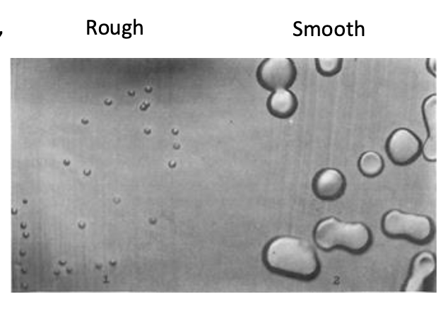
1916 Stryker: pneumococcus grown in broth gave rise to different colony morphologies.
Rough colonies were found to be avirulent, and did not possess a polysaccharide capsule. Smooth colonies were highly virulent and
had a capsule.
1928 Griffith: Rough bacteria were “transformed” to Smooth and virulent in the presence of heat-killed smooth bacteria
1944 Avery, MacLeod and McCarty: demonstrated that DNA from heat-killed bacteria was responsible for the transformation of capsule-type.
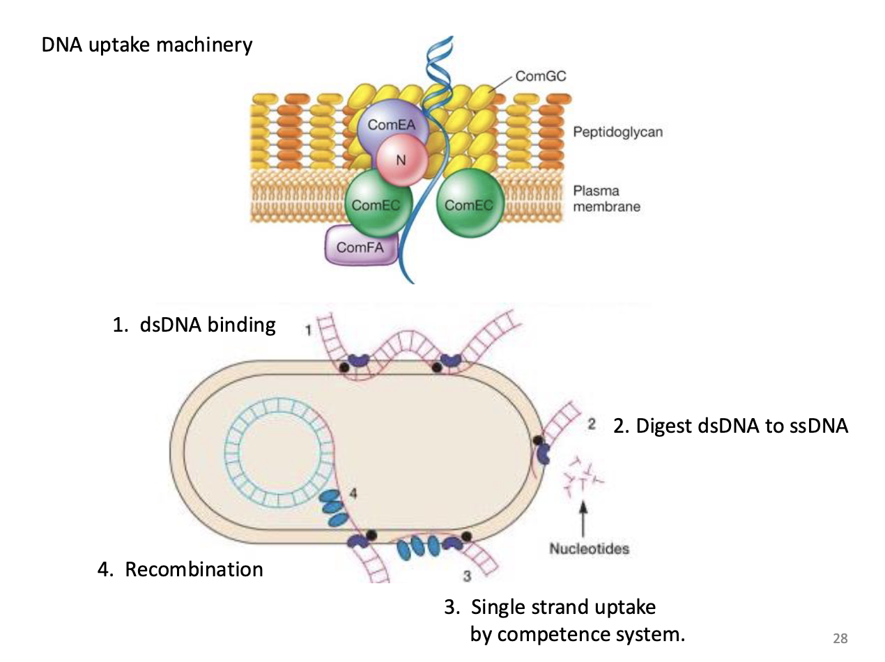
Define transformation and competence.
Transformation is the permanent genetic change of a cell/organism generally resulting in a phenotypic change.
In the case of bacteria we usually think of transformation as the uptake of exogenous DNA.
This requires the bacteria to be in a COMPETENT state for transformation
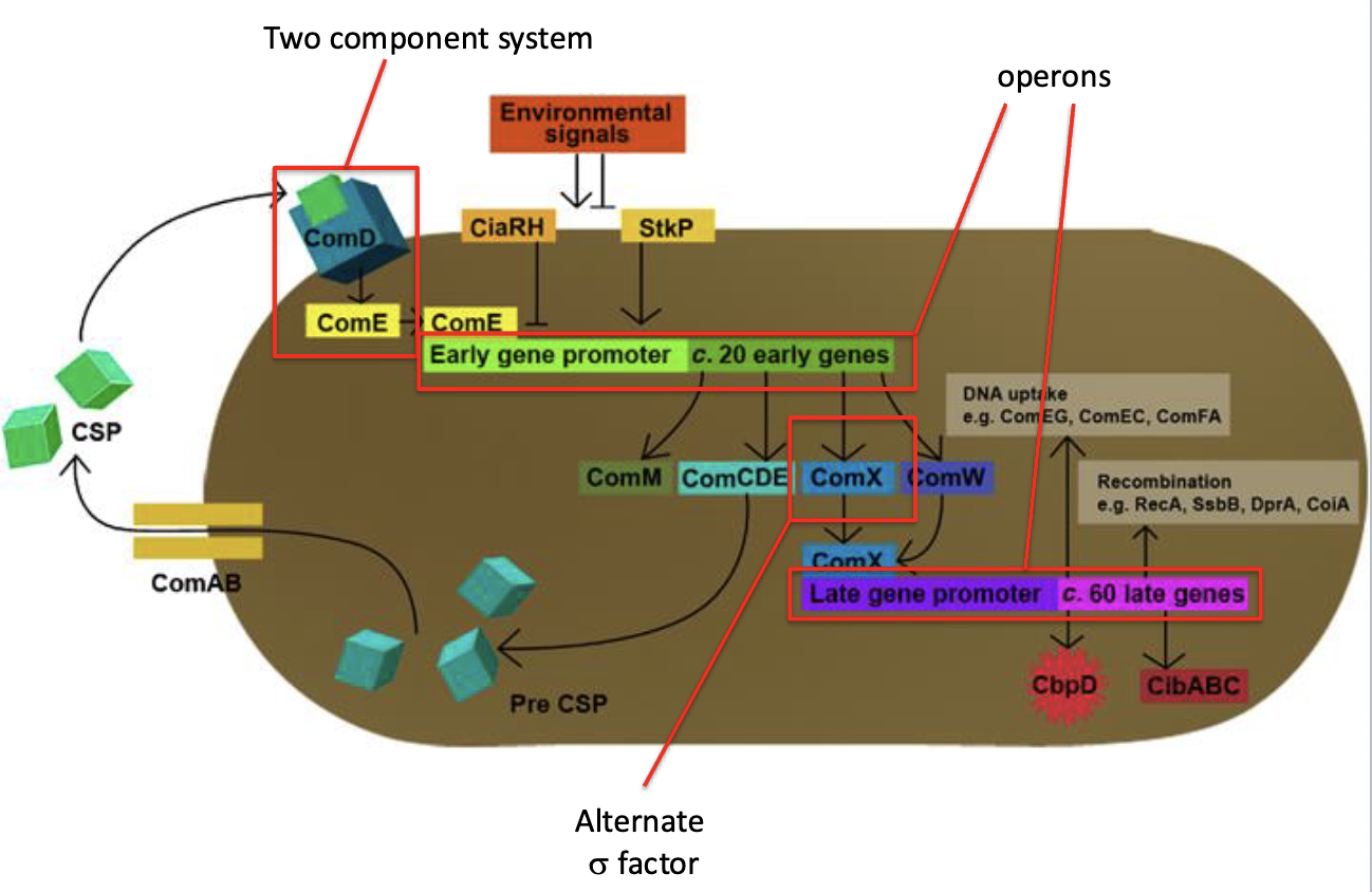
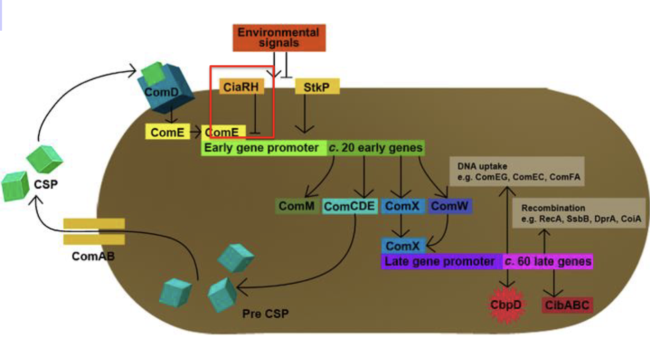
Explain how two component systems work and their involvement in competence and quorum sensing
Two-component systems (TCS) are signal transduction mechanisms bacteria use to sense and respond to environmental changes. They typically consist of two proteins:
1. HPK receives signal: in response, HPK autophosphorylates on a histidine residue. Then it transfers phosphate to an aspartate on the response regulator.
2. Phosphorylation activates DNA binding: Activators are proteins
that bind to specific DNA sequences and activate expression of downstream genes
Involvement in Competence and Quorum Sensing:
1. Competence: In bacteria like Streptococcus pneumoniae, a TCS helps regulate competence—the ability to take up external DNA.
2. Quorum Sensing: where bacteria sense population density by detecting signaling molecule called autoinducer. This typically binds to an HPK with an affinity that requires a particular
environmental concentration. When the concentration reaches a thresh-hold indicative of population density gene expression is activated
Explain of the role of activators in the competence pathway.
proteins that bind to specific DNA sequences and activate expression of downstream genes.
activators stabilize RNA polymerase interaction with promoter
Often compensates for a weak promoter
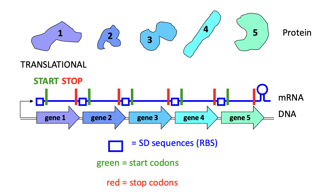
Define operon and explain the advantages in prokaryotes.
bacterial genes are often organized in operons (when multiple genes (cistrons) are expressed from a common promoter.)
Each gene has its own ribosome binding sequence, translational start, and translational stop.
Explain the role of sigma factors in transcriptional initiation and gene expression.
Activation of Early Gene operon (RESPONSE TO ENVIRONMENTAL STIMULUS) produces an alternate sigma factor
Once the RNA polymerase is correctly positioned at the promoter, the sigma factor initiate transcription by unwinding the DNA and starting RNA synthesis.
Different sigma factors recognize different promoter sequences and are activated by dif external conditions, allowing bacteria to regulate gene expression in response to changing environments way of coordinately regulating hundreds of genes of like-function throughout the genome
Explain the role of sigma factors play specifically play in the competence pathway.
Gene Activation: Specific sigma factors are activated in response to environmental signals, initiating the transcription of competence-related genes.
Regulation of Competence Genes: These sigma factors direct RNA polymerase to the promoters of competence genes, enabling the production of proteins required for DNA uptake and transformation.
Explain how DNA is taken up from the environment.
ComX turns on late gene operon
This encodes DNA uptake and recombination machinery
can cause DNA to be in the environment
Explain how the competence pathway is turned off.
The system is turned off by CiaRH, a two component system that is expressed when competence begins and eventually over-rides the competence system and represses early gene expression.
Repressors: are proteins that bind to a specific DNA sequence and inhibit gene transcription. protein binding overlaps the promoter and blocks RNA polymerase access
Explain why transformation is advantageous to the bacterial population.
For pneumococcus, the advantage is obtaining “new” capsule genes to evade an antibody response and share or pass antibiotic resistance.
Generally thought to be a mechanism of dealing with environmental pressures and repairing DNA
Explain the molecular interactions involved in each of the following steps of the influenza single-cell replication cycle: entry
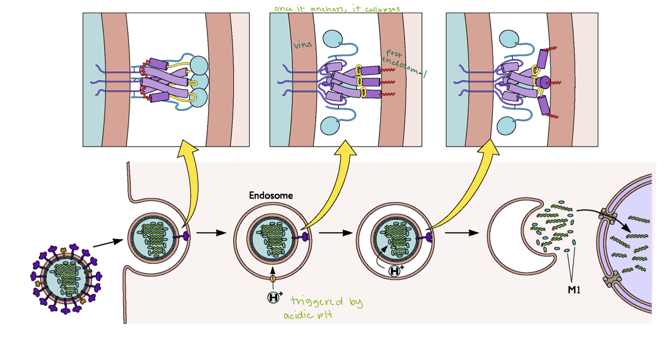
Virus particles are METASTABLE structures. Under the appropriate conditions they disassemble. Following attachment influenza virions are taken into the cell by receptor mediated endocytosis. Acidification of the endosome triggers membrane fusion as well as the activation of M2 ion channel and the disassembly of M1 and vRNPs.
Released nucleocapsid complexes are transported to the nucleus due to the presence of a nuclear localization signal (NLS) on the NP protein
What is responsible for attaching flu to the cell?
1. Viral Component: Hemagglutinin (HA)
Function: Hemagglutinin (HA) is the surface glycoprotein on the influenza virus that is responsible for binding to the host cell.
Mechanism: HA specifically recognizes and binds to sialic acid residues present on glycoproteins or glycolipids on the surface of the host cell. The type of sialic acid linkage (either α-2,6 or α-2,3) determines the virus's specificity for the host species (e.g., human or avian).
2. Host Component: Sialic Acid Receptors
Function: Sialic acids are sugars that are found on the surface of host cells, typically linked to glycoproteins or glycolipids.
Mechanism: The presence of sialic acid on the host cell surface serves as the receptor for the viral HA. The specific linkage of sialic acid to the underlying sugar chain (either α-2,6 or α-2,3) influences the virus's ability to infect different host species.
What does the viral protein look like?
Structure: Hemagglutinin (HA) is a trimeric protein with a globular head domain for receptor binding and a stem domain for membrane fusion.
How does the virus get its genetic material into the cell?
Trigger: The process is initiated by the acidification of the endosome.
Viral Protein Involved: Hemagglutinin (HA) is the key protein, specifically its receptor-binding domain and fusion peptide.
Changes Caused: The conformational change in HA due to low pH exposes the fusion peptide, facilitating membrane fusion.
Result: The viral ribonucleoprotein complexes (vRNPs) are released into the cytoplasm, allowing the virus to begin its replication cycle inside the host cell.
Explain the mechanisms of action of vancomycin
Was “drug of last resort”, now resistance is becoming more common in some Gram positives but not Pneumococcus.
Binds to (D-ala)-(D-ala) dipeptide and blocks crosslinking
Resistance occurs by replacing the terminal D-ala with
either lactate or D-serine
MAC complex
The membrane attack complex (MAC) is a key component of the complement system, role in lysis of target cells
1. Formation: The MAC is formed when complement proteins, specifically C5b, C6, C7, C8, and C9, assemble on the surface of a target cell.
2. Function: The resulting pore disrupts the integrity of the target cell membrane, leading to cell lysis and death.
3. Role in immunity: The MAC contributes to the opsonization of pathogens and enhances inflammation, promoting the recruitment and activation of immune cells.
Explain how antibody response normally occur and why
pneumococcus causes this response to be weak.
The normal antibody response occurs when immune cells (B cells) recognize antigens on pathogens. B cells are activated, differentiate into plasma cells, and produce specific antibodies that neutralize the pathogen, opsonize it for phagocytosis, or activate the complement system for clearance.
Pneumococcus (Streptococcus pneumoniae) weakens this response because it has a polysaccharide capsule that:
- Masks its antigens, making it harder for immune cells to recognize and target.
- Reduces T cell activation since the capsule is a T-independent antigen, leading to a weaker, short-lived antibody response without strong memory formation.
This allows pneumococcus to evade the immune system more effectively.
Describe the basic action of pneumolysin and its role in
pathogenesis.
Pneumolysin is a cholesterol dependent cytolysin
Basic action:
Pneumolysin binds to cholesterol in host cell membranes, ogliomerizes to form pores that lead to cell lysis and damage.
Important for invasion into bloodstream and hemolysis
At low, sublytic concentrations it can activate the classical complement pathway in the absence of antibody causing lysis of host cells and increased inflammation
Compare and contrast the two types of pneumococcal vaccines.
1. Pneumococcal polysaccharide vaccine (PPSV23):
- Contains purified polysaccharides from 23 pneumococcal serotypes.
- Induces a T-independent immune response, leading to short-lived immunity with limited memory.
- Primarily used in adults, especially those at high risk.
2. Pneumococcal conjugate vaccine (PCV13):
- Contains polysaccharides from 13 serotypes conjugated to a protein carrier.
- Induces a stronger, T-dependent immune response with better immunological memory.
- Used in infants, children, and some adults.
Contrast: PCV13 generates longer-lasting immunity and is more effective in children, while PPSV23 covers more serotypes but provides weaker, shorter-term protection.
Explain why the conjugated vaccine cause memory.
Presence of an immunogenic protein leads to T-helper cell activation and B-cell differentiation providing secreted antibody and memory
Explain the pneumococcal vaccine issues.
Expensive
Not complete in serotype coverage
PPSV23 does not provide long lasting memory
Conjugated vaccine does not work with all serotypes
PCV 7 showed increased carriage of serotype 19A which is more difficult to treat
Distribution to developing countries problematic due to cost and vaccination
identify different arthropod vectors
Hematophagous arthropods
Mosquitos (aedes aegypti, anopheles), flea, black fly, sand fly, kissing bug
Explain simple and complex transmission cycles
Identify types of questions that are important in arthropod transmission
What factors contribute to outbreak?
Population levels, location, breeding habits of vectors?
How and where do humans interact with vector?
How do environmental factors affect human, vector, and reservoir behavior?
Describe the main virus families transmitted by mosquitoes
Flaviviruses, alphaviruses - icosahedral, positive sense, enveloped
bunyaviruses - plieomorphic, negative sense, envelope
Explain the basics of mosquito behavior and anatomy and how it aids in transmission
Anatomy
1. Proboscis: Mosquitoes use their proboscis to pierce the skin and feed on blood. During this, they can inject the dengue virus into the bloodstream.
2. Salivary Glands: The virus can be present in the mosquito’s saliva, which is transferred during feeding.
Behavior
1. Feeding: Female mosquitoes require blood for egg development. If they feed on an infected person, they can carry and transmit the virus to others.
2. Active During Day: Aedes mosquitoes are most active in the early morning and late afternoon, increasing human contact and transmission chances.
3. Attraction to Humans: They are attracted to human scent and carbon dioxide, making humans their preferred hosts.
4. Multiple Feedings: Mosquitoes can bite multiple people over their lifespan, spreading the virus to various individuals.
Transmission
After feeding on an infected person, the virus incubates in the mosquito for up to two weeks, allowing it to spread to other humans during subsequent blood meals.
Explain the pathway of infection in mosquitoes
Virus taken up in blood meal
Enters midgut
Infects midgut and breeches barrier
Transmits through hemocoel to salivary glands
Infects salivary glands and grows to high titers
Explain the pattern of infection used in mosquitoes and why that is beneficial
1. Acute – high level replication with rapid (days) resolution
2. Chronic/persistent – constant replication either at low level, or high level with immune evasion
3. Latent – high level replication followed by almost complete quiescence. Can reactivate
Identify the major pathways involved in mosquito immunity
RNAi (primary antiviral defense), Toll, Imd, Jak-STAT
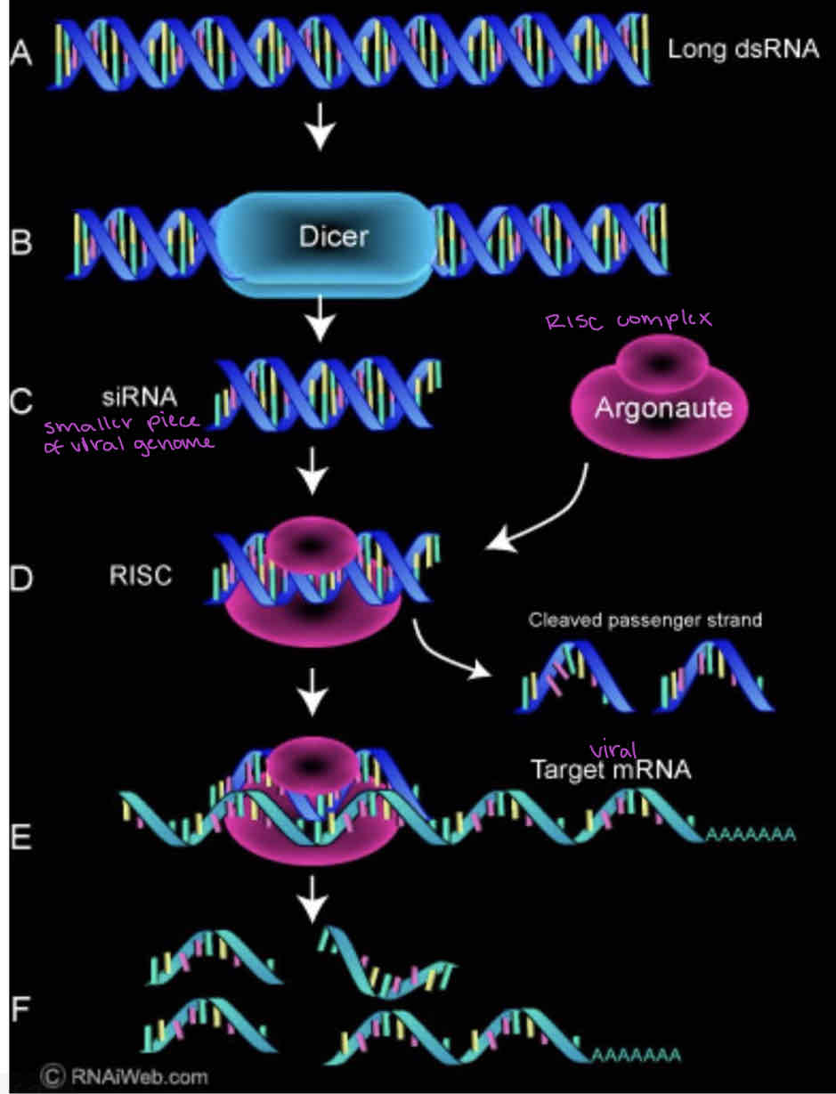
Explain how RNAi works to prevent viral infection
Dicer recognizes dsRNA and cuts it into small pieces (good trigger because dsRNA only comes from viruses)
One strand of the pieces is loaded into the RNA-induced silencing complex (RISC) (degrades and removes complementary piece)
Base pairing of the RNA strand to another RNA strand targets it for degradation by the Argonaute associated RNase activity
Explain how yellow fever spread to the Americas
Yellow fever is long endemic in central Africa, disease in native Africans is relatively mild. Due to the activities of slave traders yellow fever was imported to the Americas.
Explain how yellow fever impacted historical events such as the Louisiana Purchase and the Spanish American War
Louisiana purchase - Napoleon sends large force to crush uprising in Haiti. 27,000 French troops die of yellow fever. Due to massive losses in Haitian uprising, loss of capital from Haiti and increased need for resources, Napoleon sells Louisiana territory to US
Spanish American war - outbreak delayed military campaigns and exposed weaknesses in sanitation and medical preparedness. This experience led to improved disease control measures and spurred research into mosquito-borne illnesses, culminating in Dr. Walter Reed's discovery that mosquitoes transmitted yellow fever, which influenced later public health efforts.
explain how the mechanism of yellow fever transmission was discovered
Commission members self inoculated using mosquitoes that had bitten infected individuals.
No symptoms occurred
Used mosquitoes that had bitten an infected patient 12 days before.
Success! They became sick and died! Transmission from human to human by Aedes aegypti
identify the vector of flavivirus infection (Genus species)
Aedes aegypti
explain the factors that make a pathogen eradicable
1. Limited host range, preferably only humans or a defined vector
2. Mechanism of transmission intervention (vaccine)
3. Has been eliminated in a smaller geographic area
4. Significant public health burden
5. Makes financial sense to the countries involved
explain why yellow fever is not a good candidate for eradication
sylvatic cycle cycle involving mosquitoes, animal reservoirs (non-human primates), and widespread mosquito populations in diverse environments.
vaccine is effective but difficult to distribute in remote areas
virus's ability to persist in various settings adds to the challenge.
describe the vaccine design and efficacy for yellow fever
a live attenuated virus vaccine
highly effective, with over 90% efficacy in preventing yellow fever after a single dose. Protection begins within 10 days after vaccination and lasts for at least 10 years, often for life
explain the advantages and disadvantages of using DTT as a control method for yellow fever
Advantages: Effectiveness, Long-lasting, Low Cost
Disadvantages: Environmental Impact, Resistance, Health Concerns, Public and Regulatory Backlash
explain the advantages and disadvantages of using SIT and RIDL to control flaviviruses
Sterile Insect Technique (SIT) involves releasing sterilized male mosquitoes to mate with wild females, leading to no offspring and reducing mosquito populations over time.
Advantages:
1. targets mosquitoes without harming other species, making it environmentally friendly.
2. provides long-term mosquito population control without chemical use.
3.does not lead to resistance in mosquito populations.
Disadvantages:
1. can be expensive and logistically challenging.
2. most effective for certain mosquito species, limiting its use for controlling flaviviruses across all vector species.
3. requires large-scale releases to be effective, which may be difficult in areas with high mosquito densities or limited resources.
explain the state of yellow fever now and what factors allow the problem to persist
Urban yellow fever completely eliminated from Americas
The loss of vector control programs as a result of the ban on DDT use has led to a re-emergence of yellow fever, particularly in sub-Saharan Africa.
In addition to the loss of vector control in Africa there have been systemic failures to immunize at risk populations, mainly due to delivery of the vaccine. Political instability and warfare prevent vaccine delivery due to physical access and lack of funds
describe how Wolbachia infected Aedes is used to control flaviviruses
Wolbachia is a Gram negatvie bacterium.
Causes cytoplasmic incompatibility meaning embryos resulting from uninfected females mating with infected males die.
Infection causes decreased life span, and reduces virus replication hence reduces transmission of virus
Use Wolbachia as means to prevent Dengue transmission by introducing infected mosquitoes into natural populations.
identify the regions of the world most at risk for yellow fever infection
90% of cases occur in Africa, also some in South America
describe the factors contributing to the re-emergence of yellow fever in sub-Saharan Africa and South America
loss of vector control programs as a result of the ban on DDT
Yellow fever in South America has been a result of intrusion on the sylvatic cycle. No confirmed urban transmission, although some outbreaks suggest this is happening.
systemic failures to immunize at risk populations, mainly due to delivery of the vaccine. Political instability and warfare prevent vaccine delivery due to physical access and lack of funds
describe the hierarchical relationship between Flaviviridae family, Flavivirus genus, and species.
Flaviviridae (Family) > Flavivirus (Genus) > Dengue virus, Zika virus, West Nile virus, etc. (Species)
give examples of the major members of Flavivirus
Yellow fever, dengue, west Nile, encephalitis
compare and contrast the different host, diseases, and vectors associated with Flavivirus
describe the structure of the Flavivirus virion
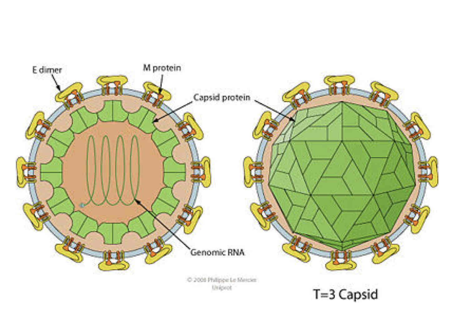
Enveloped and spherical, icosahedral geometry with T=3 symmetry
describe the viral and host proteins involved in flavivirus attachment
E protein binds to cellular receptor. No individual receptor identified, but can use integrins (connect cells together) and lectins that bind sugars.
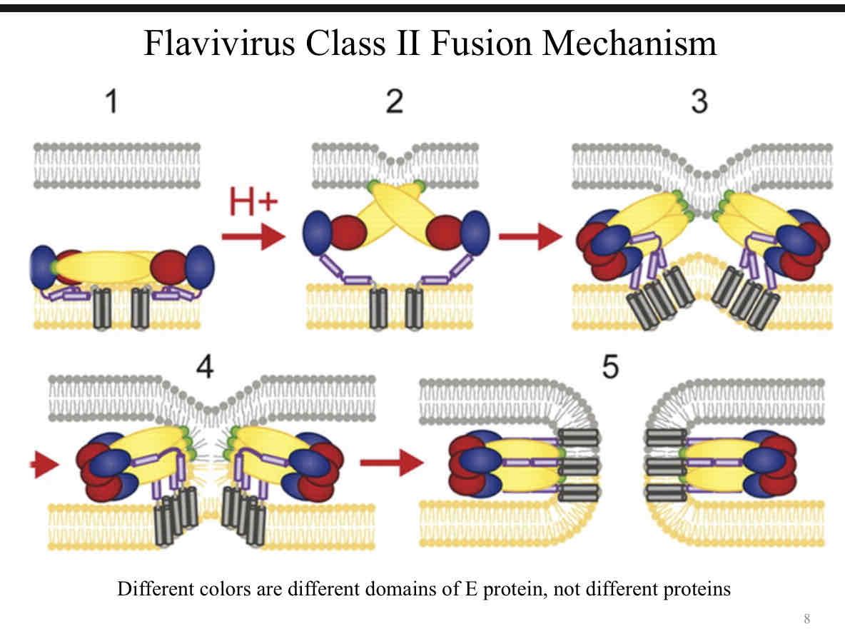
describe the class II fusion mechanism used for entry (flavivirus)
The E protein on the viral envelope binds to host cell receptors.
The virus is endocytosed into an acidic endosome.
The drop in pH triggers a conformational change in the E protein, exposing the fusion peptide.
The fusion peptide inserts into the host cell membrane, and the viral and host membranes fuse.
This fusion releases the viral genome into the host cell, where it can begin replication.
explain how the genome organization allows for immediate translation of a polypeptide
The genome has a 5' cap, which is recognized by the host’s ribosomes, allowing for translation of the viral RNA immediately after the virus enters the cell.
The genome contains a single ORF that is translated into a polyprotein which is processed by proteases into individual functional proteins.
explain the difference between the initial polypeptide and the mature viral proteins
Initial Polypeptide: a single, long chain that includes the structural (e.g., capsid, envelope) and non-structural proteins (e.g., polymerase, protease) in a precursor form.
Mature Viral Proteins: polyprotein is cleaved into individual, functional proteins that are necessary for viral assembly, replication, and release, such as the capsid protein, envelope protein, and NS (non-structural) proteins.
explain the problem between translation and replication that arises from being a positive sense genome. explain how flavivirus use the circularization motif to resolve this problem.
The problem: the same RNA is used both for translation (5 → 3) into proteins and for replication (3 →5) into new genomes. Since translation occurs as soon as the RNA enters the cell, it can compete with the need for replication
solution: using a circularization motif in their RNA. This involves the 5' and 3' untranslated regions interacting to form a circular structure. For replication, proteins bind to terminal and prevent translation
identify the viral protein responsible for replication
RNA-dependent RNA polymerase (NS5)
describe the steps and location of viral assembly and budding
1. The C (capsid) protein associates with the genomic RNA, forming the nucleocapsid. The prM (precursor membrane) and E (envelope) proteins are inserted into the ER membrane, where they undergo processing.
2. The nucleocapsid (C protein + viral RNA) interacts with the prM-E protein complex in the ER membrane, forming the immature virion.
3. The immature virion is transported from the ER to the Golgi apparatus in vesicles where the virus matures as the prM is cleaved to M by furin, and the virion acquires its final, infectious form.
5. The newly formed virions are transported to the cell surface in vesicles and are released from the cell via exocytosis.
explain the difference between viral glycoprotein modifications in the mosquito vector and the human host.
complex sugars added in vertebrates, high mannose in mosquitoes.
Humans: Modifications favor interaction with human receptors for infection.
Mosquitoes: Modifications support replication in the mosquito midgut and salivary glands.
describe the role of prM in viral infection and the importance of the cleavage of pr
The prM acts as a precursor to the M (membrane) protein. prM prevents premature fusion of the viral membrane with the host cell membrane during assembly, ensuring the virus remains in an immature, non-infectious form.
- Facilitates Viral Assembly: prM assists in the correct folding and assembly of the virus in the ER and Golgi.
importance of prM Cleavage (furin):
- Cleavage activates the virus, allowing the E protein to undergo structural changes necessary for the virus to become infectious.
describe the basic symptoms associated with yellow fever
1. Fever, chills, back pain, nausea, anorexia, mild gingival hemorrhaging, nose bleeds
2. Short remission (6-24 hours)
3. Symptoms recur with frequent vomiting (black), jaundice
4. Increased hemorrhaging
5. Death in 20-50% of cases 7-10 days following onset of symptoms
Recovery: Prolonged convalescence (months), with severe fatigue.
Explain steps involved in the initial flavivirus infection and how it spreads, paying close attention to the type of cells involved.
1. The mosquito injects the virus into the skin and, in some cases, directly into the bloodstream.
2. The virus infects Langerhans cells (immune cells in the skin.) It does this by binding to a receptor called DC-SIGN on these cells facilitated by high mannose glycans on the virus’s glycoproteins
3. The infected Langerhans cells travel to nearby lymph nodes, where they encounter macrophages. These macrophages are highly susceptible to infection by the virus.
4. From the lymph nodes, the virus enters the bloodstream, spreading throughout the body to infect various tissues where it can be picked up by other mosquitos and spread
Explain the steps involved in yellow fever pathogenesis, paying close attention to the type of cells involved and how it is resolved.
1. The virus infects Kupffer cells, which then spread the infection to nearby hepatocytes (liver cells).
2. The infected cells release cytokines, signaling for help from immune cells, including eosinophils.
3. Eosinophils, typically involved in responses to parasites or allergies, respond to the cytokine signals. Their activity can inadvertently damage liver tissue, leading to liver degradation.
4. This liver damage can cause jaundice (yellowing of the skin and eyes), a key symptom of severe yellow fever.