2. 3 - Laboratory Tests - Hematology - Blood Components
 Call Kai
 Learn
Learn Practice Test
 Spaced Repetition
 Match
Match Flashcards
 Knowt Play
1/119

Earn XP
Description and Tags
Proverbs 16:3
Name | Mastery | Learn | Test | Matching | Spaced | Call with Kai | Chat |
|---|
No analytics yet
Send a link to your students to track their progress
120 Terms
RBC
WBC
Platelets
[BLOOD COMPOSITION]
Formed Element (Solid) include ____ [3]
Proteins
Serum
[BLOOD COMPOSITION]
Plasma (Liquid) include ______ [2]
a. Erythrocytes
Red blood cells are also known as ______
a. Erythrocytes
b. Leukocytes
c. Thrombocytes
d. Lymphocytes
c. Red blood cells (RBCs)
[RED BLOOD CELLS]
These cells are responsible for transporting oxygen from the lungs to body tissues.
a. White blood cells (WBCs)
b. Platelets
c. Red blood cells (RBCs)
d. Plasma cells
a. True
[RED BLOOD CELLS]
Tissues produce energy with oxygen and release waste identified as carbon dioxide
a. True
b. False
a. True
[RED BLOOD CELLS]
Red blood cells (Erythrocytes) take the carbon dioxide waste to your lungs for you to exhale
a. True
b. False
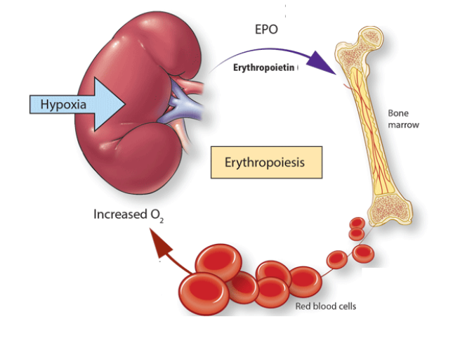
Kidneys detect reduced O2 carrying capacity of blood
↓
Kidneys respond by secreting erythropoietin (EPO) into the bloodstream
↓
Erythropoietin (EPO) stimulates erythropoiesis by the bone marrow
↓
Addition of circulating erythrocytes increases O2 carrying capacity of the blood
↓
Increased O2 carrying capacity relieves the initial stimulus that triggered EPO
[NOTES]
Erythropoiesis process:
b. Packed Cell Volume (PCV)
[RED BLOOD CELLS]
Hematocrit is also known as _______
a. Mean Corpuscular Volume (MCV)
b. Packed Cell Volume (PCV)
c. Red Blood Cell Count
d. Hemoglobin Concentration
Hematocrit (Packed Cell Volume)
[RED BLOOD CELLS]
This refers to the number of red blood cells in 100 mL of blood.
a. Hemoglobin
b. Hematocrit (Packed Cell Volume)
c. Mean Corpuscular Volume
d. Red blood cell count
b. Hematocrit (Packed Cell Volume)
[RED BLOOD CELLS]
%RBC in the blood
a. Hemoglobin
b. Hematocrit (Packed Cell Volume)
c. Mean Corpuscular Volume
d. Red blood cell count
b. Hematocrit (Packed Cell Volume)
[RED BLOOD CELLS]
Ratio of the volume occupied by RBCs to the total volume of blood
a. Hemoglobin
b. Hematocrit (Packed Cell Volume)
c. Mean Corpuscular Volume
d. Red blood cell count
Dengue
Polycythemia vera
Dehydration
Condition that INCREASES Hematocrit [3]
Blood loss
Hemolytic anemia
Iron deficiency anemia
Overhydration
Condition that DECREASES Hematocrit [4]
High Hematocrit
[RED BLOOD CELLS]
Reduced blood volume (dehydration, DI) results in ______
a. Decreased hematocrit
b. Increased hematocrit
a. Decreased hematocrit
[RED BLOOD CELLS]
Increased blood volume (overhydration, SIADH) results in _____
a. Decreased hematocrit
b. Increased hematocrit
b. Increased hematocrit
Dehydration and Diabetes Insipidus (DI) cause:
a. Decreased hematocrit
b. Increased hematocrit
a. Decreased hematocrit
Overhydration and SIADH cause:
a. Decreased hematocrit
b. Increased hematocrit
Baby RBCs
Immature RBCs
[RED BLOOD CELLS]
Reticulocytes are also known as ____ [2]
b. Reticulocytes
[RED BLOOD CELLS]
Earliest non-nucleated RBCs
a. Erythrocytes
b. Reticulocytes
c. Thrombocytes
d. Leukocytes
b. Reticulocytes
[RED BLOOD CELLS]
Its increased production can be detected in times of rapid RBC regeneration.
a. Erythrocytes
b. Reticulocytes
c. Thrombocytes
d. Leukocytes
Hemolysis
Hemorrhage
Iron deficiency anemia (IDA)
[RED BLOOD CELLS]
Reticulocytes increase in response to _____ [3]
a. True
[RED BLOOD CELLS]
Decrease RBCs → Increase Reticulocytes
a. True
b. False
b. Hemoglobin (Hgb)
[RED BLOOD CELLS]
This refers to the protein inside the RBCs carrying the oxygen from lungs to tissues.
a. Hematocrit
b. Hemoglobin (Hgb)
c. Myoglobin
d. Transferrin
b. Hemoglobin (Hgb)
[RED BLOOD CELLS]
Oxygen carriers
Responsible for the red color of blood
a. Hematocrit
b. Hemoglobin (Hgb)
c. Myoglobin
d. Transferrin
a. Heme (pigment) + globin (protein)
[RED BLOOD CELLS]
Hemoglobin is composed of:
a. Heme (pigment) + globin (protein)
b. Iron + albumin
c. Heme + albumin
d. Globin + bilirubin
TRUE
[RED BLOOD CELLS]
TRUE OR FALSE:
The hemoglobin concentration in men is normally GREATER than women due to MENSTRUATION.
● Polycythemia
● Dehydration
[RED BLOOD CELLS]
High hemoglobin indicates _____ [2]
Anemia
[RED BLOOD CELLS]
Low hemoglobin indicates _____ [1]
● Mean Cell Volume (MCV)
● Mean Cell Hemoglobin (MCH)
● Mean Cell Hemoglobin Concentration (MCHC)
[RED BLOOD CELLS]
RBC Indices [3]
b. Mean Cell Volume (MCV)
[RED BLOOD CELLS]
This refers to the average volume of a single RBC and is measured in femtoliters
a. Mean Cell Hemoglobin (MCH)
b. Mean Cell Volume (MCV)
c. Mean Cell Hemoglobin Concentration (MCHC)
d. Red Cell Distribution Width (RDW)
c. Femtoliters (fL)
[RED BLOOD CELLS]
Mean Cell Volume refers to the average volume of a single RBC and is measured in _____.
a. Grams per deciliter (g/dL)
b. Picograms (pg)
c. Femtoliters (fL)
d. Percentage (%)
Macrocytic
Normocytic
Microcytic
MCV classifies RBC size into ____ [3]
<83 fL
[RED BLOOD CELLS]
Low MCV value:
a. <83 fL
b. 83-101 fL
c. >101 fL
● Microcytic
● Iron Deficiency
● Thalassemic Disorder
[RED BLOOD CELLS]
Low MCV (<83 fL) indicates _______ [3]
b. 83-101 fL
[RED BLOOD CELLS]
Normal MCV value:
a. <83 fL
b. 83-101 fL
c. >101 fL
● Normocytic
● Acute Blood Loss
● Early Iron Deficiency
● Chronic Renal Insufficiency
[RED BLOOD CELLS]
Normal MCV indicates ______ [4]
c. >101 fL
[RED BLOOD CELLS]
High MCV value:
a. <83 fL
b. 83-101 fL
c. >101 fL
● Macrocytic
● Vitamin B12 Deficiency
● Vitamin B9 (Folate) Deficiency
● Alcohol abuse
[RED BLOOD CELLS]
High MCV indicates ____ [4]
Mean Cell Hemoglobin (MCH)
[RED BLOOD CELLS]
This refers to the average weight of hemoglobin contained in an RBC measured in picograms.
a. Mean Cell Hemoglobin (MCH)
b. Mean Cell Volume (MCV)
c. Mean Cell Hemoglobin Concentration (MCHC)
d. Red Cell Distribution Width (RDW)
b. Picograms (pg/cell)
[RED BLOOD CELLS]
Mean Cell Hemoglobin (MCH) refers to the average weight of hemoglobin contained in an RBC measured in ______.
a. Femtoliters (fL)
b. Picograms (pg/cell)
c. Grams per deciliter (g/dL)
d. Percentage (%)
Hyperchromic
Normochromic
Hypochromic
MCH classifies RBC color into ____ [3]
<27 pg/cell
[RED BLOOD CELLS]
Hypochromic MCH value:
a. <27 pg/cel
b. 27-31 pg/cell
c. >31 pg/cell
26-31 pg/cell
[RED BLOOD CELLS]
Normochromic MCH value:
a. <27 pg/cel
b. 27-31 pg/cell
c. >31 pg/cell
>31 pg/cell
[RED BLOOD CELLS]
Hyperchromic MCH value:
a. <27 pg/cel
b. 27-31 pg/cell
c. >31 pg/cell
a. True
[RED BLOOD CELLS]
MCH decreases inversely proportional as size of central pallor
a. True
b. False
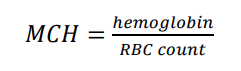
Formula for MCH:
Mean Cell Hemoglobin Concentration (MCHC)
[RED BLOOD CELLS]
This is the measure of the average concentration of hemoglobin per unit weight of RBC.
a. Mean Cell Hemoglobin (MCH)
b. Mean Cell Volume (MCV)
c. Mean Cell Hemoglobin Concentration (MCHC)
d. Red Cell Distribution Width (RDW)
b. Leukocytes
[WHITE BLOOD CELLS]
White blood cells are also known as _______
a. Erythrocytes
b. Leukocytes
c. Thrombocytes
d. Lymphocytes
Never Let Monkeys Eat Bananas
● Neutrophils (MOST ABUNDANT)
● Lymphocytes
● Monocytes
● Eosinophils
● Basophils (LEAST ABUNDANT)
[WHITE BLOOD CELLS]
Types of white blood cells in order of abundance [5]
● Lymphocytes
● Monocytes
[WHITE BLOOD CELLS]
Agranulocytes include _____ [2]
c. Monocytes
[WHITE BLOOD CELLS]
Differentiate or develop into macrophages
a. Neutrophils
b. Lymphocytes
c. Monocytes
d. Eosinophils
e. Basophils
c. Monocytes
[WHITE BLOOD CELLS]
This WBC is increased in:
Typhoid
Sub-acute Bacterial Endocarditis
Infectious Mononucleosis
Tuberculosis
a. Neutrophils
b. Lymphocytes
c. Monocytes
d. Eosinophils
e. Basophils
Typhoid
Sub-acute Bacterial Endocarditis
Infectious Mononucleosis
Tuberculosis
[WHITE BLOOD CELLS]
Monocytes are increased in ____ [4]
b. Lymphocytes
[WHITE BLOOD CELLS]
This WBC is increased in viral infections.
a. Neutrophils
b. Lymphocytes
c. Monocytes
d. Eosinophils
e. Basophils
ViraL infections
[WHITE BLOOD CELLS]
Lymphocytes are increased in _____ [1]
b. Lymphocytes
[WHITE BLOOD CELLS]
Decreases in debilitating illnesses (e.g., AIDS).
a. Neutrophils
b. Lymphocytes
c. Monocytes
d. Eosinophils
e. Basophils
NK Cells
B Cell
T Cells
[WHITE BLOOD CELLS]
Types of Lymphocytes [3]
c. B Cells (Plasma Cell)
[WHITE BLOOD CELLS]
Produce antibodies
a. T Cells
b. NK Cells
c. B Cells
d. Neutrophils
c. Plasma cells
[WHITE BLOOD CELLS]
B cells differentiate into _____ cells, which are the primary antibody-producing cells.
a. T Cells
b. NK Cells
c. Plasma cells
d. Macrophages
b. B Cells
[WHITE BLOOD CELLS]
Responsible for Humoral Immunity
a. NK Cells
b. B Cells
c. T Cells
c. T Cells
[WHITE BLOOD CELLS]
Responsible for Cell-mediated Immunity
a. NK Cells
b. B Cells
c. T Cells
CD4 cells
CD8 cells
[WHITE BLOOD CELLS]
Types of T-cells [2]
a. CD4 cells
[WHITE BLOOD CELLS]
Also known as “helper T cells”
a. CD4 cells
b. CD8 cells
b. CD8 cells
[WHITE BLOOD CELLS]
Also known as “cytotoxic T cells”
a. CD4 cells
b. CD8 cells
a. CD4 cells
[WHITE BLOOD CELLS]
They are targeted by HIV/AIDS
a. CD4 cells
b. CD8 cells
a. CD4 cells
[WHITE BLOOD CELLS]
Activate B cells and other T cells
a. CD4 cells
b. CD8 cells
Eosinophil
Basophil
Neutrophil
[WHITE BLOOD CELLS]
Granulocytes include ______ [3]
d. Eosinophils
[WHITE BLOOD CELLS]
This WBC is increased in:
Parasitic infection
Allergic Conditions (Asthma, Hay Fever)
Drug Sensitivity Reactions
a. Neutrophils
b. Lymphocytes
c. Monocytes
d. Eosinophils
e. Basophils
Parasitic infection
Allergic Conditions (Asthma, Hay Fever)
Drug Sensitivity Reactions
[WHITE BLOOD CELLS]
Eosinophils are increased in ______ [3]
e. Basophils
[WHITE BLOOD CELLS]
This WBC is increased in hypersensitivity reactions.
a. Neutrophils
b. Lymphocytes
c. Monocytes
d. Eosinophils
e. Basophils
Hypersensitivity reactions
[WHITE BLOOD CELLS]
Basophils are increased in _____ [1]
e. Basophils
Stored in mast cells
a. Neutrophils
b. Lymphocytes
c. Monocytes
d. Eosinophils
e. Basophils
e. Basophils
Releases histamine
a. Neutrophils
b. Lymphocytes
c. Monocytes
d. Eosinophils
e. Basophils
a. Neutrophils
[WHITE BLOOD CELLS]
This WBC is increased in bacterial and fungal reactions
a. Neutrophils
b. Lymphocytes
c. Monocytes
d. Eosinophils
e. Basophils
Bacterial reactions
Fungal reactions
[WHITE BLOOD CELLS]
Neutrophils are increased in ______ [2]
Thrombocytes
[PLATELETS]
Platelets are also known as _____
c. Platelets (Thrombocytes)
These are anucleate cells derived from the megakaryocytic cells in the bone marrow.
a. Red blood cells
b. White blood cells
c. Platelets (Thrombocytes)
d. Plasma cells
c. Platelets (Thrombocytes)
Important in initiating hemostasis and involved in developing non-hemostatic immune functions
a. Red blood cells
b. White blood cells
c. Platelets (Thrombocytes)
d. Plasma cells
b. Bleeding
If blood levels of platelets decrease below normal, this leads to _______
a. Clotting
b. Bleeding
c. Infection
d. Anemia
b. Thrombocytopenia
[PLATELETS]
Decreased platelets
a. Thrombocytosis
b. Thrombocytopenia
c. Leukocytosis
d. Anemia
b. Thrombocytopenia
[PLATELETS]
________-
This refers to the depressed synthesis in the marrow or consumption of formed platelets
This may be seen in pregnancy following viral infections.
a. Thrombocytosis
b. Thrombocytopenia
c. Leukocytosis
d. Anemia
b. Thrombocytopenia
[PLATELETS]
This condition is seen in pregnancy following viral infections.
a. Thrombocytosis
b. Thrombocytopenia
c. Leukocytosis
d. Anemia
c. 150,000-300,000/mm³
[PLATELETS]
Normal platelet (thrombocyte) count is:
a. 5,000-10,000/mm³
b. 50,000-100,000/mm³
c. 150,000-300,000/mm³
d. 500,000-1,000,000/mm³
● Hydralazine
● Caffeine
● Colchicine
● Vincristine
● NSAIDs
● Furosemide
● Penicillin
● Hydroxychloroquine
● Theophylline
● TCAs
📌Mnemonic: “HCCVN” “FPHTT”
[PLATELETS]
Drugs causing platelet dysfunction [8]
b. Prothrombin Time (PT)
[COAGULATION]
__________-
This tests the extrinsic and common pathways
This is most commonly employed in the monitoring of warfarin anticoagulation therapy
a. Activated Partial Thromboplastin Time (aPTT)
b. Prothrombin Time (PT)
c. Bleeding Time
d. Clotting Time
b. Prothrombin Time (PT)
[COAGULATION]
Time taken for fibrin strand formation in platelet-poor plasma
a. Activated Partial Thromboplastin Time (aPTT)
b. Prothrombin Time (PT)
c. Bleeding Time
d. Clotting Time
b. Prothrombin Time (PT)
[COAGULATION]
Measures the time it takes to form a clot
a. Activated Partial Thromboplastin Time (aPTT)
b. Prothrombin Time (PT)
c. Bleeding Time
d. Clotting Time
b. Prothrombin Time (PT)
[COAGULATION]
This is most commonly employed in the monitoring of warfarin anticoagulation therapy.
a. Activated Partial Thromboplastin Time (aPTT)
b. Prothrombin Time (PT)
c. Bleeding Time
d. Clotting Time
a. 9-12 seconds
[COAGULATION]
Normal Prothrombin Time (PT) is:
a. 9-12 seconds
b. 20-30 seconds
c. 30-40 seconds
d. 60-90 seconds
International Normalized Ratio (INR) = PT patient / PT standard
Formula for International Normalized Ratio (INR):
a. 2-3
[COAGULATION]
Target PT-INR under Warfarin in Therapy :
a. 2-3
b. 2.5
c. 3.5
b. 2.5
[COAGULATION]
Target PT-INR under Warfarin in Atrial Fibrillation:
a. 2-3
b. 2.5
c. 3.5
c. 3.5
[COAGULATION]
Target PT-INR under Warfarin in Recurrent DVT & PE:
a. 2-3
b. 2.5
c. 3.5
Activated Partial Thromboplastin Time (aPTT)
[COAGULATION]
______-
This tests the intrinsic and common pathways
This is the time (seconds) to form fibrin.
a. Activated Partial Thromboplastin Time (aPTT)
b. Prothrombin Time (PT)
c. Bleeding Time
d. Clotting Time
Activated Partial Thromboplastin Time (aPTT)
[COAGULATION]
This refers to the time (seconds) to form fibrin.
a. Activated Partial Thromboplastin Time (aPTT)
b. Prothrombin Time (PT)
c. Bleeding Time
d. Clotting Time
a. Activated Partial Thromboplastin Time (aPTT)
[COAGULATION]
Detect deficiencies in factor VIII, IX, and XI (8 ,9, 11)
a. Activated Partial Thromboplastin Time (aPTT)
b. Prothrombin Time (PT)
c. Bleeding Time
d. Clotting Time
a. Activated Partial Thromboplastin Time (aPTT)
[COAGULATION]
Most common method for monitoring unfractionated heparin (UFH) therapy
a. Activated Partial Thromboplastin Time (aPTT)
b. Prothrombin Time (PT)
c. Bleeding Time
d. Clotting Time
1.5 - 2.5 x the normal control
[COAGULATION]
Desirable APTT for optimal Heparin Therapy:
a.Bleeding
[COAGULATION]
Increase aPTT → ______
a. Bleeding
b.Coagulation
b.Coagulation
[COAGULATION]
Decrease aPTT → ______
a. Bleeding
b.Coagulation