WVU Nursing 460 Combined Exam 1 Content
 Call Kai
 Learn
Learn Practice Test
 Spaced Repetition
 Match
Match Flashcards
 Knowt Play
1/136

There's no tags or description
Looks like no tags are added yet.
Name | Mastery | Learn | Test | Matching | Spaced | Call with Kai | Chat |
|---|
No analytics yet
Send a link to your students to track their progress
137 Terms
Mental Status Exam
-a wide spectrum in order to assess the patient's mental status
-useful for if a patient is comatose, somnolent, obtunded, lethargic, confused, or alert & oriented
-changes in mental status don't have to be as drastic as alert to comatose
^they can be as simple as alert to lethargic
What is an acute change in mental status?
-occurs when a patient has a change from their baseline
Neuro
-infection, tumor, trauma, CVA, psychiatric
CV/Pulmonary
-MI, PE, hypotension
Systemic
-hypoglycemia, hyponatremia, ammonia
What are the tools of assessment for neuro status?
-Glasgow Coma Scale (GCS)
-cranial nerves (CNs)
-pupils
-vital signs
-Doll's eyes/cold calorics
Glasgow Coma Scale
(4) Eyes Open
-spontaneous (4)
-voice (3)
-tactile stimulation (2)
-no response (1)
(5) Verbal Response
-oriented (5)
-confused (4)
-inappropriate (3)
^may have safety concerns with this
-incomprehensible (2)
-none (1)
(6) Motor
-follows commands (6)
-localizes pain (5)
^can the patient find the stimulus
-withdraws to pain (4)
^pulling away from stimulus
-decorticates (3)
-decerebrates (2)
-none (1)
If a patient has gone from decorticates to decerebrates, what does this mean about their condition?
this means that a patient's condition is worsening
What is the difference between decorticate and decerebrate?
-decoriate: abnormal flexion
-decerebrate: abnormal extension
*the way I remember is "decerebrate" kind of sounds like "celebrate", and when you celebrate you extend your hands in the air
What is mild, moderate, and severe on the GCS?
-Mild: 14 to 15
^ex. patient may be confused but is still awake and alert
-Moderate: 9 to 13
-Severe: 8 or less
^we typically call these patients comatose
When can the GCS be difficult?
when we apply noxious stimuli
^students have trouble with this because it feels like they are going against their nature, but IT IS OK
Why do we apply noxious stimuli?
some patients are unable to respond to voice, so we have to check to see if they can open their eyes
What are ethical and non-ethical forms of noxious stimuli?
Ethical
-sternal rub, trapezius pinch (hard to do in obese patients), nail bed pressure
^trapezius pinch and sternal rub are the best tests for pain localization b/c they really can't withdraw when noxious stimuli is provided
^nail bed pressure is the best test for withdrawing
Non-Ethical
-nipple twisting and testicle squeezing
What cranial nerves are in charge of basic senses?
-CN I (smell)
-CN II (sight)
-CN VIII (hearing)
What cranial nerves are responsible for gaze control?
-CN III, IV, & VI
^we would be assessing the cardinal fields of gaze with these ones
What cranial nerves correspond with the face and oral cavity?
CN V, VII, & IX
What cranial nerve is involved with the tongue?
CN XII
What cranial nerve is involved with our gag reflex?
CN X
What cranial nerve is responsible for motor nerves of the neck?
CN XI
Assessing Pupils
-associated with CN III
-when assessing a patient, reactivity becomes more important than pupil size
-if a patient has pupils that aren't reacting, this could be due to increased ICP
-blown pupils: when the pupil is greater than 4 mm, this indicates brainstem compression
^could indicate impending herniation
^when one pupil is larger than the other, it could indicate the ICP is greater than 20 mmHg
Cushing's Triad
-this is the classic response to increased ICP (LATE SIGN)
Triangle Effect
-wide pulse pressure (increased systolic BP)
-bradycardia
-irregular respirations
Assessing Pulse Pressure
-pulse pressure can be indicative of a change in the patient's condition
-we calculate pulse pressure by finding the difference between the systolic and diastolic blood pressure
For example...
-if a patient has a BP of 120/70, the pulse pressure would be 50
-let's say the patient's condition changes...
-the patient has a BP now of 150/70, making the pulse pressure 80
^this is indicative that the patient's condition has worsened and something is going on
What is the most important EARLY sign of increased ICP?
-change in LOC
^this is why our MSE is so important because subtle changes can be indicative of an issue
Assessing Hypo/Hyperthermia
Hypothermia
-we tend to vasoconstrict when our bodies get cold
-this decreases the amount of blood that reaches the brain (this is bad because our brain NEEDS O2)
-secondary to lowered body temperature is shivering
^when a patient shivers, it consumes more oxygen, and a bi-product of aerobic metabolism is CO2 (CO2 is a potent vasodilator)
Hyperthermia
-we tend to vasodilate when our bodies increase in temperature
-this increases the diameter of the blood vessels, which in turn brings more blood to the brain
^this causes an increase in ICP
-as temperature increases, cerebral oxygen consumption increases
-more worrisome than hypothermia
Assessing Respirations
Hypoventilation
-this occurs when we don't blow off enough CO2
-when we don't blow it off, it can accumulate
-when it accumulates, it causes vasodilation
*hyperventilation will be discussed later
When do we assess for Doll's Eyes and Cold Calorics?
we assess these when patients become severely ill or if they are being evaluated for brain death
Assessing Doll's Eyes
-if you turn the patient in one direction, the eyes do not follow the rest of the face and they remain straight up
-if the brainstem is intact, the eyes will stay looking up, but then recover and go back to midline
*review this in the book
Assessing Cold Calorics
Normal Vestibulo-Occulo Reflex
-you take 50 ccs of cold water and slowly inject it into one of the ears
-for an intact brainstem, when the cold water is instilled, the eyes will look toward the ear that the cold water is going into
-after the 50 ccs are injected, the patient develops nystagmus
-after nystagmus, the eyes move away from the cold water
Is nystagmus just with cold water?
-it can be used for both cold and warm
Nystagmus Cold Water
-with nystagmus, eyes move away from cold water
Nystagmus Warm Water
-with nystagmus, eyes move towards the warm water
-can remember this by COWS
^Cold-Opposite, Warm-Same
Who performs the Doll's Eyes and Cold Calorics?
performed by an MD, usually neurosurgery
How critically ill is someone if you are concerned about their brainstem?
-they are severely sick
Functions of the Brainstem
-respiratory rate and rhythm (most important)
-HR and BP
-balance and coordination
-reflexes
Diagnostic Studies
CT
-this test shows the anatomy, but not the function
-most useful to reveal blood, tumor, or severe changes from increased ICP
-these are a good option because of the expediency in which they are done
^it takes longer to get the patient ready for CT than it actually takes during the CT
MRI
-useful with many neurological disorders due to the amount of detail it is able to capture
-can be difficult for patients because it is a tight space, extremely noisy, and the patient has to remain completely still for 45 minutes
-if a patient is receiving an MRI, NO METAL may go into the machine
What is Intracranial Pressure (ICP)?
-pressure exerted within the cranium by tissue, CSF, and blood
-all of this within a closed vault can exert pressure
-the skull is very bony, rigid, and does not have a lot of give to it
-normal ICP: 0 to 15
Monro-Kellie Hypothesis
- Intracranial Volume (VIC) = volume brain + volume blood + volume CSF + volume of lesion
-when any other of these components INCREASES, the other two attempt to DECREASE in order to compensate
-these mechanisms can only compensate for so long until the volume becomes too much to handle
Compensatory Mechanisms
-displacing CSF into the lumbar cistern
-reabsorbing more CSF
-compressing veins and shunting blood out of the venous sinuses
Examples of Monro-Kellie
-if there is a mass in the skull, then we will decrease the CSF or blood within the skull
-if there is an increase in blood, we will drain the CSF
Cerebral Edema
-many pathological situations cause an increase in cerebral tissue fluid
^most common cause of increased ICP
-inflammation can be caused by infection, trauma, tumor, ischemia, or hypoxia
Dangers of Hypoxia
-causes capillary leakage
-variations in the pressures causes leakage of the fluid from where it actually belongs to the outside of the vessels
Intracerebral Bleeds
-after cerebral edema, bleeding anywhere in the brain is probably the second most common cause of increased ICP
-if we have an increase in one of the components (so if we aren't able to evacuate it), then it is very difficult to mobilize brain tissue unless it herniates
Various Forms of ICP Monitoring
Fluid Filled Catheter (Intraventricular)
-it allows us to monitor ICP, remove CSF, and sample the CSF
^this can help remove one of the components in order to keep ICP within normal limits
-it has a higher risk for infection and error
Fiberoptic
-the beauty of the fiberoptic is how SMALL it is
^a camino bolt is placed on the outside of the line for securement
-this is helpful because when we are looking at cerebral edema and don't have tons of room, we can slide the small catheter in to monitor ICP
-however, CSF cannot be removed or sampled
What are issues we can face with these catheters?
because we are drilling right into the patient's skull and into the brain, we have to take extreme care by using aseptic technique anytime we are working with the line
Compensation of ICP
-the ability to compensate is LIMITED
-after the brain compensates so much, small changes in volume can cause RADICAL change in ICP
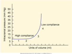
Herniation
-this can be caused by rapid changes in ICP
-if the body cannot manage the amount of blood/fluid going into the cranium, then herniation will occur
-this occurs when the brain goes out of the cranium and over the tentorium
^a fold of dura mater that covers the cerebellum and supports the occipital lobes of the cerebrum
-when it crosses over the tentorium, it creates an outpouching outside the cranial vault
Cerebral Blood Flow
-the brain needs oxygen and glucose to survive
^receives these things through cerebral blood flow
-the cerebral perfusion pressure (CPP) needs to be within 70 to 100
How do we calculate CPP?
- CPP = MAP - ICP
^any changes in blood pressure or ICP will cause changes in CPP
Practice Problem: If your MAP is 95 and the ICP is 15, then what is the CPP?
- CPP = MAP - ICP
- CPP = 95 - 15
- CPP = 80
How do we calculate MAP from a blood pressure?
2 x (DBP) + SBP / 3 = MAP
Practice Problem: If a patient has a blood pressure of 83/50, what would their MAP be?
- 2 x (50) + 83 / 3 = MAP
- 100 + 83 / 3 = MAP
- 183 / 3 = MAP
- MAP = 61 mmHg
What are the goals of treatment for increased ICP?
-improve CPP
-control ICP
*need to feed the brain and keep brainstem intact by maintaining ICP (delicate balance)
Three Major Concerns w/ Increased ICP
-hypoxia
-blood pressure
-cerebral (arterial) vasodilation
Autoregulation
-in a healthy brain, cerebral autoregulation controls CBF over a range of blood pressures
-cerebral (arterial) vessels dilate or constrict in response to pressure changes
Pressure Changes
-if BP is HIGH, cerebral blood vessels can constrict so that we don't get too much blood
-if BP is LOW, cerebral vessels can dilate so that the brain can be oxygenated and be well perfused
Impaired Autoregulation
-this occurs when the brain is injured (neurological insults often impair autoregulation)
-anything which increases HTN (coughing, sneezing, changes in HOB, suctioning, agitation) or causes cerebral vasodilation (hypoxia, hypercapnia, acidosis) increases CBF, thus INCREASING ICP
How does hypoxia, hypercapnia, and acidosis cause cerebral (arterial) vasodilation?
Hypoxia
-causes capillary leakage
-variations in the pressures causes leakage of the fluid from where it actually belongs to the outside of the vessels
-can cause vasodilation to try and get more O2 to the brain
Hypercapnia
-CO2 is a potent vasodilator, thus causes cerebral vasodilation
Acidosis
-impacts transport channels and effects the way electrolytes move
-when electrolytes don't move properly, it causes vasodilation
Notice!: A patient needs suctioning to avoid hypoxia. However, suctioning increases ICP. How do we balance?
-we will not perform sustained actions of suctioning or agitation
-let's think about this as well with a patient whom we are bathing
^don't keep them turned for long periods of time
^try using the side that doesn't have pain
Hypoxia
-between 30 to 180 seconds of oxygen deprivation, you may lose consciousness
-at the one-minute mark, the brain cells begin to die
-at three minutes, neurons suffer more extensive damage, and lasting damage becomes more likely
*this is why it is so important to act quickly
Blood Pressure Control
-this can be tricky
-an acutely injured brain often needs increased CPP to maintain perfusion
-relatively high BP may be necessary, but we don't want to increase CPP so much that it increases ICP or makes an existing brain bleed more extensive than it already is
Why is hypotension dangerous to the brain?
-the brain receives 20% of cardiac output and is responsible for 15% of the body's oxygen demand
-we have to perfuse the brain!
Cerebral Arterial Vasodilation
-increased PaCO2 is the most potent cerebral vasodilator
-this can ultimately lead to increased ICP
Hyperventilation
-decreases PaCO2 which causes cerebral vasoconstriction, thus decreasing CBF and ICP
^was a past intervention that was used
Treatment for Increased ICP
Sedatives and Analgesia
-these are good options if the patient becomes agitated or is in pain
Neuromuscular Blockade
-if a patient becomes extremely sensitive to agitation we can actually sedate and paralyze them
^DO NOT FORGET TO SEDATE
-this causes 0 movement, which decreases agitation, thus preventing increases in ICP
Diuretics
-mannitol, 3% NS, lasix
^mannitol: crosses the blood brain barrier
^3% NS: hypertonic saline; helps draw fluid out of the blood vessel and decreases cerebral edema
Decadron
-for tumors
^depends on size and aggressiveness
-works as an anti-inflammatory
Prevent and Tx Hyperthermia
-hyperthermia requires high amounts of oxygen consumption
Prevent and Tx Seizures
-can increase agitation
-requires high amounts of oxygen consumption
CSF Drainage
-manual way of helping the patient autoregulate
^we can decrease one the components
Controlled Hypothermia
-causes vasoconstriction and decreases cerebral blood flow
Nursing Care of a Patient w/ Increased ICP
-q 1 neuro checks
^do not FUDGE your assessments; be timely and accurate about this
-positioning HOB up, neck midline, and no knee bend
^facilitates drainage of the head
^if the head is tilted, it is obstructing blood flow to the head
^if we have flexed knees or hips, this can cause abdominal HTN
-prevent hypoxia/hypercapnia
-control hypo/hypertension
-prevent/tx hyperthermia
-prevent/tx seizures
^common practice is the administration of KEPPRA
-sedation/pain control
-paralytics
-No Valsalva
^increases ICP
^start patients on pretty aggressive bowel regimen (don't want to overdue it on the stool softeners; delicate balance)
^typically on senokot, colace, miralax
^if it progresses to worsening constipation, we use mag citrate or enemas
-plan activities to avoid prolonged increased ICP
Care of the Comatose Patient
-this patient is the most vulnerable and it is up to the nurse to guard them of all safety measures
-we need to maintain the dignity of the patient as well
^watch what you say!
-we need to also think of if the patient is possibly a candidate for organ donation, because now your care is affecting 5 to 7 patients
Cerebral Vascular Accident
-two types: ischemic and hemorrhagic
-common causes: HTN and emboli
Ischemic
-occlusive: have a clot that blocks blood flow distal from the clot
-thrombotic: caused by a narrowing blood vessel (MOST CVAs)
-embolic: where a thrombus has dislodged from its origin, travels, and blocks off an artery
What are the different causes of an embolic ischemic stroke?
Atrial Fibrillation
-when the atrium doesn't contract well, the blood can become static and clots can develop, break free, and travel to the brain
Patent Foramen Ovale
-clots can exchange through here as well
Atherosclerotic Plaques
-occur when they are on the carotid
-portions can break off and travel up to the brain
Hemorrhagic
-when the blood escapes from the blood vessels
-intracerebral hemorrhage (ICH): a collection of blood within the brain
-subarachnoid hemorrhage (SAH): generally aneurysms and AV malformations can cause these
^can rupture and cause bleeding in the brain
How do SAH manifest?
a profound headache described as the "Worst Headache Ever!"
What are some risk factors for CVAs?
-hypertension (HTN)
-insulin dependent diabetes melitus (IDDM)
-high cholesterol
*these mimic the risk factors of coronary artery disease
Pathophysiology of both CVAs
-occurs when there is interrupted perfusion to the brain (there is an issue with the pathway)
^ex 1. when we have a ruptured vessel, the blood cannot continue along its usual pathway, thus causing it to collect resulting in hypoperfusion
^ex 2. a clot will block off an area distal to the clot
-when there is not enough blood, we decrease oxygen to the brain, and this causes ischemia
^think back to MSE: if the brain is not getting enough oxygen, then we will see changes in their mental status
-if the brain is not receiving enough O2, then 2 million brain cells die per minute
Clinical Manifestations for Ischemic Strokes
Focal Deficits
-facial asymmetry
-hemiparesis: weakness of one side of the body, or part of it, due to an injury in the motor area of the brain
-hemiplegia: paralysis of one side of the body, or part of it, due to an injury in the motor area of the brain
-aphasia: inability to express oneself or to understand language
^receptive or expressive
-dysarthria: defects of articulation due to neurologic causes (difficulty speaking)
^usually will have marbled, garbled speech
-hemianopsia: blindness of half of the field of vision in one or both eyes
Hemianopsia
Helpful Tip
-if a patient is neglecting half of their plate they may be experiencing hemianopsia

Cincinatti Scale
Facial Droop
-have the patient smile
Arm Drift
-hold both hands out straight for 10 seconds
^they can have a pronator drift if the are having a stroke
Abnormal Speech
-have patient say, "You can't teach an old dog new tricks."
*an abnormality in any one of these three = 72% chance of a CVA
BEFAST
-balance
^CN 8
-eyes
^CN III, IV, VI
-face
^CNV, VII, IX
-arms
-speech
^CN XII: seeing if the tongue is midline, but also w/ articulation
-time
*she emphasized this for the exam
Clinical Manifestations of Hemorrhagic Stroke
-sudden, severe decrease in LOC
-remember, SAH has that SEVERE headache
-rapid increase in ICP
^this may cause the patient to lose LOC and airway quickly
Additional Information of Hemorrhagic Strokes
-a severe intracranial hemorrhage is almost always fatal
-arteriovenous malformations (AVM's) and aneurysms offer more hope, but 33% die even with aggressive treatment
If a patient loses their airway and LOC quickly due to hemorrhagic stroke, what should be implemented?
-intubation
-seizure tx
-control of BP
-etc.
Tissue Plasminogen Activator (TPA)
-clot dissolving medication
-only for ISCHEMIC
^if we gave this for hemorrhagic it would cause the patient to bleed out even more
What are the TPA rules?
-must be treated within 4.5 hours from the onset of symptoms
^must have a reliable witness to verify the onset of symptoms
-contraindicated if symptoms upon arising from sleep
^not sure when the onset of symptoms occurred
-must have a head CT completed prior to administration of TPA, in order to rule out a bleed
-if previous trauma or damage to the brain is shown on the CT, then TPA is withheld
If TPA is used, the nursing care involves careful monitoring for _________?
changes in LOC
^if we administer TPA and have a sudden loss of consciousness, then the ischemic could have flipped to a hemorrhagic
Treatment for Hemorrhagic Stroke
PREVENTION
-need to clip the aneurysm(s) and/or repair AVM(s)
-we want to find and tx before they rupture
Coils and Glue
-you can thread a catheter through the groin and start shooting in metal
-the body recognizes it as foreign, and it clots it off
Nimodipine
-Calcium Channel Blocker (CCB)
-works as a smooth muscle relaxer, but also decreases the incidence of vasospasm
^vasospasms irritate the blood vessels, cause ischemia, which can increase ICP
HHH Therapy
-Hypervolemic Hypertensive Hemodilution therapy
-we will see high rates of volume drive up blood pressures and cause dilution
-this dilution decreases coagulability, which decreases the chances of clots being formed
What do we need to be mindful of if a patient has an aneurysm clipped?
REBLEED
-we want to make sure that the patient is not having a rebleed because this can turn into a sentinel event
Airway
-how we get air into the lungs
-when we are talking about the airway, we are most concerned with patency
^in our patients who are in critical condition or even patients in emergent situations, one of the first things we will assess for is airway obstruction
-the tongue is a pretty good culprit of airway obstruction, especially in patients who have an altered level of consciousness
-a sure-fire way to maintain patency of the airway is through intubation
Endotracheal Tube (ETT)
-we can see that the endotracheal tube is placed within the airway
-the esophagus is on the posterior side and the trachea on the anterior
-the tube is a foot to a foot and a half long
-it passes through the mouth and then through the vocal cords
-a balloon is inflated to separate the upper and lower airway
^the balloon is not completely impervious to secretions, so we need to keep the HOB elevated
-our job is to make sure that the tube has the correct placement
^in the lungs, not the abdomen
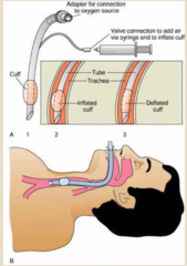
Breathing
-air travels through the trachea and then it makes its way to the alveoli
-carina: tracheal bifurcation for the right and left lungs
^the right main stem is a little bit straighter than the left one, so if the ETT is pushed too far then it can exclusively enter the right tract
^if this happens, only one lung is being oxygenated (this is why it is important to listen to the lung sounds of intubated patients)
Non-Rebreathers
-the reservoir bag needs to expand fully for it to be effective for the patient
-if we don't hook this up properly, the patient is essentially breathing into a paper bag
-the mask has a one-way valve, so when the patient exhales the valves are going to open which allows for the CO2 to escape
^when the patient takes a breath in, the valves close so they aren't breathing in CO2
-we NEVER want to withhold 100% oxygen within an emergency
^patients end up in critical situations when they are not oxygenated, so putting a patient on 100% initially is not a bad thing to do

Assessment of Respiratory Status
Level of Consciousness
-how are they behaving on the continuum of the mental status spectrum?
-are they restless and agitated, somnolent, or are they completely out of it?
Effects of Hypoxia (Initial v Progressive)
-initial: restless behavior
^example in class of a man cursing and ripping his BiPAP off due to hypoxia
-progressive: compensatory mechanisms stop working and CO2 rises, causing somnolent behavior
Respiratory Effort
-breathing takes work
-are they using their accessory muscles or are they gasping?
-normal breathing effort: ease to our breathing pattern
-if someone is working to breathe, regardless of how they are satting, they are still in respiratory distress
Vital Signs
-tachypnea
^if I'm not getting enough oxygen or if I'm trying to breathe off more CO2, then my RR will INCREASE
-tachycardia
^my HR is going to increase b/c part of the heart's job is to circulate oxygen throughout the body
Cyanosis
-this is a LATE sign of severe respiratory distress
What are the signs and symptoms of EARLY respiratory distress?
-anxiety, tachycardia, tachypnea
-working to breathe
-cyanosis suggest PROFOUND hypoxia
^this is typically a later sign, but it is still something that we should be assessing for in the early stages
-diaphoresis and somnolence indicate hypercapnia and respiratory acidosis
^elevated CO2 is associated with respiratory acidosis
*DO NOT wait for your patients to become cyanotic and diaphoretic
What are the different breath sounds?
-wheezes
-rhonci
-rales
Wheezes
-they are associated with musical tendencies
-they are not exclusively with inspiration or expiration, but they often occur with expiration
-associated with an inflammatory process
^inflammation of the airways makes it a little difficult for air to pass through, which causes that musical sound
Edema of the Airway
-1 mm of edema in the airway can cause significant changes to our breathing
1mm Effect on Infants
-this can be scary for our infants b/c their airways are already pretty small
-when 1mm is present, it can almost render the whole airway
1mm Effect on Adults
-it is not as severe as when it happens to infants, but nevertheless it is a narrowing of the airway
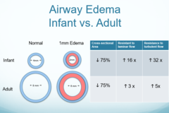
What lung sounds might we hear when the EARLY part of our airway is narrowed?
-if there is narrowing in the EARLY part of the airway, then you aren't going to have a lot of breath sounds in the distal portions of the lungs
^the nurse would report this as diminished breath sounds, or (if the inflammation is severe) the nurse may report absence which is not good
^if we are able to open this up, we are able to see better airway movement with our patients
Rhonci
-have a coarse sound to them
-associated with sputum accumulation
^sputum collects in the alveoli, which causes a barrier for gas exchange
-tx: get the patient to cough or if they are unable to cough, suction the patient's secretions out
Rales
-crackling sound
^nurses describes this as if you place hair by your ear and rub it together
-associated with fluid build-up in the lungs
^may be pulmonary edema
How do we treat each of the breath sounds listed?
Wheezes
-bronchodilator or steroid
^use a bronchodilator FIRST, wait 5 minutes and then use the steroid
Rhonchi
-have the patient cough or provide suctioning
Rales
-ask the patient to perform deep breathing exercises
-diuretics
Jaw Thrusting
this is a very quick and easy way to open the airway for a spontaneously breathing patient with decreased LOC, in order to relieve obstruction

What things are required for adequate cellular respiration?
1. Patent Airway
-this deals with the airway (A) portion of our ABCs
2. Adequate Ventilation
-this deals with the breathing (B) portion of our ABCs
-we need to ask ourselves
^are we delivering oxygenated air to the patient?
^are they receiving it properly?
3. Diffusion of Gases
-the alveoli are what facilitate gas exchange
^we need to optimize the situation so that gasses can diffuse through the alveolar membrane and be picked up by the pulmonary capillaries
4. Adequate Pulmonary Perfusion
-all of our pathologies are going to affect one or all of these things
^if one of these systems gets interrupted, then we are hampering the ability of oxygen to get to the cells
What is the main goal of cellular respiration?
getting oxygen to the cells
Shunts
-essentially wasted blood flow
-multiple types of shunts
^adequate blood flow, poor ventilation
^poor blood flow, adequate ventilation
^poor blood flow, poor ventilation
What are the effects of each type of shunt?
Adequate Blood Flow, Poor Ventilation
-ex. atelectasis or mucous plug
-this blocks the alveoli, which blocks the flow of air into the alveolar unit
-the blood is there, but there is an inability to release CO2 and receive O2
Poor Blood Flow, Adequate Ventilation
-ex. pulmonary embolism
-this blocks off pulmonary circulation
-we have no mechanisms for deoxygenated blood to come in contact with the alveoli
Poor Blood Flow, Poor Ventilation
-ex. pulmonary embolism with a blocked alveolar unit
What is ventilation and perfusion?
ventilation is air going in and perfusion is the blood that is going past the alveoli
Dead Space
-wasted ventilation
^adequate ventilation, but no perfusion
Pulse Oximetry
-a little bit of oxygen is dissolved in the plasma, but a lot of it is combined with hemoglobin
-saturation of arterial oxygen (SaO2) tells us the percentage of Hgb that is bound to oxygen
What does pulse oximetry tell us about CO2?
-doesn't tell us much
-we have End Tidal CO2 monitors
^special attachment on the ventilator tubing that tells us what a patient's PaCO2 is
^we then can adjust on the ventilator settings based on the patient's PaCO2
OHD Curve
-95% may seem good but we need to be cognizant of early signs of oxygen decline
-this could be a sign that our PaO2 is diminishing, causing a decrease in our oxygen saturation
-if you see declines in your patient, you need to address it

Normal ABG Values
-pH: 7.35 to 7.45
-PO2: 80 to 100
-CO2: 35 to 45
-HCO3: 22 to 26
Respiratory Alkalosis
-pH = > 7.45
-PaCO2 = < 35
-why?: fundamental cause is HYPERventilation
^with a high respiratory rate we are blowing off a lot of CO2
Example
-a patient may have a high respiratory rate because they are in pain or they have anxiety. If we tx the primary problem, then the second issue will fix itself
^if we address pain levels, we can slow RR
^if we address anxiety, we can slow RR