Chapter 4 - Peripheral Arterial
 Call Kai
 Learn
Learn Practice Test
 Spaced Repetition
 Match
Match Flashcards
 Knowt Play
1/114

There's no tags or description
Looks like no tags are added yet.
Name | Mastery | Learn | Test | Matching | Spaced | Call with Kai | Chat |
|---|
No analytics yet
Send a link to your students to track their progress
115 Terms
A normal spectral waveform of the brachial artery is
(A) Triphasic
(B) Biphasic
(C) Monophasic
(D) Both A and B
(D) Both A and B
The normal spectral waveform of the brachial artery is high resistant and can either be triphasic or biphasic. The only difference between the two is the third component of the waveform that can be lost with age due to decrease in the elasticity of the arteries.
Which of the following is a pathology specific to the upper extremity arteries?
(A) Thoracic outlet syndrome
(B) Carotid body tumor
(C) Raynaud’s syndrome
(D) Embolus
(A) Thoracic outlet syndrome
Thoracic outlet syndrome is specific to the upper extremities. Carotid body tumors only occur at the carotid bifurcation, and the other two choices can occur in the upper or lower extremities.
The type of flow pattern in which the velocities increase toward the center of the vessel and is found in most of the normal peripheral arteries is known as
(A) Turbulent
(B) Plug
(C) Laminar
(D) Occluded
(C) Laminar
Laminar blood flow is the normal flow found in most of the peripheral arteries and consists of a parabolic velocity profile which means that flow increases toward the center of the vessel and decreases toward the vessel walls
What blood vessel is most likely to have a high resistive waveform?
(A) Common femoral artery
(B) Common carotid artery
(C) Renal artery
(D) Hepatic artery
(A) Common femoral artery
The common femoral is a peripheral artery and should have high resistant flow in normal patients.
The dorsalis pedis artery is a continuation of which artery?
(A) Anterior tibial artery
(B) Posterior tibial artery
(C) Popliteal artery
(D) Common femoral artery
(A) Anterior tibial artery
The dorsalis pedis artery is a continuation of the anterior tibial artery .
All of the following are true regarding spectral broadening EXCEPT
(A) Spectral broadening is commonly associated with stenosis
(B) Spectral broadening refers to the filling of the spectral window with disturbed flow
(C) Spectral broadening refers to a decrease in bandwidth with disturbed flow
(D) Spectral broadening occurs with turbulent blood flow
(C) Spectral broadening refers to a decrease in bandwidth with disturbed flow
Spectral broadening does not refer to a decrease in bandwidth with disturbed flow. It refers to an increase in bandwidth with disturbed flow.
What is the first branch originating from the aortic arch that is only present on the right side?
(A) Subclavian artery
(B) Brachiocephalic artery
(C) Lateral thoracic artery
(D) Internal mammary artery
(B) Brachiocephalic artery
The brachiocephalic artery is the first branch origination from the thoracic aortic arch and is only present on the right side.
Which artery is a continuation of the superficial femoral artery as it passes through the adductor canal below the knee?
(A) Popliteal artery
(B) Tibioperoneal trunk
(C) Profunda femoris artery
(D) Posterior tibial artery
(A) Popliteal artery
The popliteal is a continuation of the superficial femoral artery as it passes through the adductor canal below the knee into the popliteal fossa.
Which artery runs along the medial aspect of the lower leg and courses posterior to the medial malleolus?
(A) Peroneal artery
(B) Anterior tibial artery
(C) Dorsalis pedis artery
(D) Posterior tibial artery
(D) Posterior tibial artery
The posterior tibial artery runs along the medial aspect of the lower leg and courses posterior to the medial malleolus.
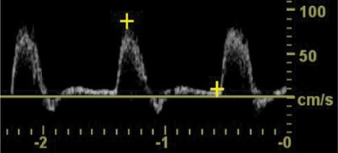
The waveform shown in Figure 4–47 is
(A) Monophasic
(B) Biphasic
(C) Triphasic
(D) Preocclusive
(C) Triphasic
The waveform shown in Figure 4–47 is a typical triphasic waveform.

The waveform shown in Figure 4–48 was obtained from the superficial femoral artery and demonstrates
(A) Increased peak systolic velocity
(B) A triphasic waveform
(C) Laminar flow
(D) Flow reversal
(A) Increased peak systolic velocity
The peak systolic velocity is increased in this monophasic waveform. Also note the spectral broadening present. Not enough information is given to determine if there is reversal of flow or not; based on the spectral Doppler the flow is all going in one direction

The waveform shown in Figure 4–49 suggests
(A) Normal laminar arterial flow
(B) Poststenotic turbulent flow
(C) Multi-phasic flow
(D) Distal occlusion
(B) Poststenotic turbulent flow
The waveform shown in Figure 4–49 is a classic poststenotic waveform demonstrating an ill-defined border, spectral broadening and some reversal of flow.

The waveform shown in Figure 4–50 suggest that
(A) There is an occlusion just proximal to this area
(B) There is an occlusion just distal to this area
(C) There is a critical stenosis at this area
(D) This is a normal waveform
(B) There is an occlusion just distal to this area
The waveform shown in Figure 4–50 is a monophasic waveform with no diastolic flow and diminished peak systolic flow suggesting a distal occlusion.
A patient is sent to your laboratory to have his lower extremity bypass graft evaluated with duplex. While performing the examination you notice that the velocity at the proximal anastomosis is 80 cm/s. Y ou continue the examination and obtain a pulse wave Doppler signal about 1 cm distal to the proximal anastomosis and you get a velocity of 210 cm/s. What do these findings suggest?
(A) Normal flow present within this bypass
(B) 1% to 19% narrowing of this bypass
(C) 20% to 49% narrowing of this bypass
(D) 50% to 99% narrowing of this bypass
(D) 50% to 99% narrowing of this bypass
A doubling in peak systolic velocity between adjacent segments is consistent with a hemodynamically significant stenosis greater than 50%. The velocities obtained within these segments more than doubles going from 80 to 210 cm/s.
The following is a type of lower extremity bypass
(A) Brachial artery to cephalic vein
(B) Common femoral artery to popliteal reversed saphenous vein
(C) Cephalic vein to brachial vein
(D) Femoral vein to internal iliac vein
(B) Common femoral artery to popliteal reversed saphenous vein
Reversed vein grafts are common lower extremity bypasses. None of the other choices are lower extremity bypass grafts.
Which statement is true about the symptom of claudication?
(A) Exercise-induced pain caused by too much blood flow to the legs
(B) Varies each time regarding distance the patient is able to walk
(C) Always relieved by rest
(D) Only occurs in the calves
(C) Always relieved by rest
Claudication is pain in the lower extremities as a result of hypoxia that is reproducible and induced by exercise. After a patient rests, they are then able to walk the same distance before the pain reoccurs
Rest pain can be relieved by
(A) Standing
(B) Elevating legs
(C) Exercise
(D) Taking deep breaths
(A) Standing
Ischemic rest pain can be relieved by lowering the legs dependently , such as standing or dropping the legs off the side of the bed. The arterial inflow is so poor, gravity is needed to get the blood to the feet.
The peak systolic blood pressure in an area of a limb distal to a significant obstruction or stenosis
(A) Will increase
(B) Will decrease
(C) Will remain the same
(D) Will double
(B) Will decrease
The peak systolic blood pressure in an area of a limb distal to a significant obstruction or stenosis will decrease.
Which is the most common cause of peripheral arterial disease of the lower extremities?
(A) Arteritis
(B) Arterial spasm
(C) Atherosclerosis
(D) Embolism
(C) Atherosclerosis
Atherosclerosis is the most common cause of peripheral arterial disease of the lower extremities.
The most common site of atherosclerosis in the lower extremities is
(A) The bifurcation of the common iliac arteries
(B) The origin of the profunda femoris artery
(C) The trifurcation of the tibial arteries
(D) The distal superficial femoral artery through the adductor canal
(D) The distal superficial femoral artery through the adductor canal
The most common site of atherosclerosis in the lower extremities is the distal superficial femoral artery through the adductor canal.
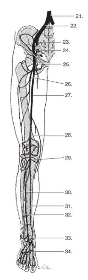
Match the structures in Fig. 4–51 with the names of the arteries - #21
(A) Common femoral artery
(B) Popliteal artery
(C) Aorta
(D) Superficial femoral artery
(E) Internal iliac artery
(F) Profunda femoris artery
(G) External iliac artery
(H) Common iliac artery
(I) Tibioperoneal trunk
(J) Dorsalis pedis artery
(K) Anterior tibial artery
(L) Peroneal artery
(M) Plantar arch
(N) Posterior tibial artery
(C) Aorta
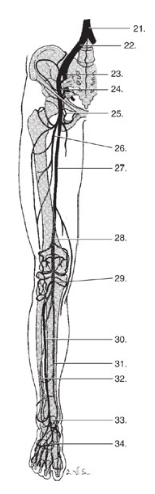
Match the structures in Fig. 4–51 with the names of the arteries - #22
(A) Common femoral artery
(B) Popliteal artery
(C) Aorta
(D) Superficial femoral artery
(E) Internal iliac artery
(F) Profunda femoris artery
(G) External iliac artery
(H) Common iliac artery
(I) Tibioperoneal trunk
(J) Dorsalis pedis artery
(K) Anterior tibial artery
(L) Peroneal artery
(M) Plantar arch
(N) Posterior tibial artery
(H) Common iliac artery

Match the structures in Fig. 4–51 with the names of the arteries - #23
(A) Common femoral artery
(B) Popliteal artery
(C) Aorta
(D) Superficial femoral artery
(E) Internal iliac artery
(F) Profunda femoris artery
(G) External iliac artery
(H) Common iliac artery
(I) Tibioperoneal trunk
(J) Dorsalis pedis artery
(K) Anterior tibial artery
(L) Peroneal artery
(M) Plantar arch
(N) Posterior tibial artery
(E) Internal iliac artery

Match the structures in Fig. 4–51 with the names of the arteries - #24
(A) Common femoral artery
(B) Popliteal artery
(C) Aorta
(D) Superficial femoral artery
(E) Internal iliac artery
(F) Profunda femoris artery
(G) External iliac artery
(H) Common iliac artery
(I) Tibioperoneal trunk
(J) Dorsalis pedis artery
(K) Anterior tibial artery
(L) Peroneal artery
(M) Plantar arch
(N) Posterior tibial artery
(G) External iliac artery
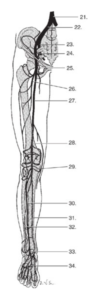
Match the structures in Fig. 4–51 with the names of the arteries - #25
(A) Common femoral artery
(B) Popliteal artery
(C) Aorta
(D) Superficial femoral artery
(E) Internal iliac artery
(F) Profunda femoris artery
(G) External iliac artery
(H) Common iliac artery
(I) Tibioperoneal trunk
(J) Dorsalis pedis artery
(K) Anterior tibial artery
(L) Peroneal artery
(M) Plantar arch
(N) Posterior tibial artery
(A) Common femoral artery

Match the structures in Fig. 4–51 with the names of the arteries - #26
(A) Common femoral artery
(B) Popliteal artery
(C) Aorta
(D) Superficial femoral artery
(E) Internal iliac artery
(F) Profunda femoris artery
(G) External iliac artery
(H) Common iliac artery
(I) Tibioperoneal trunk
(J) Dorsalis pedis artery
(K) Anterior tibial artery
(L) Peroneal artery
(M) Plantar arch
(N) Posterior tibial artery
(F) Profunda femoris artery

Match the structures in Fig. 4–51 with the names of the arteries - #27
(A) Common femoral artery
(B) Popliteal artery
(C) Aorta
(D) Superficial femoral artery
(E) Internal iliac artery
(F) Profunda femoris artery
(G) External iliac artery
(H) Common iliac artery
(I) Tibioperoneal trunk
(J) Dorsalis pedis artery
(K) Anterior tibial artery
(L) Peroneal artery
(M) Plantar arch
(N) Posterior tibial artery
(D) Superficial femoral artery
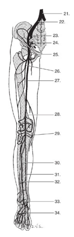
Match the structures in Fig. 4–51 with the names of the arteries - #28
(A) Common femoral artery
(B) Popliteal artery
(C) Aorta
(D) Superficial femoral artery
(E) Internal iliac artery
(F) Profunda femoris artery
(G) External iliac artery
(H) Common iliac artery
(I) Tibioperoneal trunk
(J) Dorsalis pedis artery
(K) Anterior tibial artery
(L) Peroneal artery
(M) Plantar arch
(N) Posterior tibial artery
(B) Popliteal artery

Match the structures in Fig. 4–51 with the names of the arteries - #29
(A) Common femoral artery
(B) Popliteal artery
(C) Aorta
(D) Superficial femoral artery
(E) Internal iliac artery
(F) Profunda femoris artery
(G) External iliac artery
(H) Common iliac artery
(I) Tibioperoneal trunk
(J) Dorsalis pedis artery
(K) Anterior tibial artery
(L) Peroneal artery
(M) Plantar arch
(N) Posterior tibial artery
(I) Tibioperoneal trunk
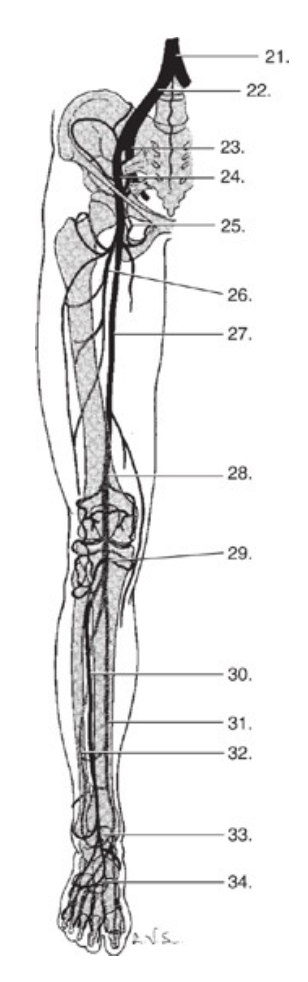
Match the structures in Fig. 4–51 with the names of the arteries - #30
(A) Common femoral artery
(B) Popliteal artery
(C) Aorta
(D) Superficial femoral artery
(E) Internal iliac artery
(F) Profunda femoris artery
(G) External iliac artery
(H) Common iliac artery
(I) Tibioperoneal trunk
(J) Dorsalis pedis artery
(K) Anterior tibial artery
(L) Peroneal artery
(M) Plantar arch
(N) Posterior tibial artery
(K) Anterior tibial artery

Match the structures in Fig. 4–51 with the names of the arteries - #31
(A) Common femoral artery
(B) Popliteal artery
(C) Aorta
(D) Superficial femoral artery
(E) Internal iliac artery
(F) Profunda femoris artery
(G) External iliac artery
(H) Common iliac artery
(I) Tibioperoneal trunk
(J) Dorsalis pedis artery
(K) Anterior tibial artery
(L) Peroneal artery
(M) Plantar arch
(N) Posterior tibial artery
(N) Posterior tibial artery

Match the structures in Fig. 4–51 with the names of the arteries - #32
(A) Common femoral artery
(B) Popliteal artery
(C) Aorta
(D) Superficial femoral artery
(E) Internal iliac artery
(F) Profunda femoris artery
(G) External iliac artery
(H) Common iliac artery
(I) Tibioperoneal trunk
(J) Dorsalis pedis artery
(K) Anterior tibial artery
(L) Peroneal artery
(M) Plantar arch
(N) Posterior tibial artery
(L) Peroneal artery

Match the structures in Fig. 4–51 with the names of the arteries - #33
(A) Common femoral artery
(B) Popliteal artery
(C) Aorta
(D) Superficial femoral artery
(E) Internal iliac artery
(F) Profunda femoris artery
(G) External iliac artery
(H) Common iliac artery
(I) Tibioperoneal trunk
(J) Dorsalis pedis artery
(K) Anterior tibial artery
(L) Peroneal artery
(M) Plantar arch
(N) Posterior tibial artery
(J) Dorsalis pedis artery

Match the structures in Fig. 4–51 with the names of the arteries - #34
(A) Common femoral artery
(B) Popliteal artery
(C) Aorta
(D) Superficial femoral artery
(E) Internal iliac artery
(F) Profunda femoris artery
(G) External iliac artery
(H) Common iliac artery
(I) Tibioperoneal trunk
(J) Dorsalis pedis artery
(K) Anterior tibial artery
(L) Peroneal artery
(M) Plantar arch
(N) Posterior tibial artery
(M) Plantar arch
An obese patient is sent to your laboratory to be evaluated for arterial disease of the lower extremities. When performing their PVR and segmental pressure study you notice that their thigh PVR waveform indicates moderate disease but the pressure index at this level is 1.14.
How do you explain this?
(A) The disease is not severe enough to lower the pressure
(B) The technologist had to have made a mistake
(C) It is most likely the result of cuff artifact
(D) This is fine because a pressure of 1.14 is in the range of moderate disease
(C) It is most likely the result of cuff artifact
Pulse volume recordings in general are more accurate than segmental pressures because they are not affected by the limitations of segmental pressures such as cuff artifact or calcified vessels. An obese patient will most likely have falsely elevated pressures due to the cuff size being too small for the limb. In an obese patient with underlying peripheral arterial disease these elevated pressures may appear to be at a normal level. If the cuff size were appropriate for the limb in this same patient it would result in a much lower pressure.
If a patient has a steal from an A VF graft with no underlying atherosclerotic disease the pre-graft compression digital PPG waveforms obtained during non-invasive arterial testing would show
(A) Normal blood flow to the digits
(B) Reduced blood flow to the digits
(C) No difference in blood flow to the digits
(D) Increased blood flow to the digits
(B) Reduced blood flow to the digits
The pre-graft compression study in a patient with an A VF steal will have reduced blood flow to the arm and hand because the fistula is stealing that blood flow. When the graft is manually compressed during the post study there should be an increase in blood flow in order to confirm the diagnosis of a steal.
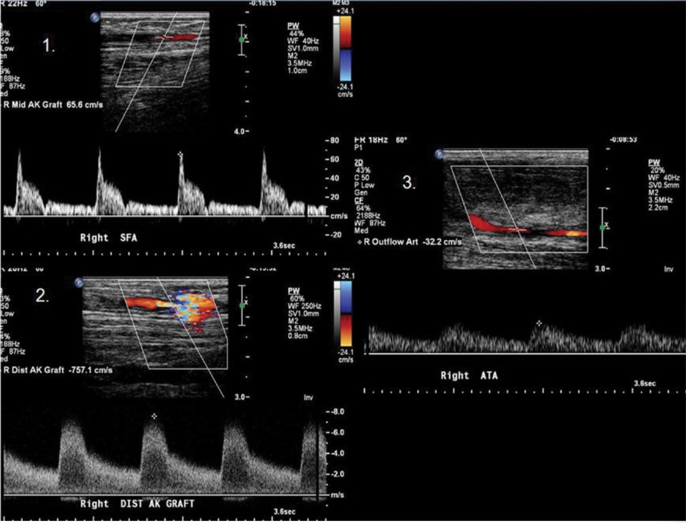
The images in Figure 4–52 were obtained from a patient in order 1, 2, and 3 from proximal to distal in a lower extremity bypass graft. What do these findings suggest?
(A) Normal
(B) 1% to 19% stenosis
(C) 20% to 49% stenosis
(D) 50% to 99% stenosis
(D) 50% to 99% stenosis
Spectral Doppler waveform number two shows a focal increase in velocity compared to number one resulting in a Vr > 3 which is consistent with a >75% stenosis. Spectral Doppler waveform number three demonstrates tardus parvus flow supplying further evidence of a critical stenosis.

The waveform shown in Figure 4–53 was obtained in the lower extremity. How is this waveform best described?
(A) Normal monophasic waveform of the lower extremity
(B) Abnormal monophasic waveform found distal to a stenosis
(C) Abnormal monophasic waveform found proximal to a stenosis
(D) Abnormal monophasic waveform found at a stenosis
(B) Abnormal monophasic waveform found distal to a stenosis
The waveform shown in Figure 4–53 is a classic tardus parvus waveform that is found distal to a significant stenosis.
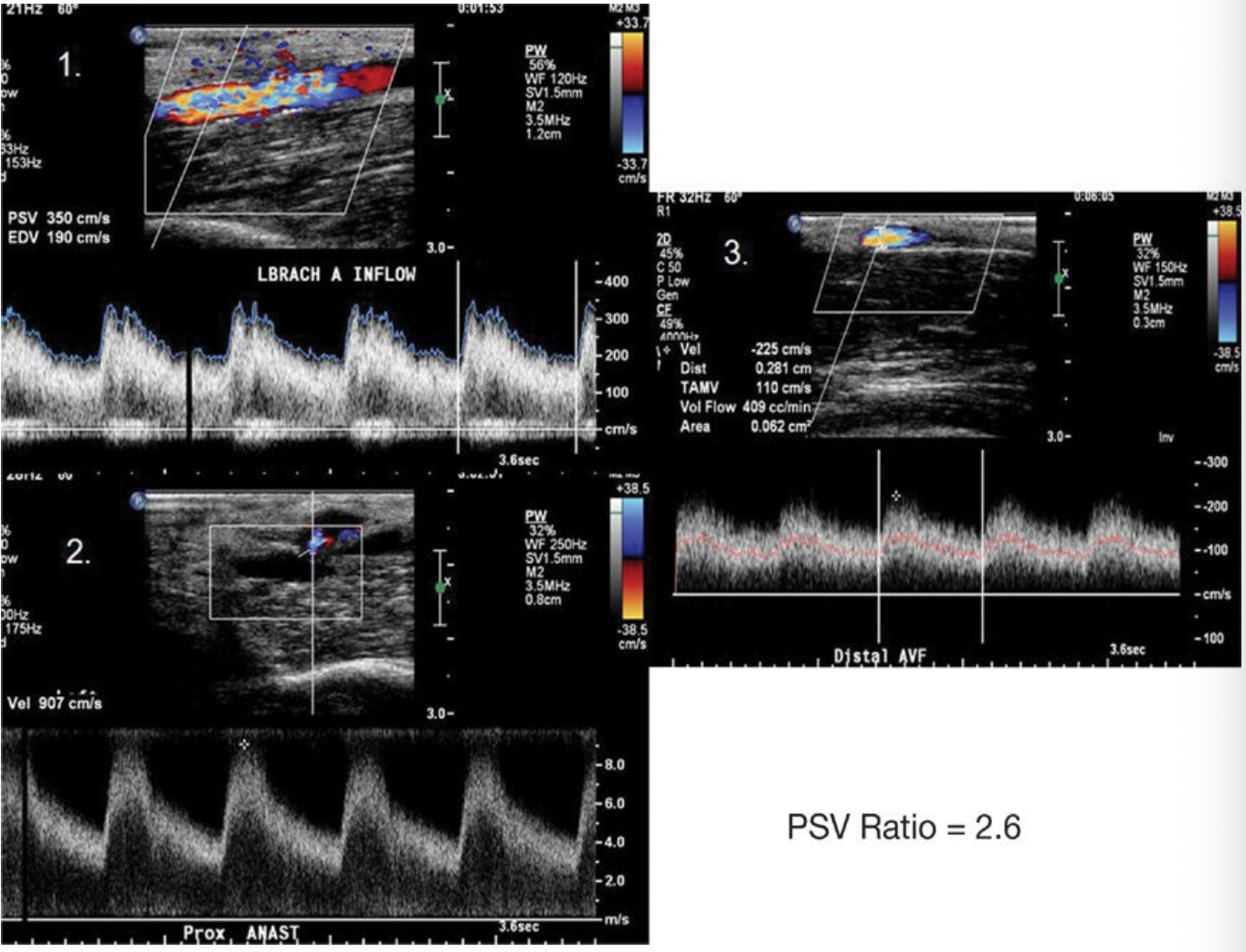
The images in Figure 4–54 were obtained from a patient in order 1, 2, and 3 from proximal to distal in an upper extremity arterial venous fistula. What is your diagnosis based on these findings?
(A) Normal
(B) <50% stenosis at the anastomosis
(C) >50% stenosis at the anastomosis
(D) Occlusion at the anastomosis
(C) >50% stenosis at the anastomosis
There is an increase in velocity at the anastomosis site demonstrated in the spectral Doppler waveform number two resulting in a PSV ratio >2 which is consistent with a >50% stenosis at the anastomosis site. The spectral Doppler waveform number three demonstrates decreased volume flow (<500 mL/min) as a result of the stenosis.

The spectral Doppler waveform shown in Figure 4–55 is characteristic for which abnormality?
(A) AVF steal
(B) Stenosis
(C) Pseudoaneurysm
(D) Vasospasm
(C) Pseudoaneurysm
The waveform shown in Figure 4–55 is a classic to-and-fro waveform that is found within the neck of a pseudoaneurysm.
What is the procedure in which a balloon-tipped catheter is advanced to the level of a focal stenotic lesion under fluoroscopic guidance and a balloon is inflated to push the plaque up against the walls of the artery in an effort to restore normal blood flow to the limb?
(A) CT angiography
(B) Angioplasty
(C) Digital subtraction angiography
(D) MR angiography
(B) Angioplasty
Angioplasty is the procedure in which a balloon-tipped catheter is inflated within a stenotic lesion in an attempt to restore normal blood flow to an area.
The brachial artery bifurcates just below the antecubital fossa into which two arteries?
(A) Subclavian and axillary arteries
(B) Deep and superficial palmar arteries
(C) Brachiocephalic and subclavian arteries
(D) Radial and ulnar arteries
(D) Radial and ulnar arteries
The brachial artery bifurcates just below the antecubital fossa into the radial and ulnar arteries. The radial artery runs along the lateral side of the forearm while the ulnar artery runs along the medial side.
Noninvasive physiologic testing that involves placing a PPG on one of the digits of each hand recording waveforms and pressures while the arms are moved through a series of positions is used to evaluate for which syndrome?
(A) Subclavian steal syndrome
(B) Thoracic outlet syndrome
(C) Raynaud’s syndrome
(D) Fibromuscular dysplasia
(B) Thoracic outlet syndrome
Noninvasive testing for thoracic outlet syndrome involves performing physiologic testing with the use of pulse volume recordings or photoplethysmography while moving the patient’s arms in several positions in an attempt to reduce the perfusion to the hand. The symptomatic position should be attempted; however, there are many common positions used during this examination such as the Adson maneuver with arms abducted out to sides, the costoclavicular maneuver with chest pushed forward and shoulders back, military position with elbow pointing to the rear and arms almost upright with palms facing forward, the hands straight up 180 degrees, and the arms straight out and abducted toward the rear.
In a blood vessel with a nonhemodynamically significant stenosis, if the diameter decreases what happens to the flow volume?
(A) Flow volume increases
(B) Flow volume decreases
(C) Flow volume remains unchanged
(D) Volume increases but velocity decreases
(C) Flow volume remains unchanged
As the diameter of a blood vessel decreases, the flow remains a constant as long as the stenosis is not hemodynamically significant
Which statement is accurate related to a vessel that has a hemodynamically significant stenosis?
(A) There is an increased pressure gradient across the segment
(B) There is a decreased pressure gradient across the segment
(C) There is an increase in velocity at area of stenosis
(D) There is a decrease in velocity at area of stenosis
(E) Both A and C
(F) Both A and D
(E) Both A and C
In a normal vessel the velocity of blood flow and the pressure do not change significantly . When a hemodynamically significant stenosis is present within an artery there is an increase in the pressure gradient across the segment as well as an increase in the velocity of blood flow within the stenosis. The turbulent flow that exists past the stenosis caused the pressure distal to the stenosis to drop resulting in a pressure that is now lower than the pressure proximal to the stenosis. The difference between these two pressures is what is known as a pressure gradient and is what increases with a stenosis due to the change in pressures.
Which surgical procedure has the highest potential for problems such as arteriovenous fistulas and retained valves?
(A) Aorta-femoral bypass
(B) In-situ saphenous vein bypass
(C) Reversed saphenous vein bypass
(D) Synthetic bypass
(B) In-situ saphenous vein bypass
An in-situ saphenous vein bypass is created by leaving the native vein in its normal anatomical position, removing the internal valves and ligating the accessory branches, and finally attaching each end to the proximal and distal native arteries. This type of surgical bypass has the highest potential for a missed branch and retained valves because the vein is not being completely removed and reversed, unlike the reversed saphenous vein bypass in which the vales do not have to be removed. There would be no concern for missed branches or retained valves with an artificial bypass graft.

The waveform shown in Figure 4–56 is a characteristic waveform for which condition?
(A) Raynaud’s syndrome
(B) Thoracic outlet syndrome
(C) Takayasu’s arteritis
(D) Moyamoya disease
(A) Raynaud’s syndrome
The waveform shown in Figure 4–56 is a classic peaked pulse waveform where the dicrotic notch is higher than normal and is commonly seen in patients with Raynaud’s disease.
An abnormal connection between an artery and a vein that can occur spontaneously or as a result of trauma is known as
(A) Anastomosis
(B) Pseudoaneurysm
(C) Arteriovenous fistula
(D) Dissection
(C) Arteriovenous fistula
An arteriovenous fistula occurs when there is an abnormal connection between a native artery and vein resulting in a high volume jet of flow passing between both systems. They can occur spontaneously or traumatically .
What is the name of the most widely utilized ratio used to evaluate overall perfusion to the lower extremity arteries?
(A) Resistive index
(B) Pulsatility index
(C) Ankle-brachial index
(D) Pressure gradient index
(C) Ankle-brachial index
The ankle-brachial index or ABI is the most widely utilized ratio used to evaluate overall perfusion to the lower extremity arteries and is calculated by taking the ankle pressure and dividing it by the higher of the two brachial pressures.
Which of the following may be used to diagnosis the severity of a stenotic lesion with duplex?
(A) Peak systolic velocity
(B) Resistive index
(C) Pulsatility index
(D) Ankle-brachial index
(A) Peak systolic velocity
Peak systolic velocity , end diastolic velocity , and peak systolic velocity ratios are all used to determine the severity of a stenosis. RI and PI are indices used to quantify the distal bed. The ankle- brachial index is not performed with duplex sonography .
All of the following are physical findings that can be associated with obstructive arterial disease of the lower extremities EXCEPT
(A) Shiny tight appearance to the skin
(B) Hair loss to the foot
(C) Ulcer on the heel
(D) Palpable pulse (grade 3)
(D) Palpable pulse (grade 3)
A palpable grade 3 pulse indicates normal perfusion to a lower extremity therefore is not an indication of an obstruction.
Which of the following is NOT a common risk factor associated with arterial occlusive disease?
(A) Tobacco use
(B) Hypertension
(C) Diabetes
(D) Hypolipidemia
(D) Hypolipidemia
Tobacco, hypertension, diabetes, and hyperlipidemia are all risk factors for cardiovascular disease. Hypolipidemia is a decrease in the amount of blood lipids.
While reviewing the segmental pressures on a patient you notice that the patient has a right thigh pressure of 155 mm Hg and a right calf pressure of 90 mm Hg. The patient’s brachial pressure is 120 mm Hg. Based on just these findings, what level of disease does this patient have?
(A) Aortoiliac
(B) Femoral-popliteal
(C) Distal small vessel
(D) Multilevel
(B) Femoral-popliteal
A pressure gradient >30 mm Hg indicates that there is disease present at or above the cuff with the lower pressure. A pressure gradient >30 mm Hg between the thigh and calf cuffs indicates femoral popliteal disease.
What is the first component that is lost on a PVR waveform when arterial disease exists?
(A) Anacrotic limb
(B) Catacrotic limb
(C) Dicrotic limb
(D) Bicrotic limb
(C) Dicrotic limb
The dicrotic limb is the first component that is lost in a PVR waveform when any arterial disease is present.
Which finding suggests that an arterial venous fistula is mature and ready for hemodialysis use?
(A) Diameter of the draining vein ≥ 4 mm
(B) PSV ratio <4
(C) Flow volume ≥350 mL/min
(D) Continuous flow in the venous side of the fistula
(A) Diameter of the draining vein ≥ 4 mm
In order for an arteriovenous fistula to be considered mature and ready for hemodialysis use by duplex the draining vein should be at least 4 mm in size, there should be no narrowing at the anastomosis site that is confirmed by a ratio of <2, and the volume flow should be at least 500 mL/min (ideally the volume flow should be >800 mL/min).

Figure 4–57 shows a resting noninvasive arterial examination of a 76-year-old patient with intermittent claudication bilaterally. Based on the examination data below what is the extent of this patient’s disease?
(A) Aorta and bilateral severe iliac occlusive disease
(B) Right mild femoral popliteal and mild tibial artery occlusive disease and left moderate iliac occlusive disease
(C) Right mild iliac and moderate femoral popliteal occlusive disease and left mild femoral popliteal occlusive disease
(D) Right severe iliac and femoral popliteal occlusive disease and left mild iliac occlusive disease
(C) Right mild iliac and moderate femoral popliteal occlusive disease and left mild femoral popliteal occlusive disease
On the right there is a decrease in the high thigh pressure (Should be 30 mm Hg greater than the higher brachial with the four-cuff technique) with slight rounding and decrease in amplitude of the PVR waveform consistent with mild iliac disease. There is also a decrease in pressure of more than 30 mm Hg between the low thigh and calf cuffs with a dampened PVR waveform at the calf level consistent with moderate femoral popliteal disease. On the left there is a decrease in pressure of more than 30 mm Hg between the low thigh and the calf cuff with an absent dicrotic notch on the PVR waveform consistent with mild femoral popliteal disease.

Figure 4–58 shows segmental pressures and pulse volume recording waveforms from a patient with ischemic type symptoms of the left hand. Based on this data what is the location of the disease present?
(A) Left subclavian and/or axillary arteries
(B) Left radial artery
(C) Left ulnar artery
(D) Left digits
(A) Left subclavian and/or axillary arteries
Figure 4–61 demonstrates a 60 mm Hg pressure gradient between the two brachial cuffs as well as a dampened PVR waveform at the same level. A pressure gradient >15 mm Hg between arms or adjacent cuffs indicates that there is disease present at or above the cuff with the lower pressure. The 60 mm Hg pressure gradient between arms in this patient indicates that there is disease present within the left subclavian and/or axillary arteries

Figure 4–59 shows a segmental pressure and pulse volume recording examination. Based on these findings what level of disease can be ruled out?
(A) Left femoral popliteal disease
(B) Left iliac disease
(C) Left small vessel disease
(D) Aortoiliac disease
(D) Aortoiliac disease
Aortoiliac disease can be ruled out in this patient because the right high thigh systolic pressure and PVR waveform are normal. If there were significant aorta disease it would affect both the right and left inflow to the lower extremities.

Based on the analog Doppler waveform in Figure 4–60 what level of disease is present on the patient’s right side?
(A) Right iliac disease
(B) Right femoral popliteal disease
(C) Right tibial disease
(D) There is no disease present on the right side
(C) Right tibial disease
The analog Doppler waveforms are multiphasic and bidirectional down to the popliteal level. Below that level in the posterior tibial and dorsalis pedis arteries the waveforms are monophasic and unidirectional indicating tibial disease
Blue toes are an indication of which type of acute arterial obstruction?
(A) Popliteal entrapment
(B) Raynaud’s syndrome
(C) Compartment syndrome
(D) Embolus
(D) Embolus
Blue toes are a classic sign of an embolism that traveled through the blood steam to end up lodged within the smaller arteries of the digits
A reversed saphenous vein bypass graft involves
(A) Harvesting the saphenous vein, removing the valves, and connecting to the arteries in its normal anatomical orientation
(B) Leaving the saphenous vein in its normal anatomical position, removing valves, ligating any branches, and connecting to the arteries
(C) Harvesting the saphenous vein, reversing its normal anatomical orientation, and connecting to the arteries
(D) Harvesting the saphenous vein, reversing its normal anatomical orientation, removing the valves, and connecting to the arteries
(C) Harvesting the saphenous vein, reversing its normal anatomical orientation, and connecting to the arteries
A reversed saphenous vein graft involves harvesting the saphenous vein, reversing its normal anatomical orientation and connecting to the arteries. The anatomical reversing of the vein eliminates the need to remove the internal valves making them open in the correct direction of flow.
The analog Doppler signal distal to an area of severe stenosis in the lower extremities will likely be
(A) Triphasic
(B) Monophasic
(C) Biphasic
(D) Aphasic
(B) Monophasic
A normal analog Doppler signal within a peripheral artery should be triphasic or biphasic with bidirectional flow because of the distal resistance that exists normally in a peripheral arterial system. When significant disease is present the distal arterioles dilate resulting in a decrease in resistance changing the waveform to monophasic with flow continuous in one direction.
A normal toe pressure is ______ of the higher brachial pressure.
(A) 10% to 30%
(B) 40% to 60%
(C) 60% to 80%
(D) Equal
(C) 60% to 80%
A normal toe pressure is 60% to 80% of the higher brachial pressure.

Match the structures in Fig. 4–61 with the names of the arteries - #64
(A) Radial artery
(B) Common carotid artery
(C) Ulnar artery
(D) Innominate artery
(E) Brachial artery
(F) Axillary artery
(G) Subclavian artery
(D) Innominate artery
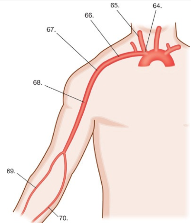
Match the structures in Fig. 4–61 with the names of the arteries - #65
(A) Radial artery
(B) Common carotid artery
(C) Ulnar artery
(D) Innominate artery
(E) Brachial artery
(F) Axillary artery
(G) Subclavian artery
(B) Common carotid artery
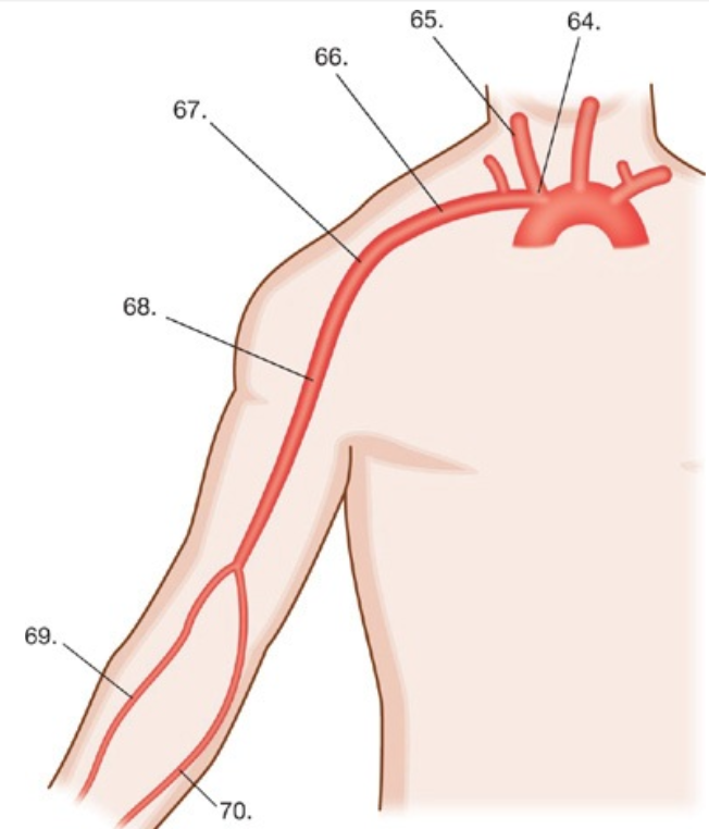
Match the structures in Fig. 4–61 with the names of the arteries - #66
(A) Radial artery
(B) Common carotid artery
(C) Ulnar artery
(D) Innominate artery
(E) Brachial artery
(F) Axillary artery
(G) Subclavian artery
(G) Subclavian artery

Match the structures in Fig. 4–61 with the names of the arteries - #67
(A) Radial artery
(B) Common carotid artery
(C) Ulnar artery
(D) Innominate artery
(E) Brachial artery
(F) Axillary artery
(G) Subclavian artery
(F) Axillary artery
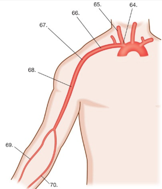
Match the structures in Fig. 4–61 with the names of the arteries - #68
(A) Radial artery
(B) Common carotid artery
(C) Ulnar artery
(D) Innominate artery
(E) Brachial artery
(F) Axillary artery
(G) Subclavian artery
(E) Brachial artery

Match the structures in Fig. 4–61 with the names of the arteries - #69
(A) Radial artery
(B) Common carotid artery
(C) Ulnar artery
(D) Innominate artery
(E) Brachial artery
(F) Axillary artery
(G) Subclavian artery
(A) Radial artery
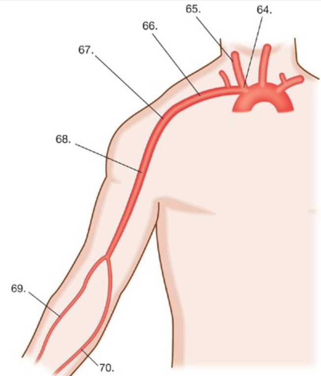
Match the structures in Fig. 4–61 with the names of the arteries - #70
(A) Radial artery
(B) Common carotid artery
(C) Ulnar artery
(D) Innominate artery
(E) Brachial artery
(F) Axillary artery
(G) Subclavian artery
(C) Ulnar artery
When using the four-cuff technique during a segmental pressure examination which of the following is consistent with a normal finding?
(A) The high thigh pressure is 30 mm Hg higher than the higher brachial pressure
(B) The high thigh pressure is equal to the higher brachial pressure
(C) The high thigh pressure is 30 mm Hg lower than the brachial pressure
(D) The high thigh pressure is 30 mm Hg higher than the lower brachial pressure
(A) The high thigh pressure is 30 mm Hg higher than the higher brachial pressure
When using the four-cuff technique during a segmental pressure examination the high thigh pressure is typically elevated due to the size of the limb. This higher pressure is reproducible and should be at least 30 mm Hg greater than the higher brachial pressure to be considered normal.
In relation to arteries and veins, which statement is true?
(A) Arteries have thinner walls than veins
(B) Arteries have internal valves
(C) Arteries have a much higher internal pressure
(D) Arteries are more readily collapsible
(C) Arteries have a much higher internal pressure
Both arteries and veins have three distinct layers to their vessel walls. The innermost layer is known as the intima, the middle layer is the media, and the outer layer is the adventitia. Even though they have the same three layers, arterial walls are thicker than vein walls and have a much higher internal pressure than the veins as well. In addition to these differences, veins also have internal valves that help promote flow back to the heart that are not present in the arteries. Veins should be easily collapsible, while the rigid walls of the arteries make them much harder to compress.
What type of pulse wave waveform is characteristic for the diagnosis of a pseudoaneurysm?
(A) Poststenotic waveform within neck of pseudoaneurysm
(B) Triphasic waveform within the pseudoaneurysm
(C) Monophasic waveform within the neck of the pseudoaneursym
(D) To-and-fro waveform within the neck of the pseudoaneursym
(D) To-and-fro waveform within the neck of the pseudoaneursym
A to-and-fro waveform within the neck of the pseudoaneursym is a classic finding used for the diagnosis.
Which statement is accurate related to blood pressure cuff artifact?
(A) If the width of the blood pressure cuff is <20% the diameter of the limb it can result in a falsely lowered peak systolic pressure
(B) If the width of the blood pressure cuff is >20% the diameter of the limb it can result in a falsely elevated peak systolic pressure
(C) If the width of the blood pressure cuff is <20% the diameter of the limb it can result in a falsely elevated peak systolic pressure
(D) Blood pressure cuff size has no effect on peak systolic pressure
(C) If the width of the blood pressure cuff is <20% the diameter of the limb it can result in a falsely elevated peak systolic pressure
Cuff artifact can occur when the width of the blood pressure cuff is either too large or too small for the limb. If the width of the cuff is <20% the diameter of the limb it will result in a falsely elevated peak systolic pressure. If the width of the cuff is too large it can also cause the peak systolic pressure to be falsely lower.
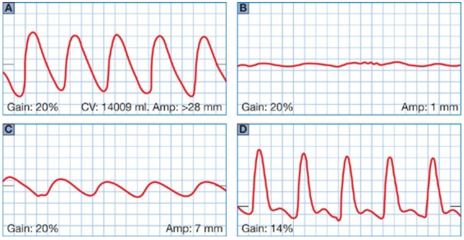
Figure 4–62 shows PVR waveforms with various categories of disease. Identify the category of disease by matching them to the correct corresponding letters of each waveform
______ Normal
______ Mild
______ Moderate
______ Severe
(D) Normal
(A) Mild
(C) Moderate
(B) Severe
Which syndrome causes displacement and compression of the vessels due to the medial head of the gastrocnemius muscle?
(A) Thoracic outlet syndrome
(B) Popliteal entrapment syndrome
(C) Adductor canal compression syndrome
(D) Compartment syndrome
(B) Popliteal entrapment syndrome
Popliteal entrapment syndrome occurs when the head of the gastrocnemius muscle compresses the popliteal artery resulting in a decrease in perfusion to the lower extremity when the calf muscle is contracted. The evaluation with noninvasive testing includes obtaining pressures and waveforms with the calf muscle relaxed and then repeated with the calf muscle contracted. If popliteal entrapment is present there will be a reduction in blood flow during contraction.
A dampened waveform with a delay to peak systole is obtained in the common femoral artery. The cause is most likely due to which of the following?
(A) Distal occlusion
(B) Pseudoaneurysm
(C) Aortoiliac disease
(D) Arteriovenous fistula
(C) Aortoiliac disease
A dampened waveform with a delay to peak systole (tardus parvus) seen in the common femoral artery indicates that significant proximal disease is present, most likely in the aortoiliac region.
A patient is sent to your laboratory for a lower extremity arterial study with and without exercise for thigh claudication symptoms. Her baseline ABI’s at rest were 0.96 on the right and 0.97 on the left. Post-exercise her ABI’s drop to 0.79 on the right and 0.75 on the left. They return to baseline values after 15 minutes. Based on these findings what level of disease does this patient have?
(A) Single level
(B) Multilevel
(C) The patient has no significant disease
(D) Femoral-popliteal
(B) Multilevel
The normal response to exercise is an increase in the ABIs post-exercise. A reduction in the ABIs indicates that disease is present. The greater the pressure drop the more severe the disease. The recovery time also provides information about the level of disease. If the recovery time exceeds 12 minutes that typically indicates that multiple levels of disease are present.
Which penile-brachial index (PBI) is consistent with vasculogenic impotence?
(A) <0.80
(B) <0.65
(C) <0.75
(D) <0.90
(B) <0.65
A penile-brachial index (PBI) of <0.65 is consistent with vasculogenic impotence.
During a penile duplex examination what is the normal post-injection response of the cavernous arteries?
(A) Decrease in diameter
(B) Increase in peak systolic velocity
(C) Decrease in end diastolic velocity
(D) Change to multiphasic flow
(B) Increase in peak systolic velocity
The normal response of the cavernous arteries during a penile duplex examination post-injection is that they will typically double in diameter size, have an increase in peak systolic velocity of at least 30 cm/s, and will have increased diastolic flow.
All of the following parameters must be met during a noninvasive upper extremity arterial examination to confirm the diagnosis of thoracic outlet syndrome EXCEPT
(A) Position dependent
(B) Symptomatic
(C) Complete loss of flow
(D) Reduction of flow
(C) Complete loss of flow
There are various degrees of thoracic outlet syndrome not all of which will result in a complete loss of blood flow to the hand; however in order to confirm the diagnosis there must be a reduction in blood flow that is position dependent and it must be accompanied by symptoms
Submerging a patient’s hands in ice water resulting in a more than 20% drop in peak systolic pressure is an examination performed for which syndrome?
(A) Thoracic outlet syndrome
(B) May–Thurner syndrome
(C) Superior vena cava syndrome
(D) Raynaud’s syndrome
(D) Raynaud’s syndrome
Raynaud’s syndrome is commonly diagnosed using a cold immersion study which involves submerging a patient’s hands in ice water that results in a more than 20% drop in peak systolic pressure.
What is the main advantage of an orthograde saphenous vein bypass graft over a reversed saphenous vein bypass?
(A) The internal valves do not have to be removed
(B) No size discrepancy at both anastomosis sites
(C) No issue with missed accessory branches
(D) They have a lower rate of failure
(B) No size discrepancy at both anastomosis sites
One of the main advantages of an orthograde saphenous vein bypass over a reversed saphenous vein bypass is that there is no size mismatch at the proximal and distal anastomosis sites. In general the saphenous vein is larger in caliber at the proximal portion and small at the distal end. By leaving the vein in this anatomical position it allows the larger end to be connected to the larger proximal arteries of the leg and the smaller end to the smaller tibial arteries. This does however now require the removal of the internal valves that does not have to occur with a reserved saphenous vein bypass.
Retrograde flow in the native artery proximal to the distal anastomosis of a lower extremity bypass graft is consistent with what finding?
(A) It is a normal finding
(B) Distal resistance
(C) Proximal disease
(D) Arteriovenous fistula
(A) It is a normal finding
Retrograde flow in the native artery proximal to the distal anastomosis of a lower extremity bypass graft is a normal finding that can often be providing collateral flow to the proximal portion of the limb and beneficial to the patient.
Which of the following is a type of arteriovenous fistula that can be used for hemodialysis?
(A) Cephalic artery to radial artery
(B) Basilic vein to cephalic vein
(C) Cephalic vein to brachial artery
(D) Basilic vein to brachial vein
(C) Cephalic vein to brachial artery
There are many types of arteriovenous fistulas that can be used for hemodialysis. Some of the more common are the cephalic vein to either the brachial or radial arteries and the basilic vein to either the brachial or radial arteries.
What is the typical normal volume flow through an arteriovenous fistula used for hemodialysis?
(A) >200 mL/min
(B) >400 mL/min
(C) >600 mL/min
(D) >800 mL/min
(D) >800 mL/min
The flow volume through an arteriovenous fistula needs to be high in order for the fistula to be used successfully during hemodialysis. The typical volume flow through a normal mature arteriovenous fistula is >800 mL/min and is often in the 1000 mL/min range.
Which imaging modality is considered the “gold standard” for peripheral artery imaging?
(A) Conventional angiography
(B) CT angiography
(C) MR angiography
(D) Arterial duplex
(A) Conventional angiography
There are many ways to image peripheral arteries; however, conventional angiography is still considered the “gold standard” when it comes to correlation of results.
The effect of the absence of contrast around areas of disease during an angiography study is known as
(A) Digital subtraction
(B) Cine film
(C) Filling defect
(D) Run off
(C) Filling defect
A “filling defect” is the effect that makes a vessel appear narrowed where plaque has replaced the lumen.

Figure 4–63 shows an angiogram of the distal thigh just above the knee. What pathology is identified in this image?
(A) Normal angiogram of the distal thigh
(B) Critical stenosis of the distal superficial femoral artery
(C) Segmental occlusion of the distal superficial femoral artery with collateral flow
(D) Fibromuscular dysplasia of the distal superficial femoral artery
(C) Segmental occlusion of the distal superficial femoral artery with collateral flow
This is an angiogram of the lower extremity demonstrating an occlusion of the distal superficial femoral artery with a large collateral vessel.

The findings in Figure 4–64 are consistent with what diagnosis?
(A) Normal peripheral arterial flow
(B) Hemodynamically significant stenosis
(C) Arteriovenous fistula
(D) Pseudoaneurysm
(B) Hemodynamically significant stenosis
The duplex image shows an elevated peak systolic velocity with aliasing on color Doppler and spectral broadening all findings that are consistent with a hemodynamically significant stenosis

A 76-year-old patient presented with a bruit in the right groin area status post cardiac catheterization. The images in Figure 4–65 were obtained for the patient’s right groin. What do these findings most likely represent?
(A) Pseudoaneurysm
(B) Dissection
(C) Arteriovenous fistula
(D) Hemodynamically significant stenosis
(C) Arteriovenous fistula
The waveform seen in Figure 4–65 shows high peak systolic and high diastolic flow that is consistent with a traumatic arteriovenous fistula. These findings are also consistent with the patient’s clinical presentation. A bruit heard in the area of a recent puncture site following a cardiac catheterization is a common presentation of these findings.

Figure 4–66 demonstrates the results of pre- and postsystolic pressures during an exercise examination. What do these findings suggest?
(A) Normal response to exercise
(B) Single-level peripheral arterial disease
(C) Multilevel peripheral arterial disease
(D) Mild peripheral arterial disease
(A) Normal response to exercise
Both the right and left ankle peak systolic pressures increased following exercise, which is the normal response in the absence of disease.

The segmental pressures shown in Figure 4–67 indicate which level of disease?
(A) Right iliac disease and left femoral popliteal disease
(B) Right femoral popliteal disease and left iliac disease
(C) Right tibial disease and left iliac disease
(D) Bilateral multilevel disease
(B) Right femoral popliteal disease and left iliac disease
The segmental pressures have a significant drop in systolic pressure (>30 mm Hg) between the right thigh and calf indicating femoral popliteal disease as well as a reduced systolic pressure and pressure index at the left thigh level indicating left iliac disease.

Match the PVR components in Figure 4–68 with the correct term - #94
(A) Catacrotic limb
(B) Dicrotic limb
(C) Anacrotic limb
(C) Anacrotic limb

Match the PVR components in Figure 4–68 with the correct term - #95
(A) Catacrotic limb
(B) Dicrotic limb
(C) Anacrotic limb
(A) Catacrotic limb

Match the PVR components in Figure 4–68 with the correct term - #96
(A) Catacrotic limb
(B) Dicrotic limb
(C) Anacrotic limb
(B) Dicrotic limb
What is the condition that involves a congenital narrowing of the thoracic aorta where it arches down toward the abdomen?
(A) Takayasu’s arteritis
(B) Thoracic outlet syndrome
(C) Thromboangiitis obliterans
(D) Coarctation of the aorta
(D) Coarctation of the aorta
Coarctation of the aorta is a congenital narrowing of the thoracic aorta where it arches down toward the abdomen
What is the second most common area for atherosclerosis to develop in the lower extremities?
(A) Aorta and iliac arteries
(B) Distal femoral artery
(C) Posterior tibial artery
(D) Profunda femoris artery
(A) Aorta and iliac arteries
The aorta and iliac arteries are the second most common area of atherosclerotic disease in the lower extremities, with the distal superficial femoral artery as the most common site.
A 55-year-old male with severe uncontrolled hypertension is having symptoms of right lower extremity numbness and loss of sensation. While performing a lower extremity duplex examination a linear echogenic structure is visualized in the external iliac artery that appears to be mobile with cardiac pulsations. What is most likely the diagnosis?
(A) Atherosclerosis
(B) Arteriovenous malformation
(C) Arterial dissection
(D) Thromboangiitis obliterans
(C) Arterial dissection
A finding of an echogenic linear structure that appears to move within the lumen of a blood vessel is consistent with an arterial dissection that can be caused by trauma or severe hypertension. Severe hypertension if untreated can cause an increased strain on the blood vessel walls causing them to separate resulting in a dissection.
What anatomical structure serves as the landmark for where the external iliac artery becomes the common femoral artery in the lower extremities?
(A) Adductor canal
(B) Profunda femoris
(C) Inguinal ligament
(D) Arcuate ligament
(C) Inguinal ligament
The inguinal ligament is the anatomical structure stretching from the anterior, superior border of the iliac crest to the pubic bone and serves as the landmark for where the external iliac artery becomes the common femoral artery in the lower extremities.