Muscle Anatomy - A&P
1/74
There's no tags or description
Looks like no tags are added yet.
Name | Mastery | Learn | Test | Matching | Spaced |
|---|
No study sessions yet.
75 Terms
Epimysium
Dense irregular connective tissue encasing entire skeletal muscle, integrating with tendons to transmit contractile force and reduce friction between adjacent muscles.
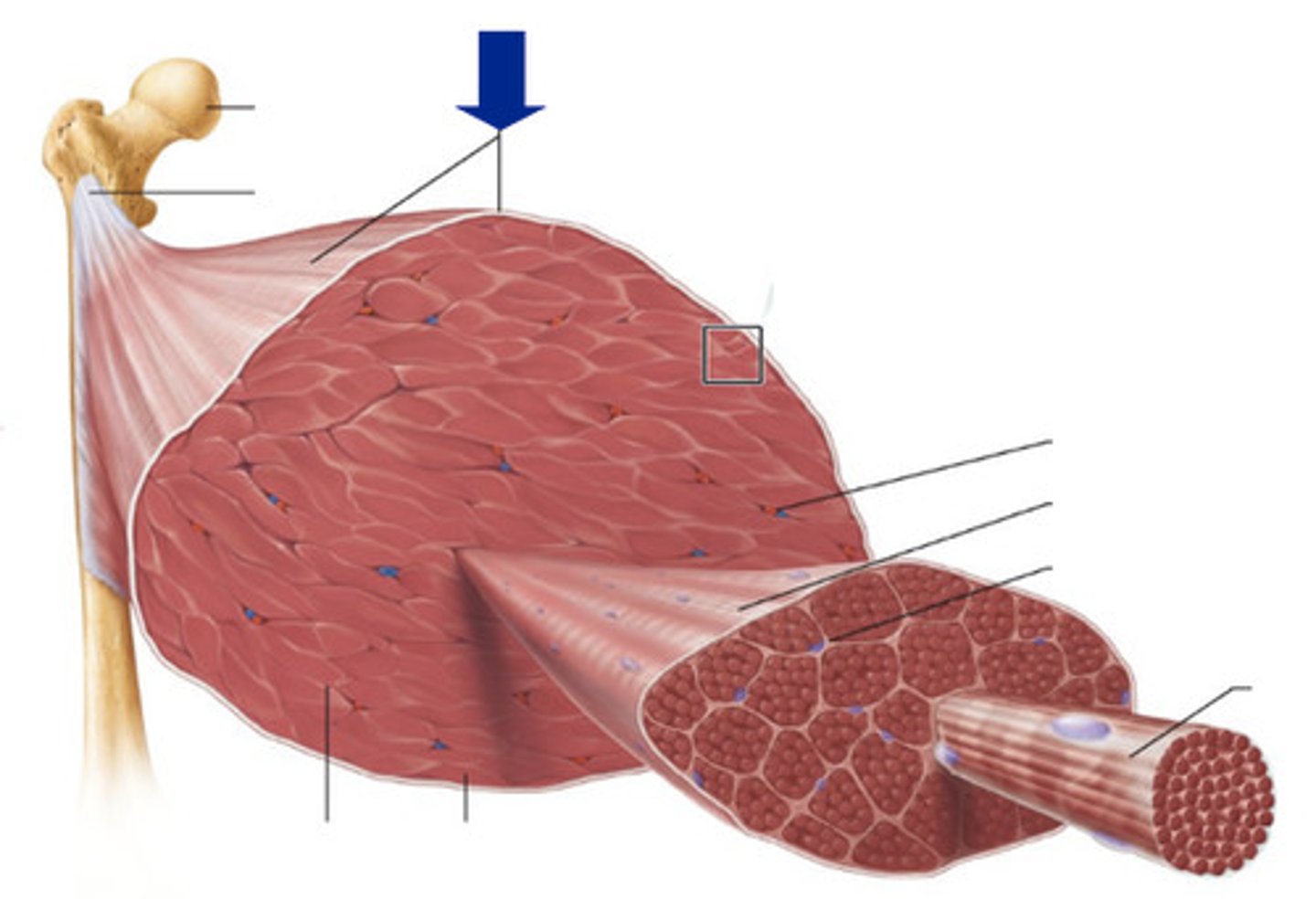
Fasicles
Bundles of skeletal muscle fibers encased by perimysium, serving as structural and functional subunits within a muscle to coordinate contraction.
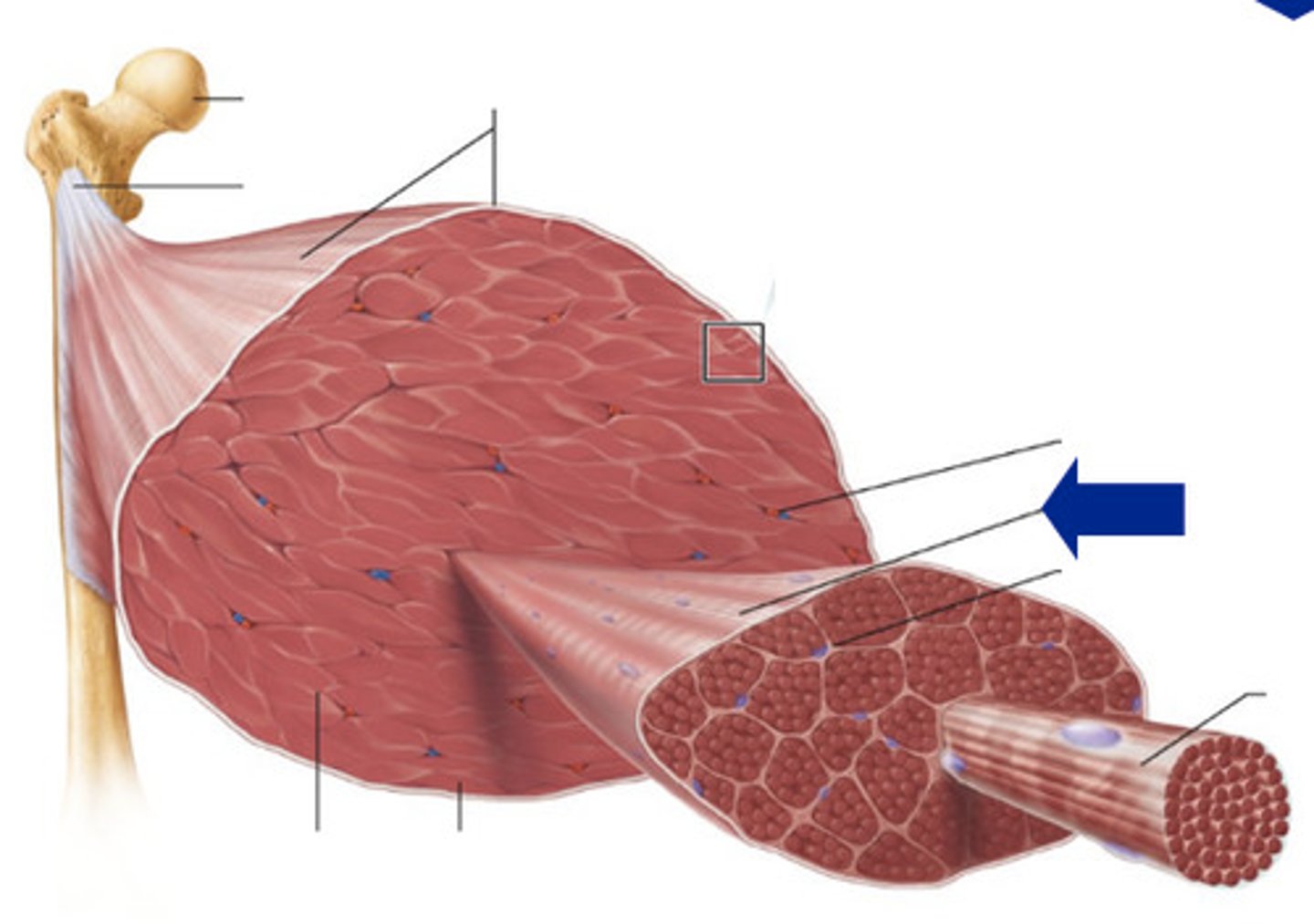
Perimysium
Connective tissue surrounding a fascicle.
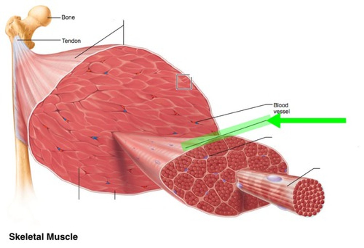
Muscle Fiber
A single muscle cell.
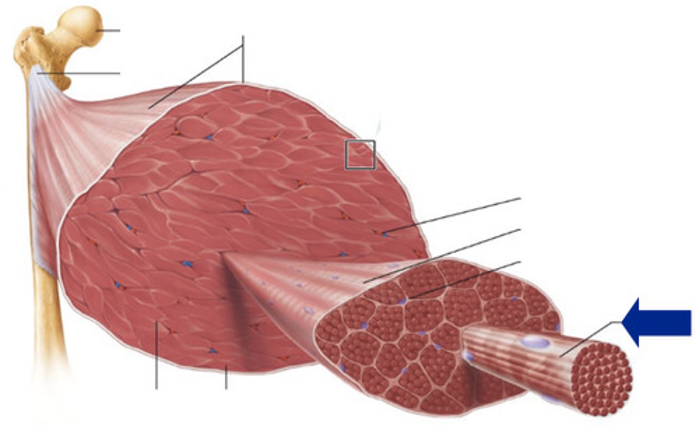
Endomysium
Connective tissue surrounding a muscle cell.
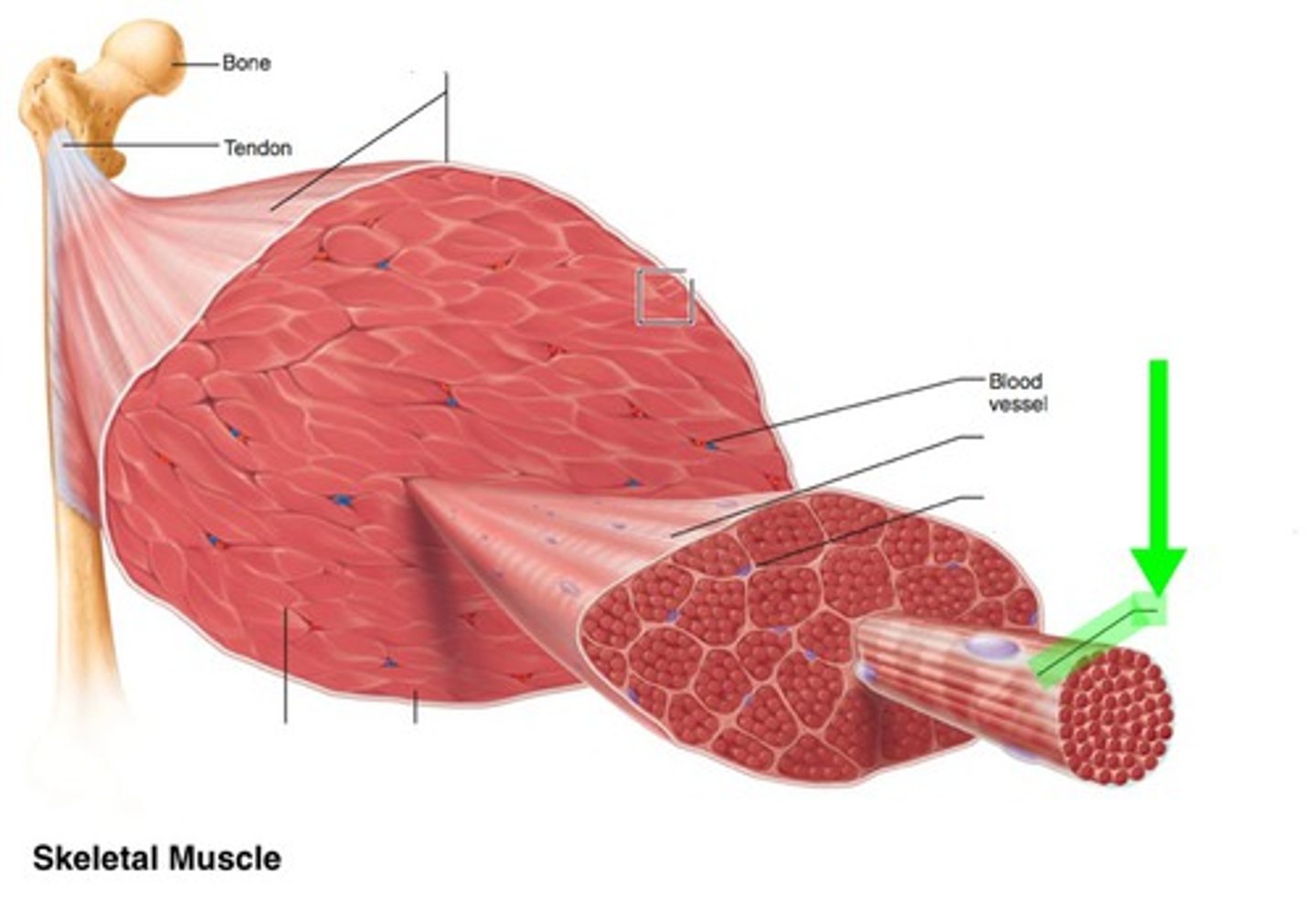
Tendon
Connects muscle to bone.,
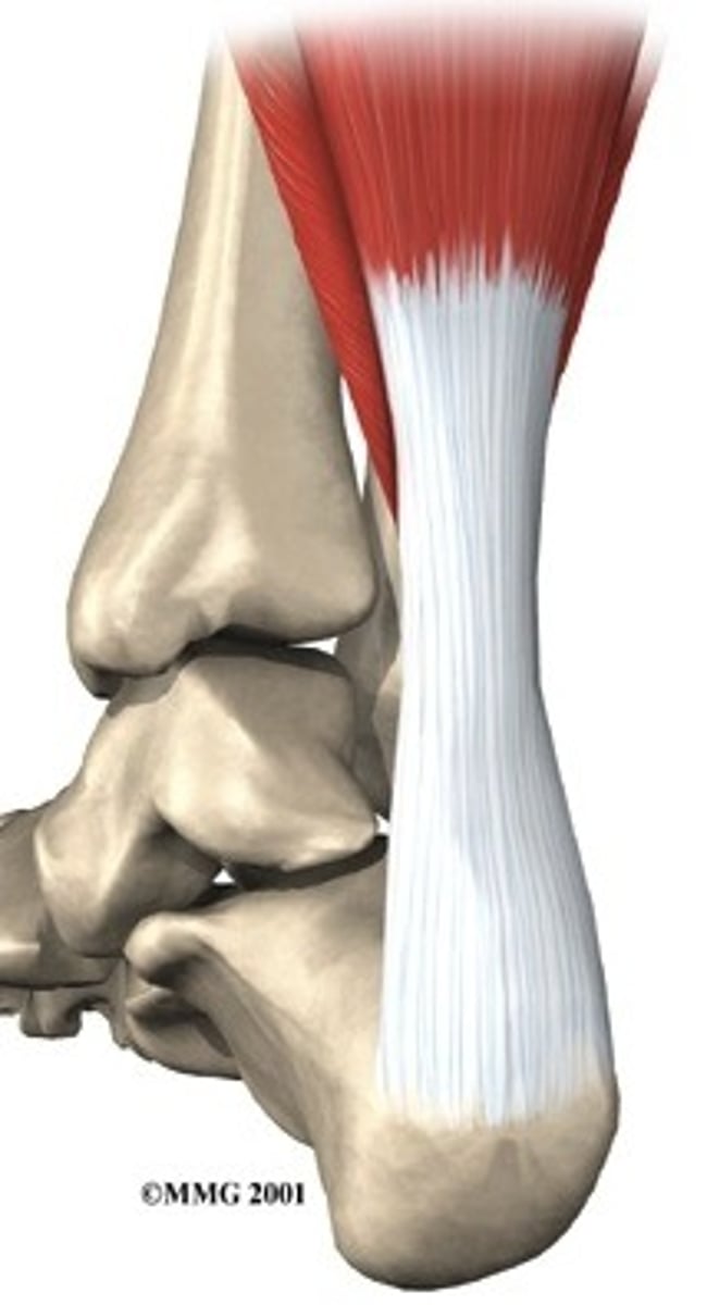
Aponeurosis
Strong sheet of tissue that acts as a tendon to attach muscles to bone.
Origin
Attachment of a muscle that remains relatively fixed during muscular contraction.
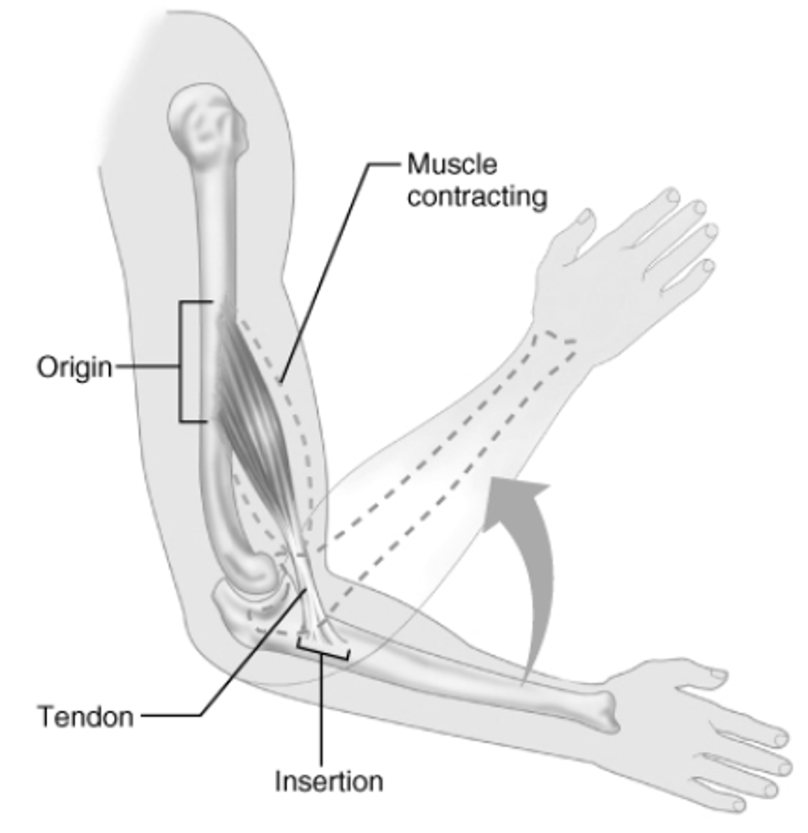
Insertion
The attachment of a muscle tendon to a moveable bone or the end opposite the origin.
Action
The specific movement a muscle produces at a joint, determined by its origin, insertion, and line of pull.
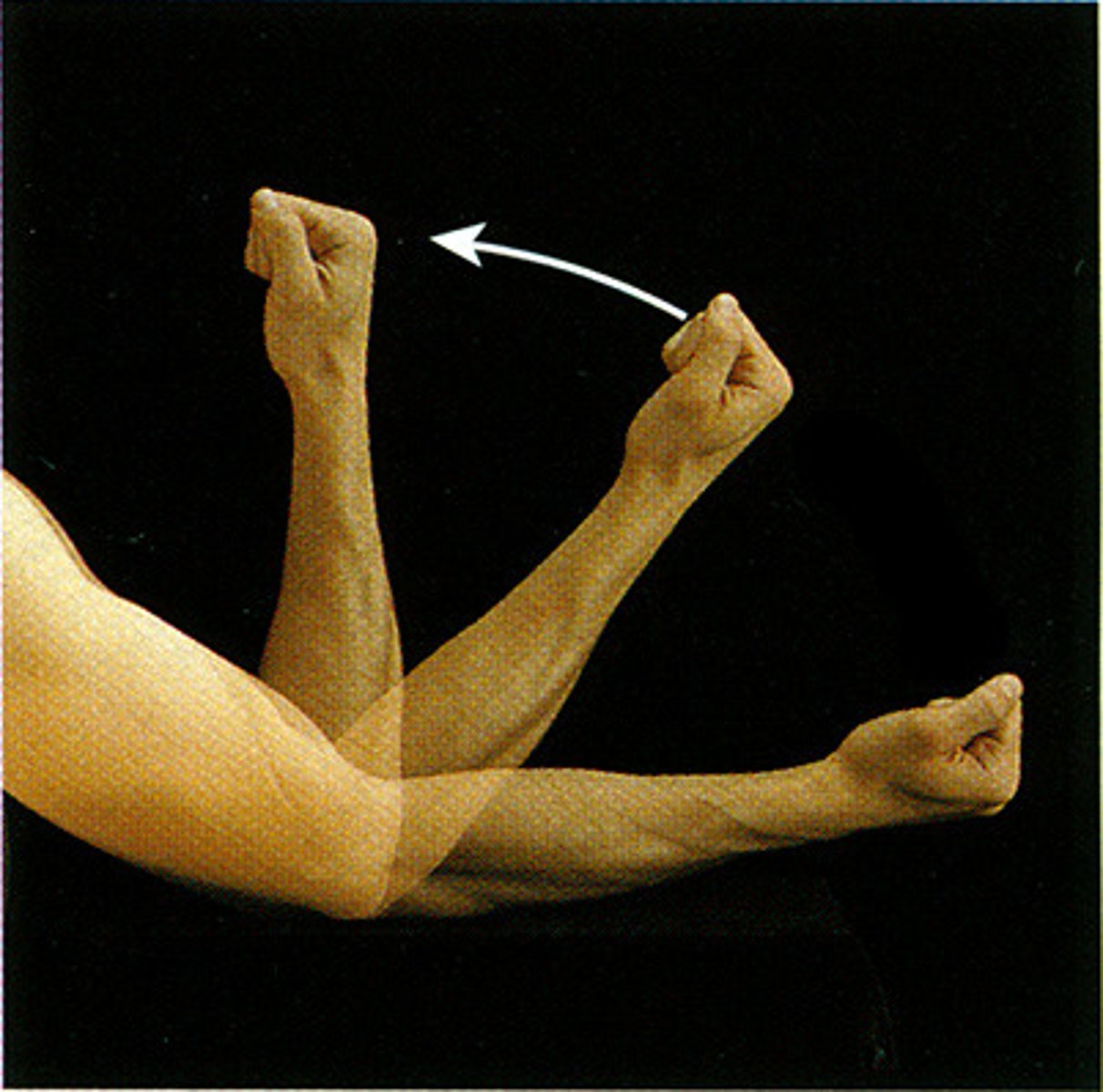
Flexion
Decreases the angle of a joint.
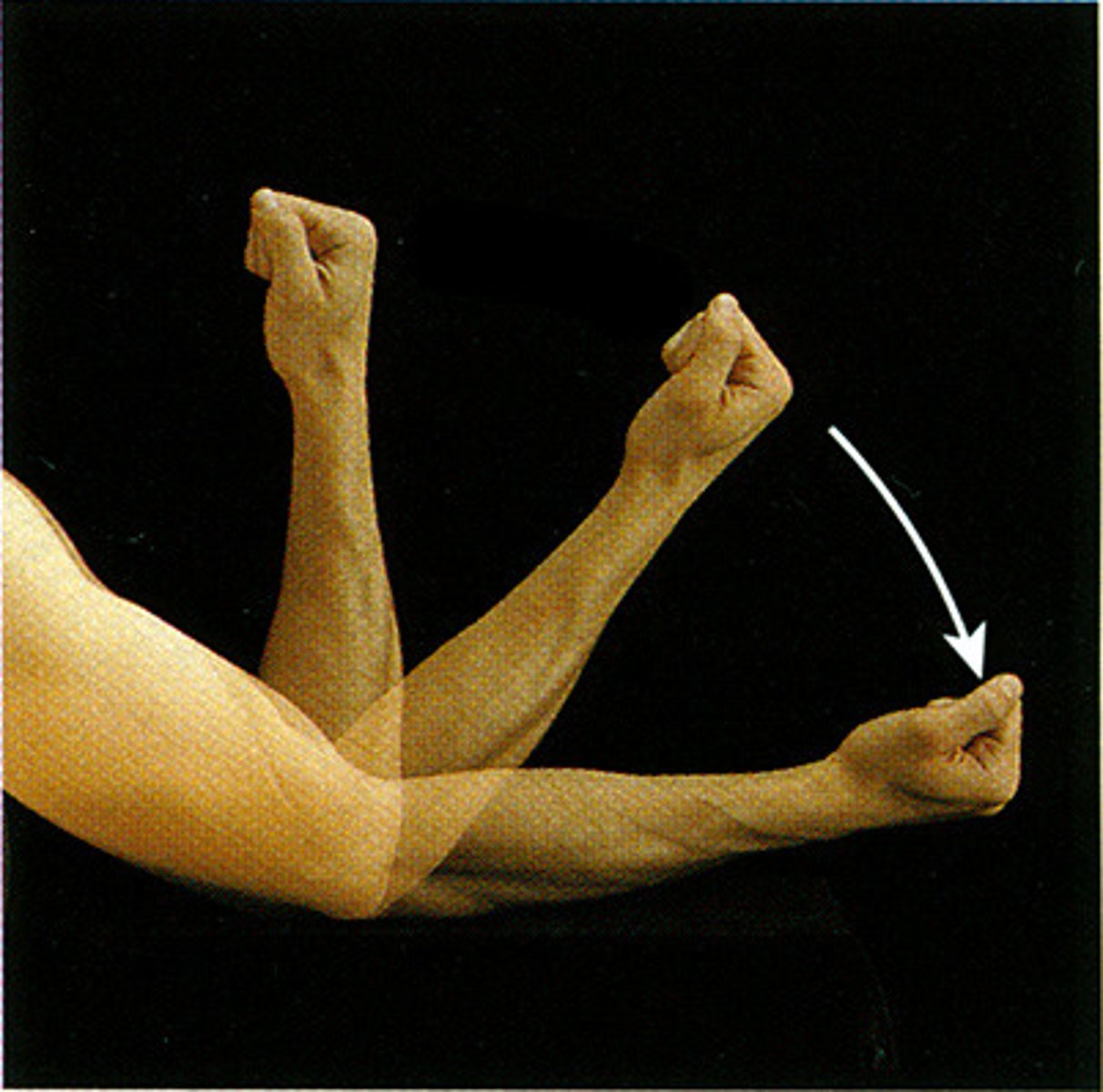
Extension
Increases the angle of a joint.
Hyperextension
Extension beyond anatomical position.
Abduction
Movement away from the midline of the body.
Adduction
Movement toward the midline of the body.
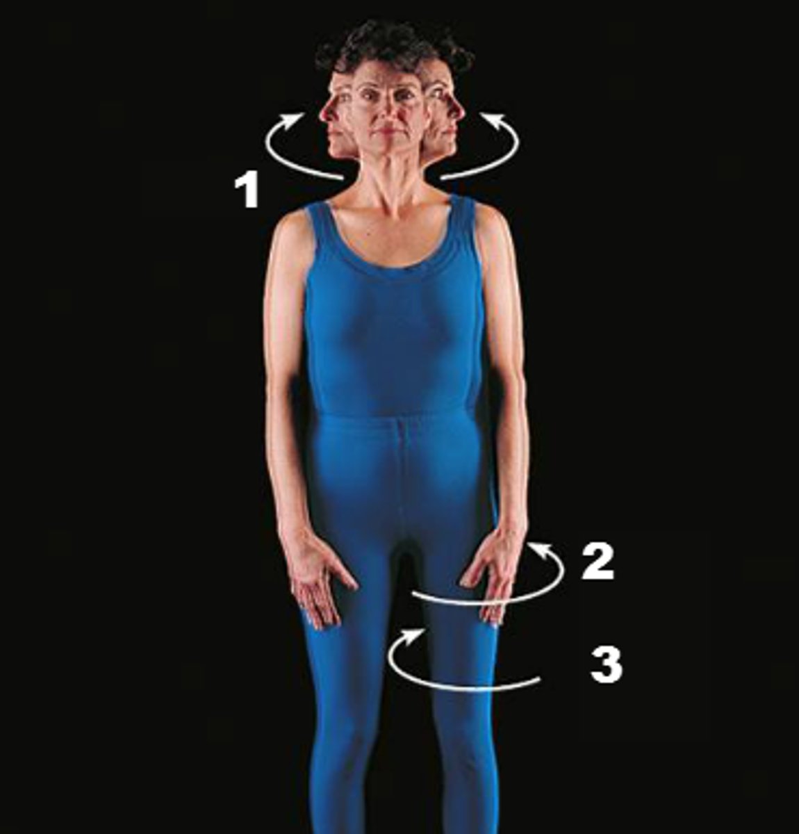
Rotation
Movement of a bone around its longitudinal axis within a joint, without lateral displacement.
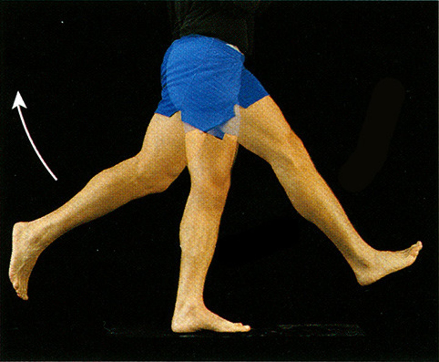
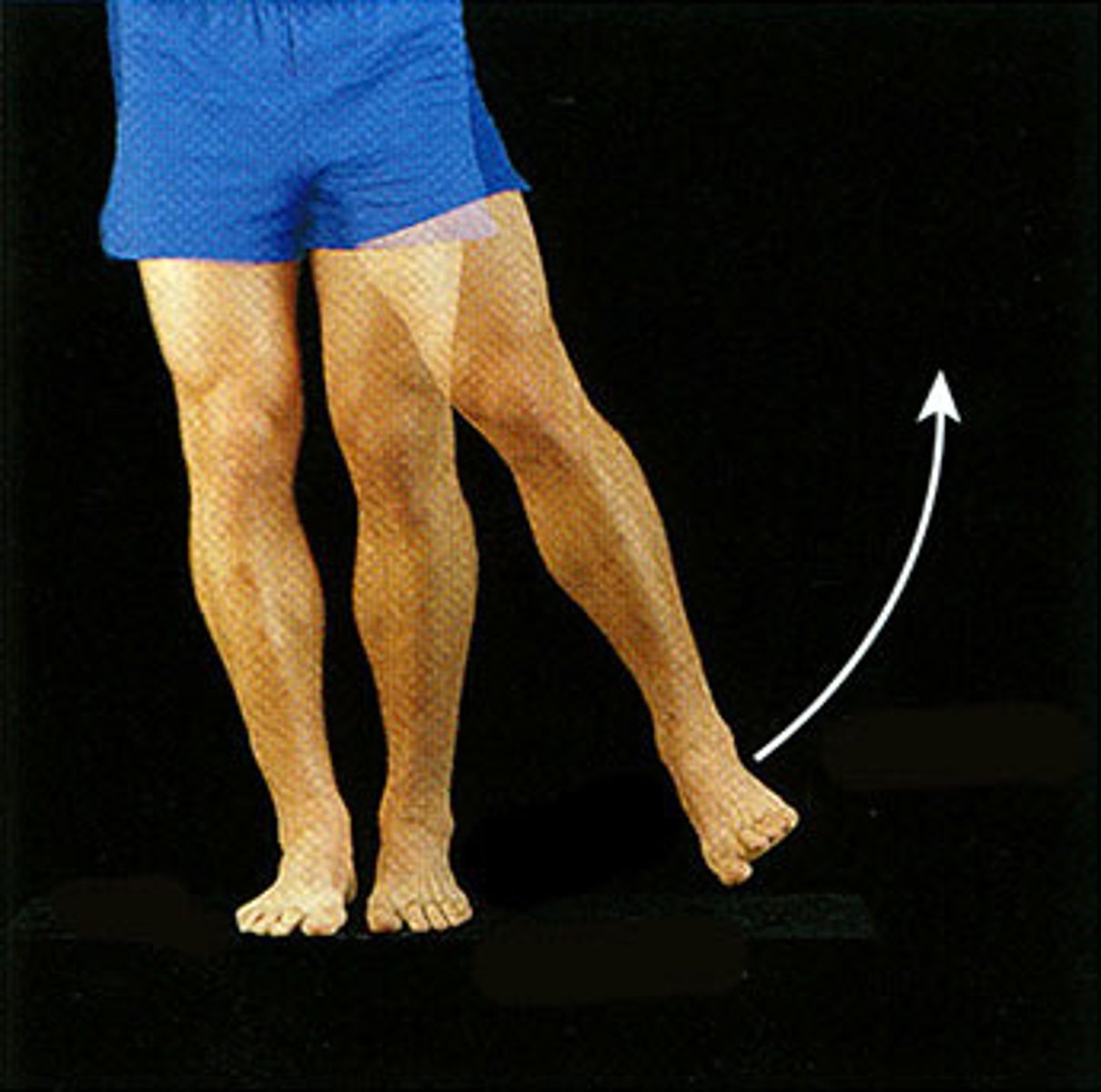
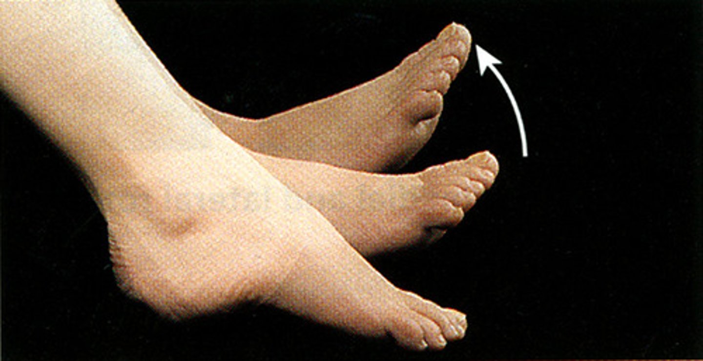
Dorsiflexion
Bending of the foot or the toes upward.
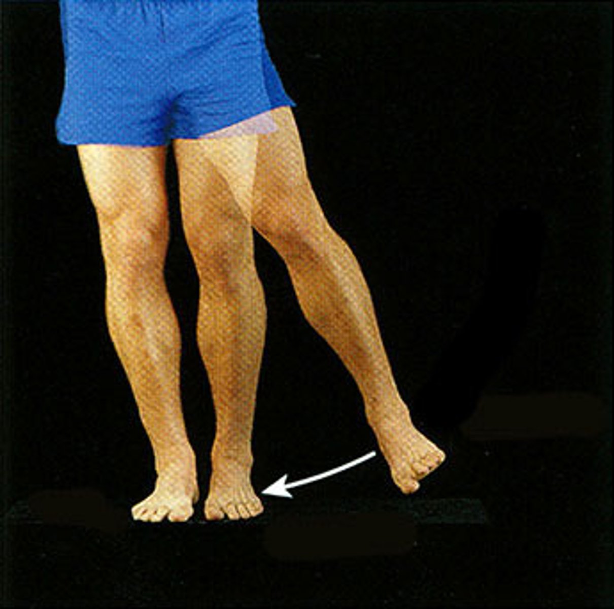
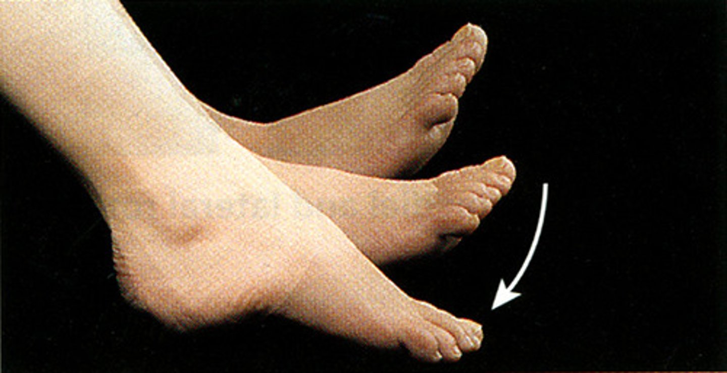
Plantar Flexion
Bending of the sole of the foot by curling the toes toward the ground.
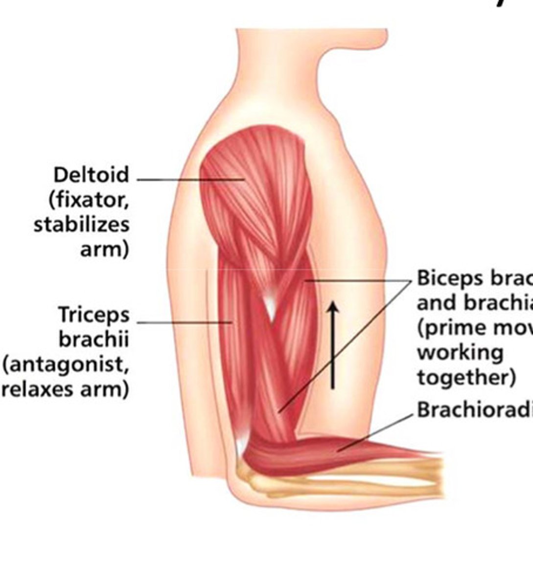
Agonist
Contraction that generates movement.
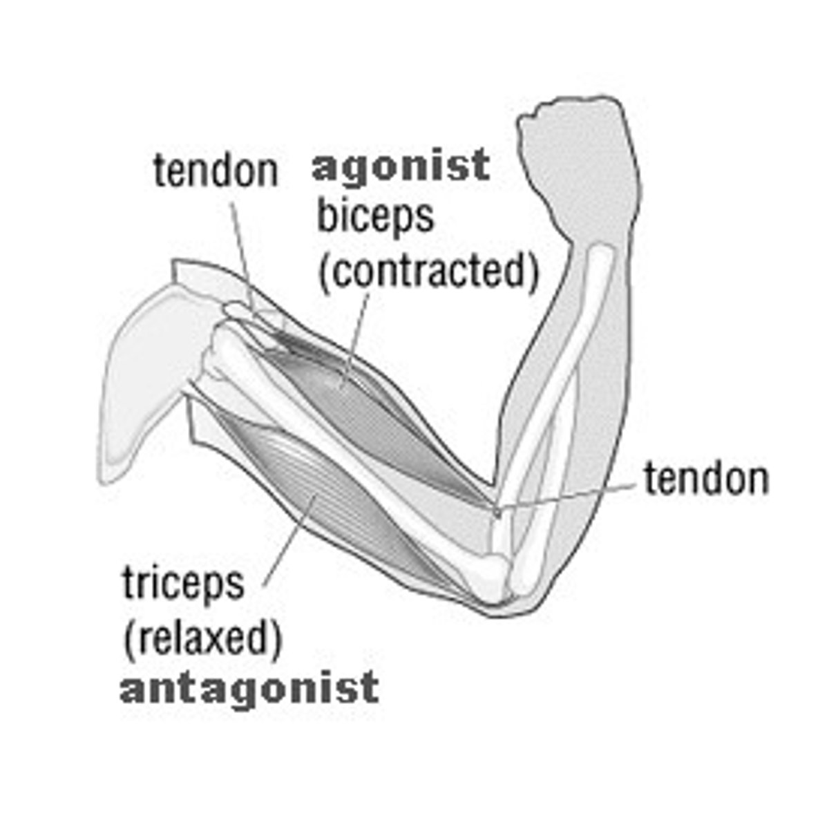
Antagonist
Relaxation require for movement.

Synergist
Contracts with the agonist to aid movement.
Fixator
Synergist that stabilizes joints.
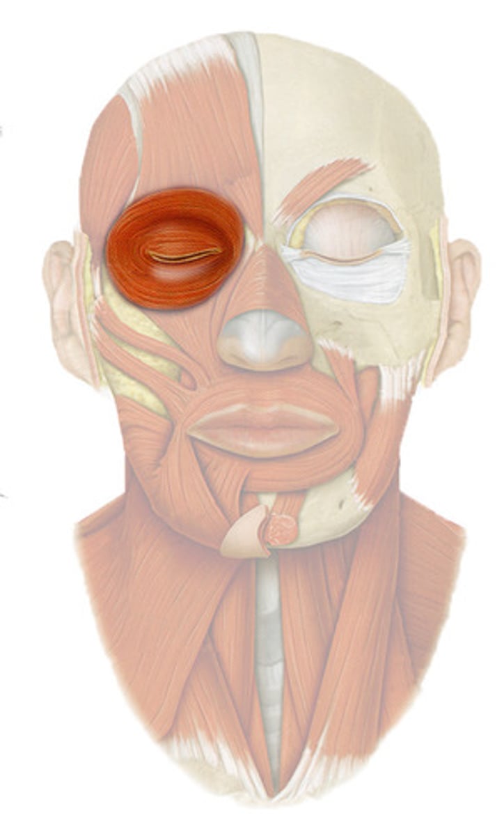
Orbicularis Oculi
Closes the eyelids.
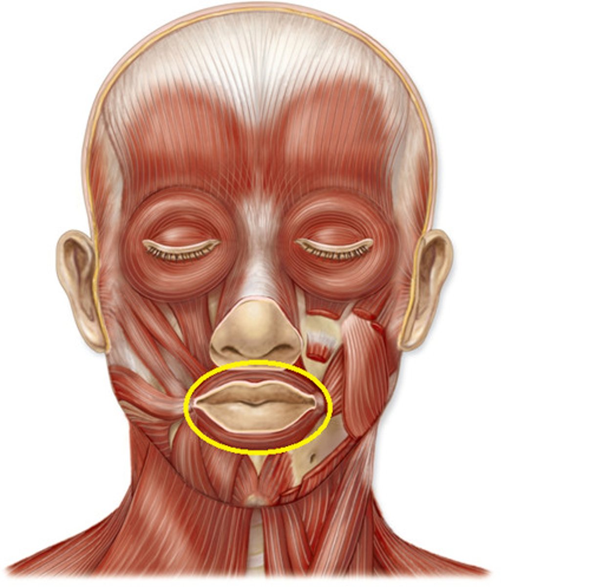
Orbicularis Oris
Closes lips.
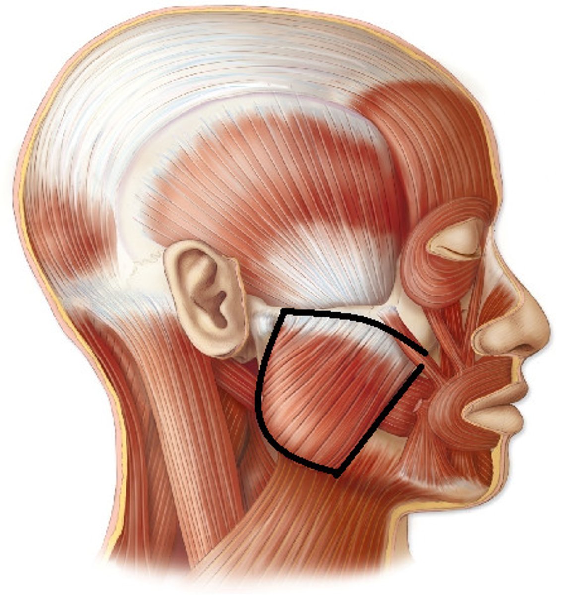
Masseter
Elevates the mandible to close the jaw.
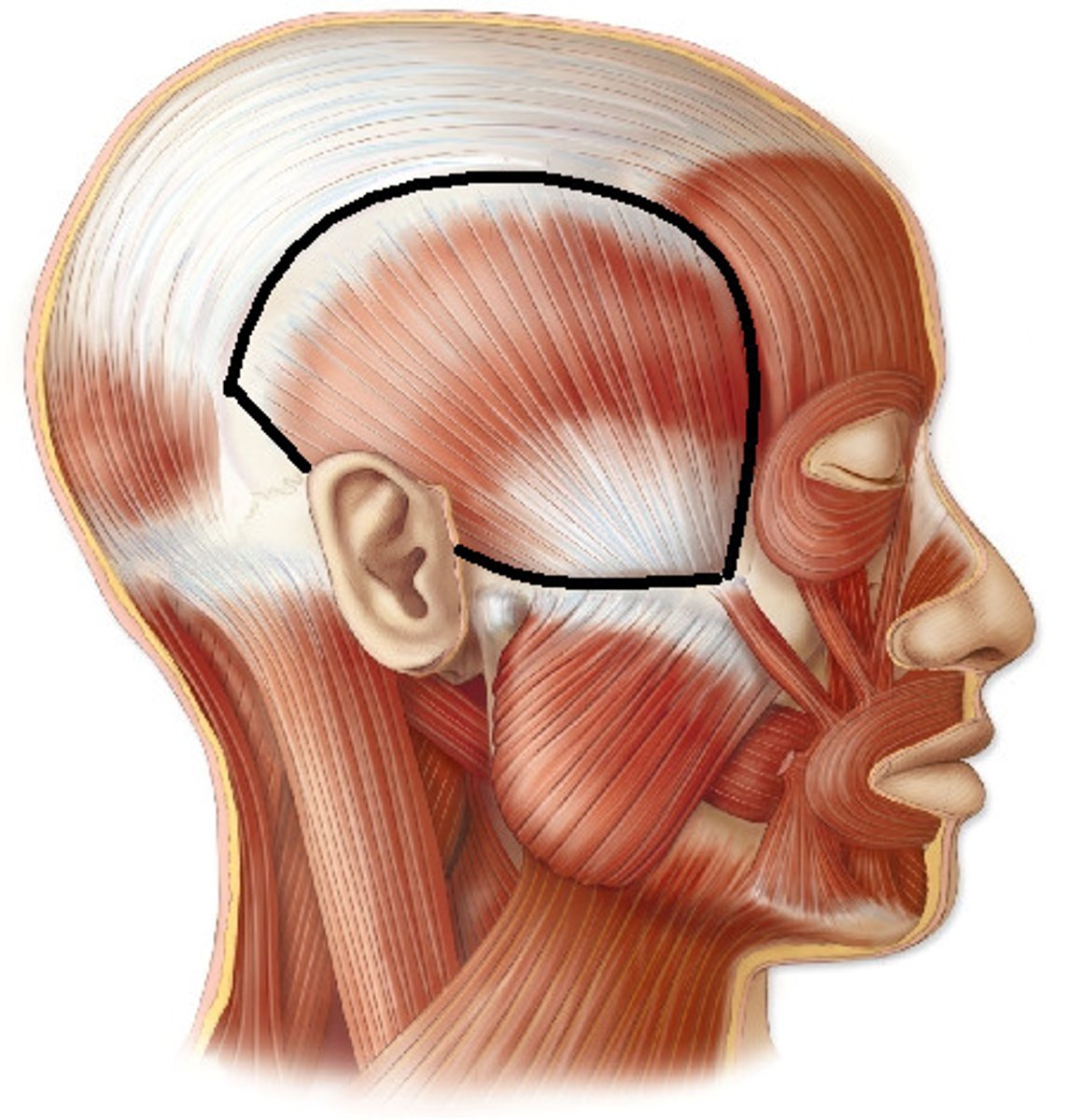
Temporalis
Elevates and retracts the mandible.
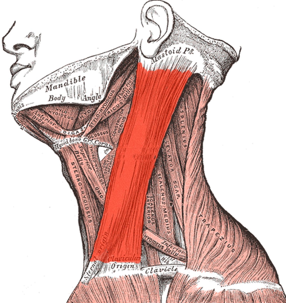
Sternoclei Domastoid
Rotates the head to the opposite side and tilts it to the same side; flexes neck.
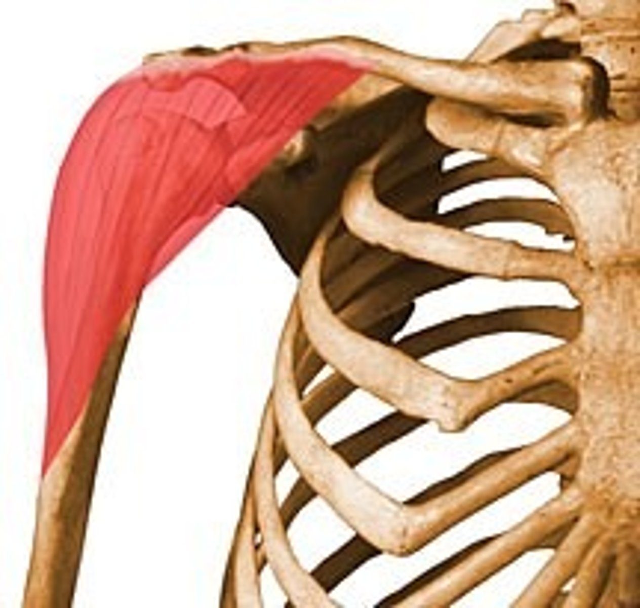
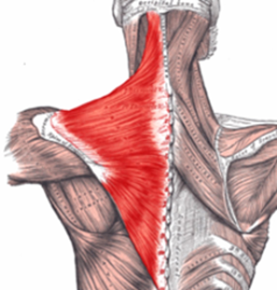
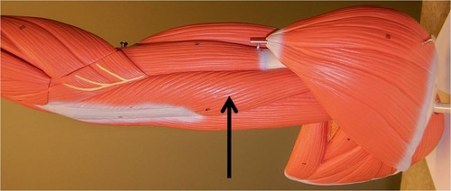
Trapezius
Elevates, depresses, retracts, and rotates the scapula; rotates the arm.
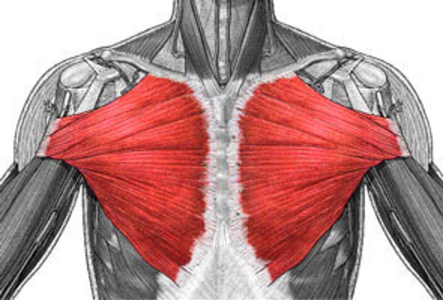
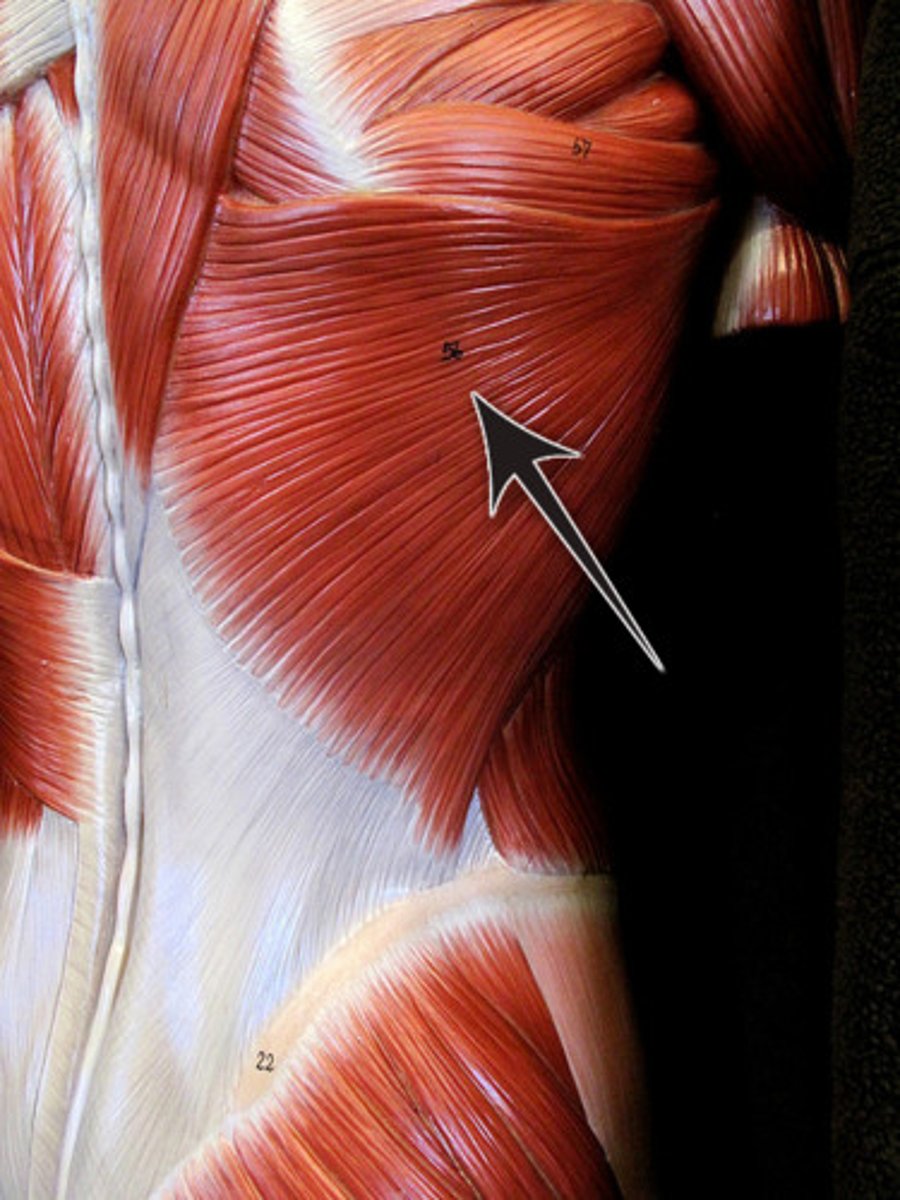
Pectoralis Major
Adducts and flexes humerus.
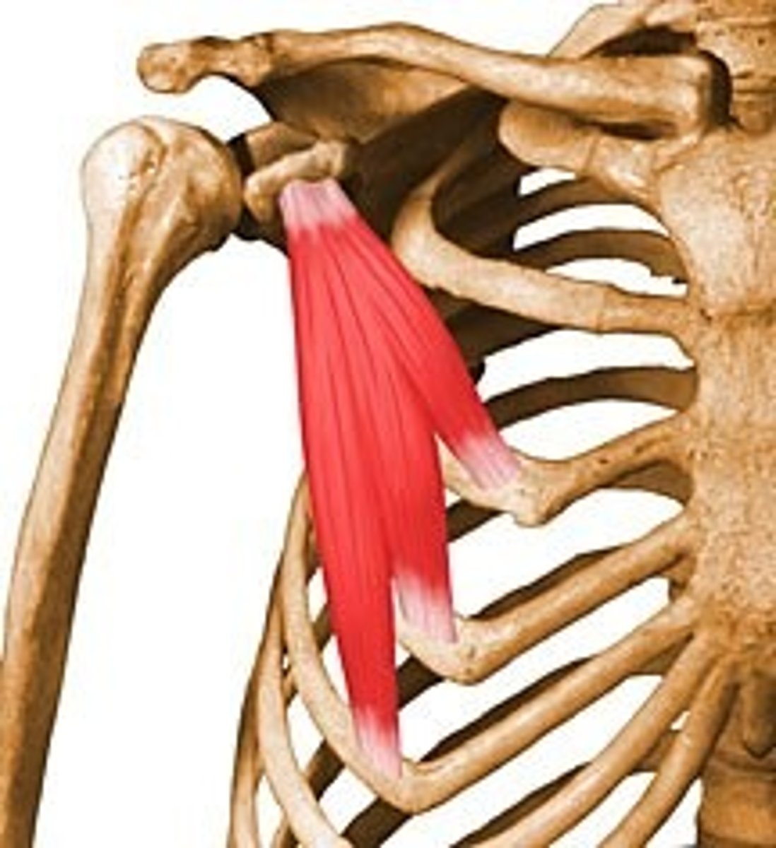
Pectoralis Minor
Protracts and depresses scapula.
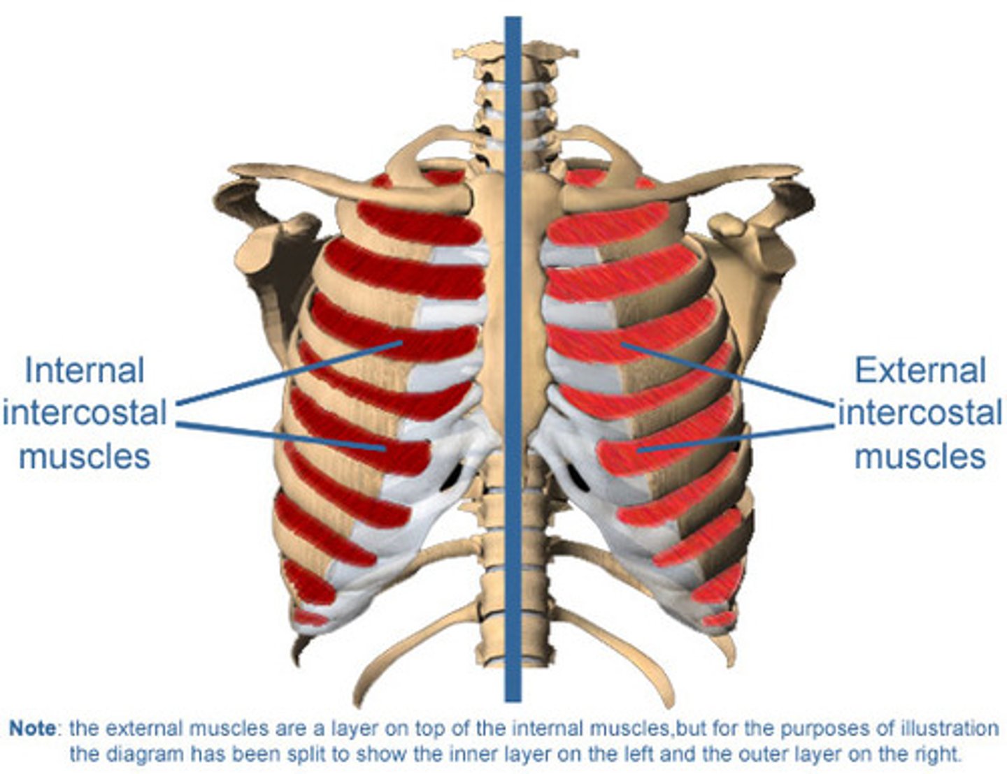
Internal Intercostals
Depresses ribs during forced expiration.

External Intercostals
Elevates ribs during inspiration.
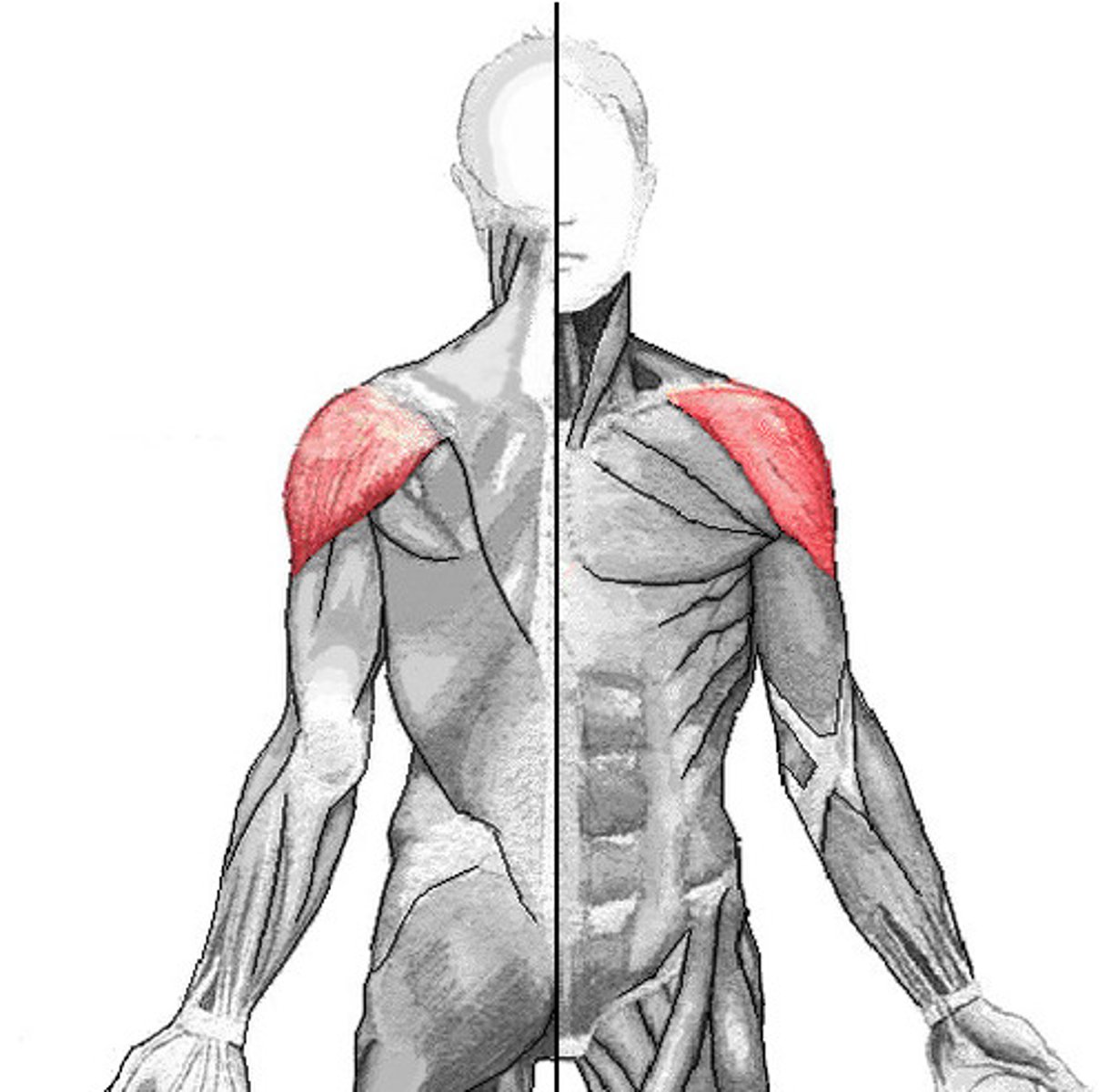
Deltoid
Flexes, abducts, and extends the arm; medially and laterally rotates the arm.
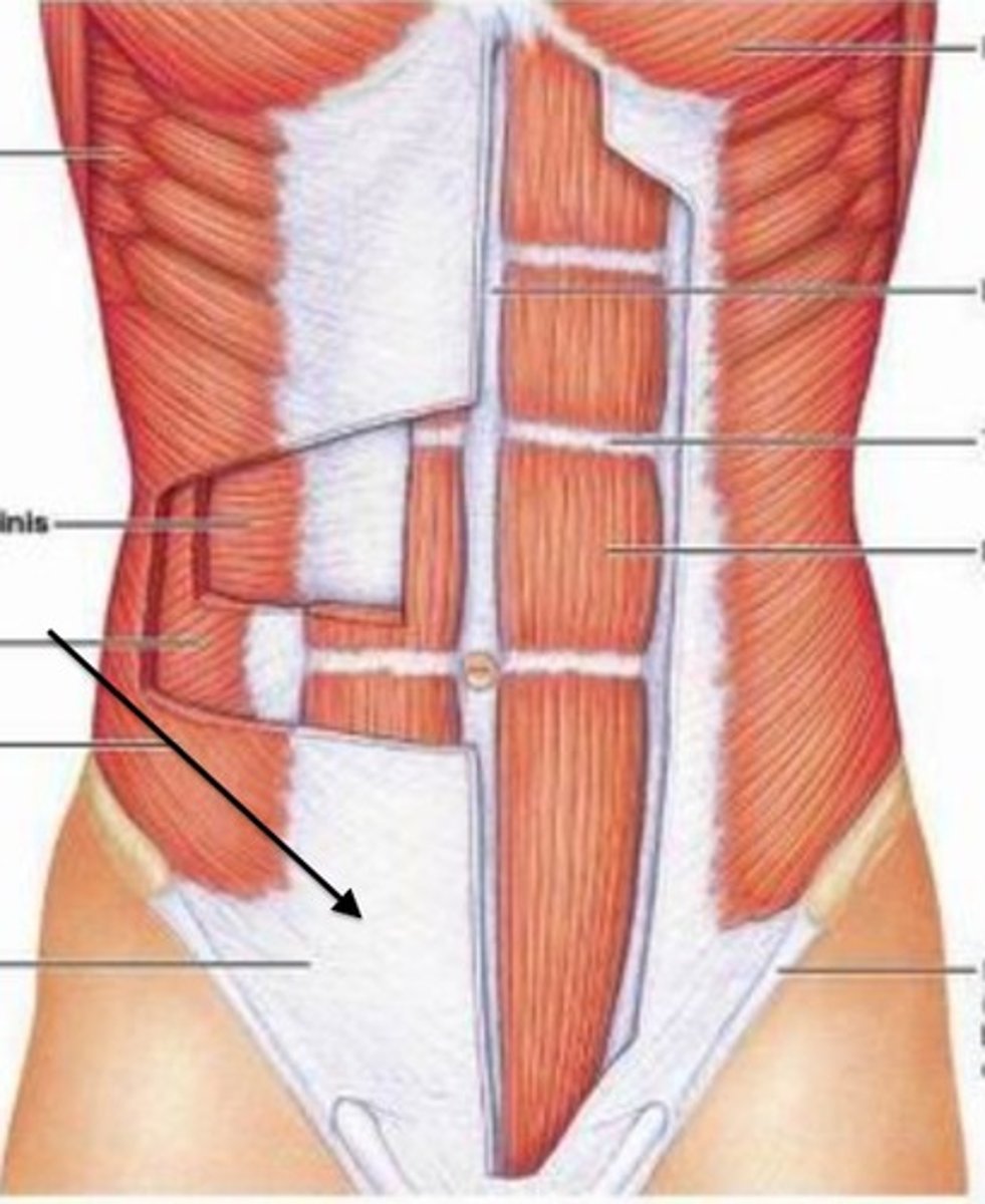
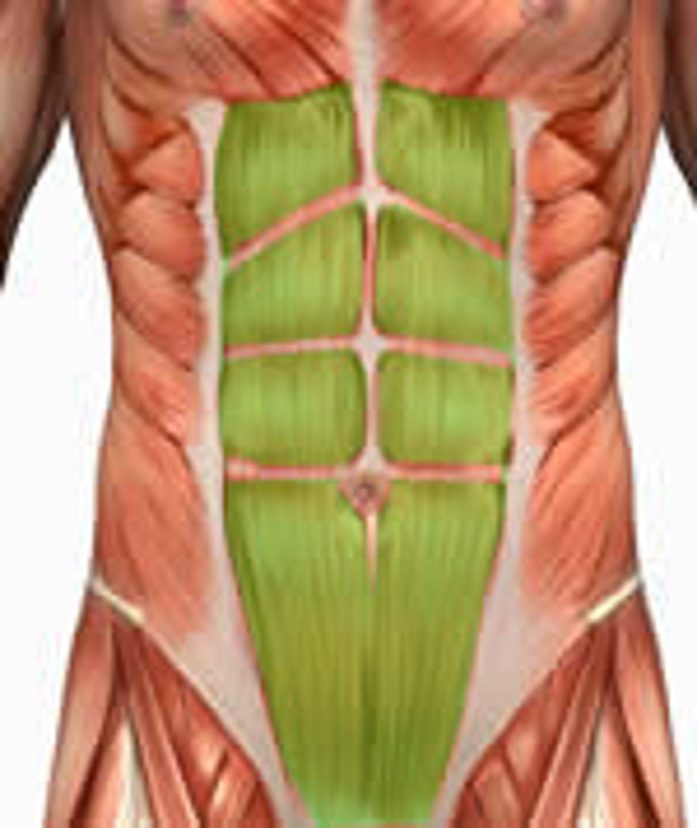
Rectus Abdominus
Flexes the vertebral column; compresses abdominal contents.
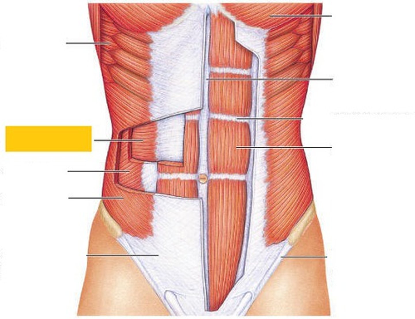
External Oblique
Contralateral trunk rotation; lateral flexion; bilateral trunk flexion.
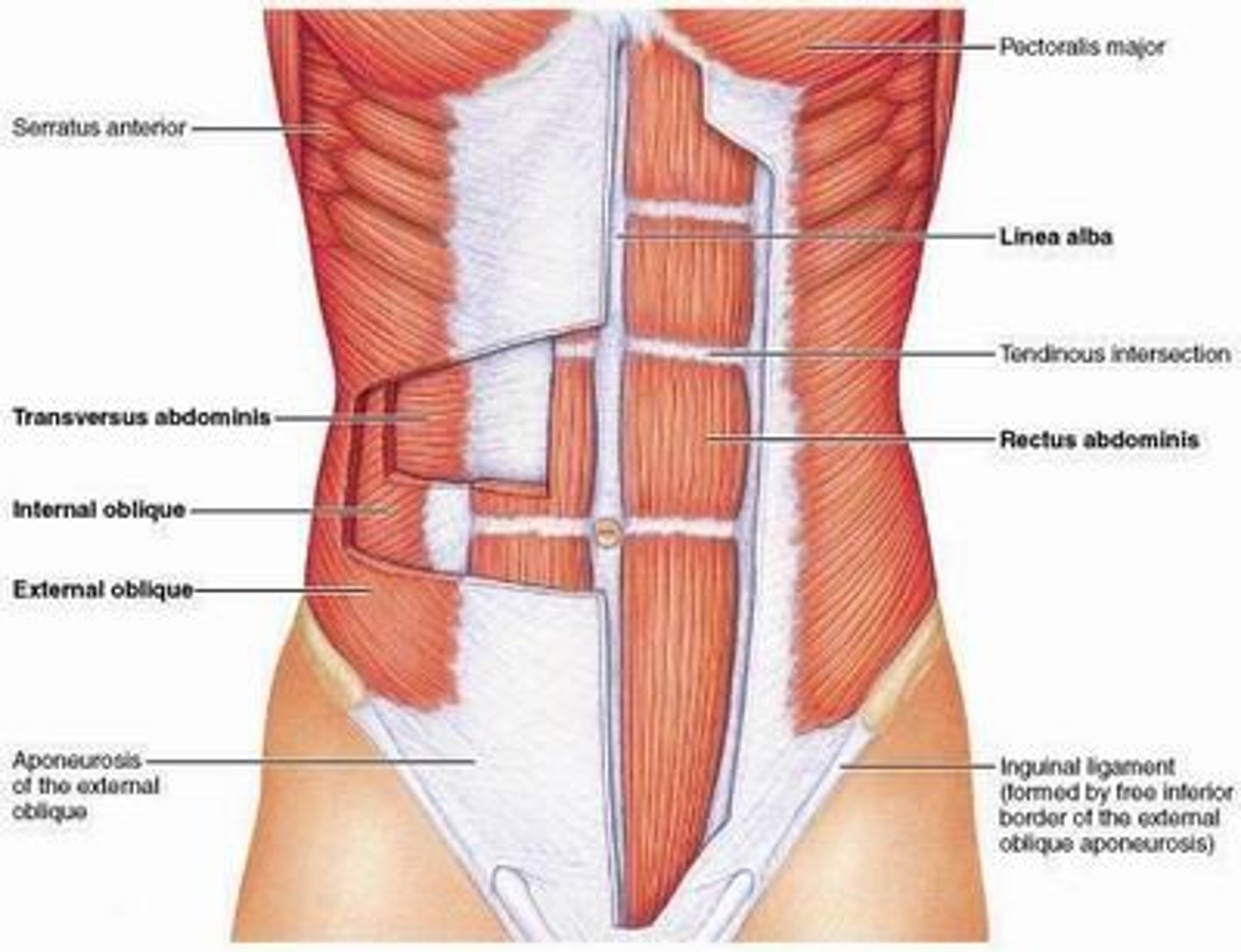
Internal Oblique
Ipsilateral trunk rotation; lateral flexion; bilateral trunk flexion.

Transversus Abdominis
Compresses abdominal contents; stabilizes the trunk.
Latissimus Dorsi
Extends, adducts, and medially rotates the arm; draws the shoulder downward and backward.
Humerus and scapula. (Origin)
Origin of Triceps Brachii
Olecranon Process (Insertion)
Insertion of Triceps Brachii
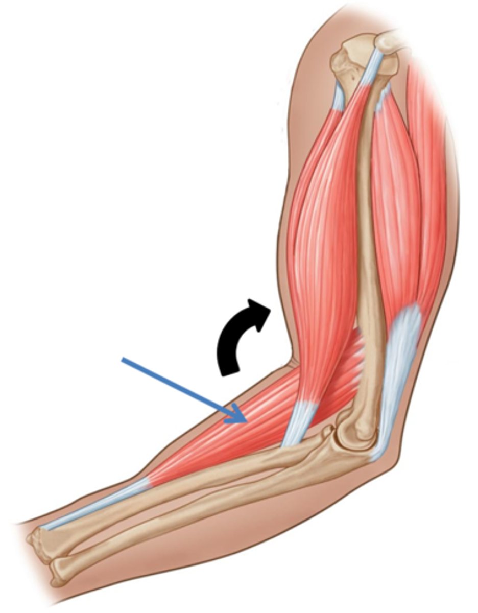
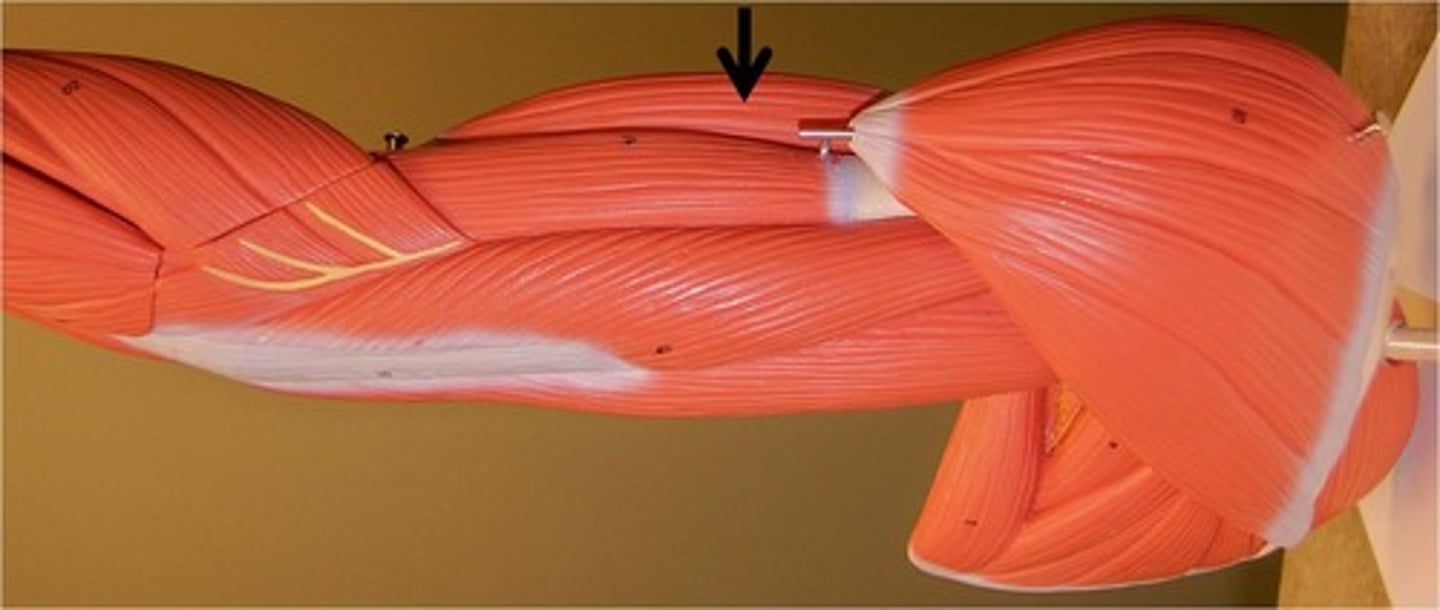
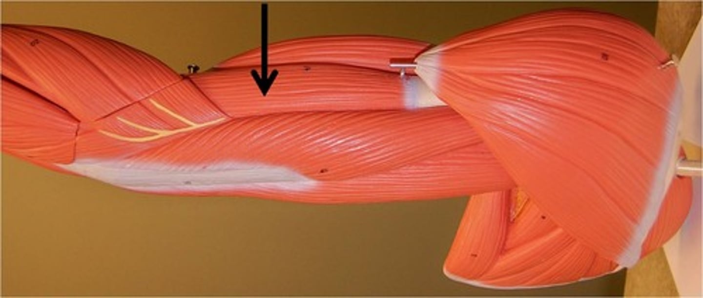
Triceps Brachii
Extends forearm.
Scapula. (Origin)
Origin of Biceps Brachii
Radial tuberosity. (Insertion)
Insertion of Biceps Brachii
Biceps Brachii
Flexes forearm.
Humerus (anterior distal shaft). (Origin)
Origin of Brachialis
Coronoid process of ulna. (Insertion
Insertion of Brachialis
Brachialis
Elbow flexion.
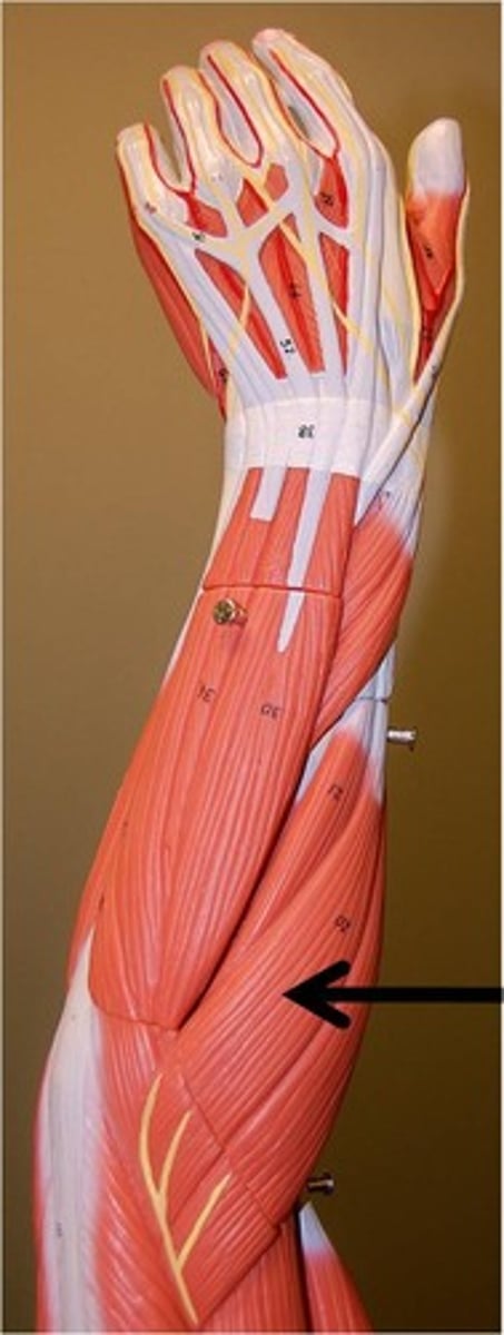
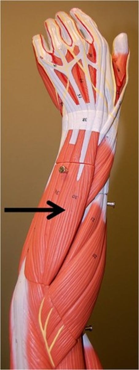
(ECRL) Extensor Carpi Radialis Longus
Extends wrist and abducts hand.
Extensor Digitorum
Extends fingers and the wrist.
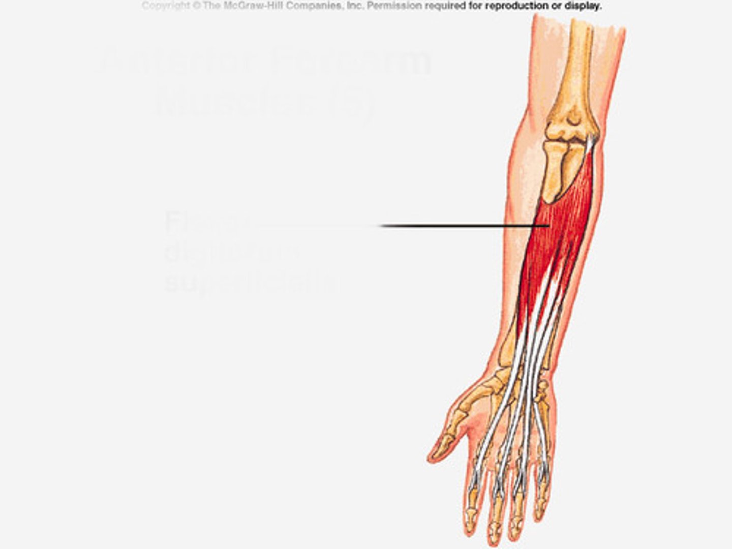
Flexor Digitorum Superficialis
Flexes wrist and fingers.
Flexor Carpi Radialis (FCR)
Flexes wrist and abducts hand.

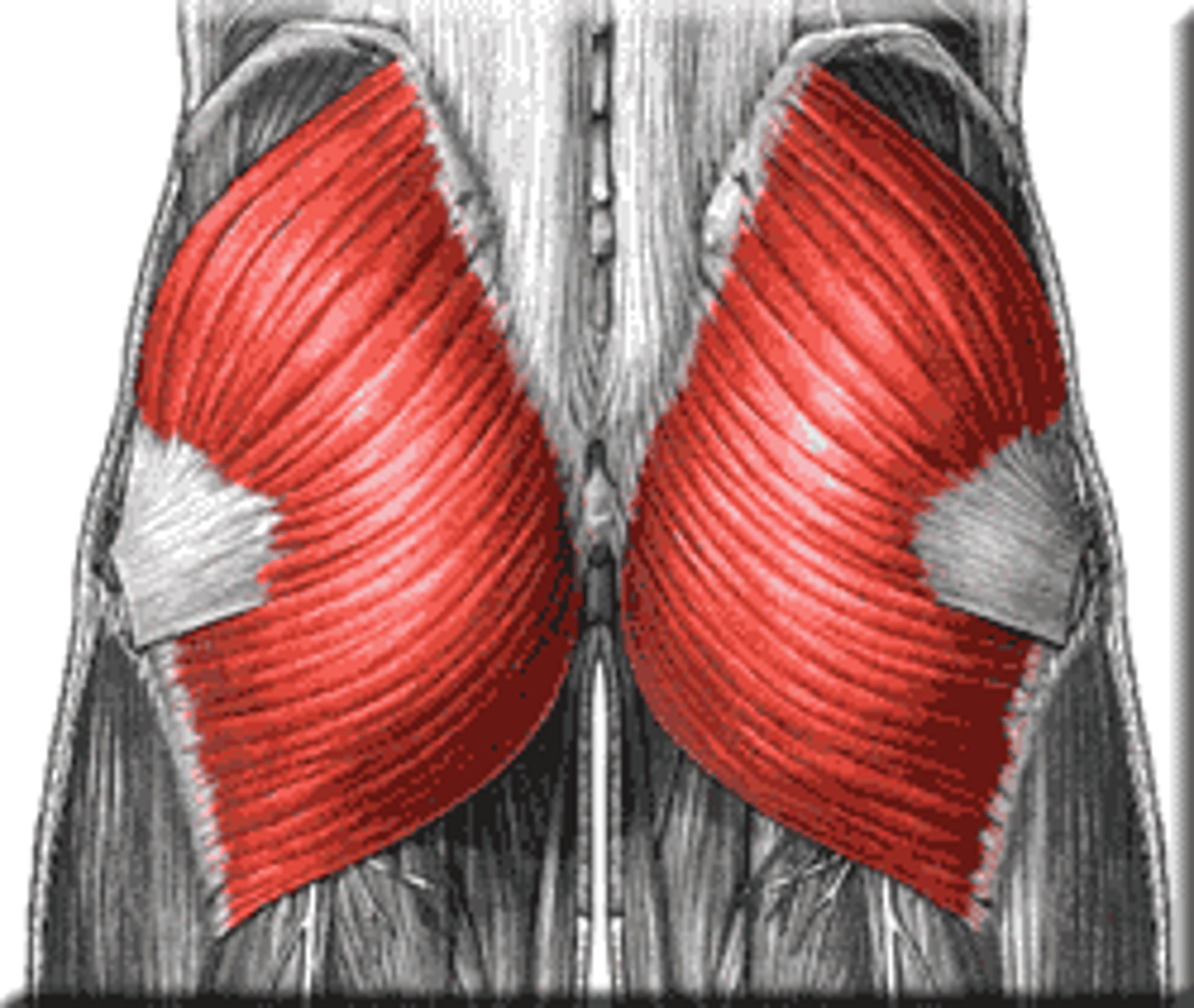
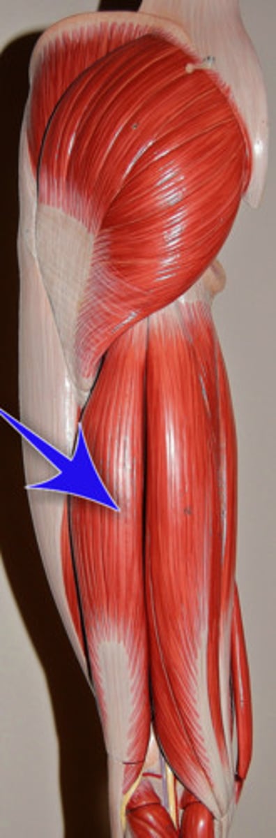
Gluteus Maximus
Extends thigh.
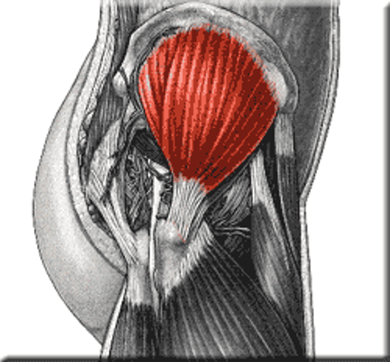
Gluteus Medius
Abducts and medially rotates thigh.
Sartorius
Flexes, abducts, and laterally rotates thigh at the hip; flexes knee.
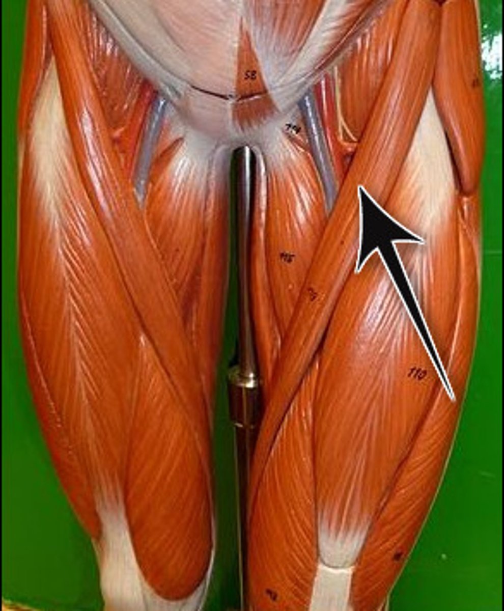
Gracilis
Adducts thigh.
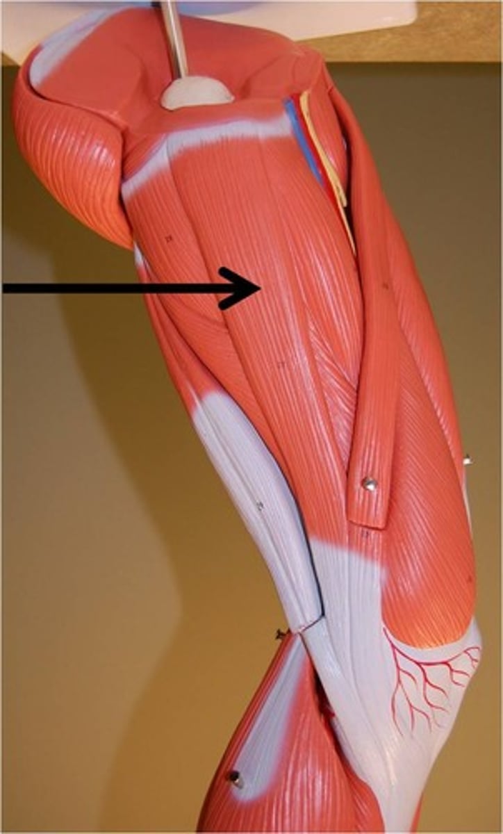
Quadriceps Femoris
Extends leg.
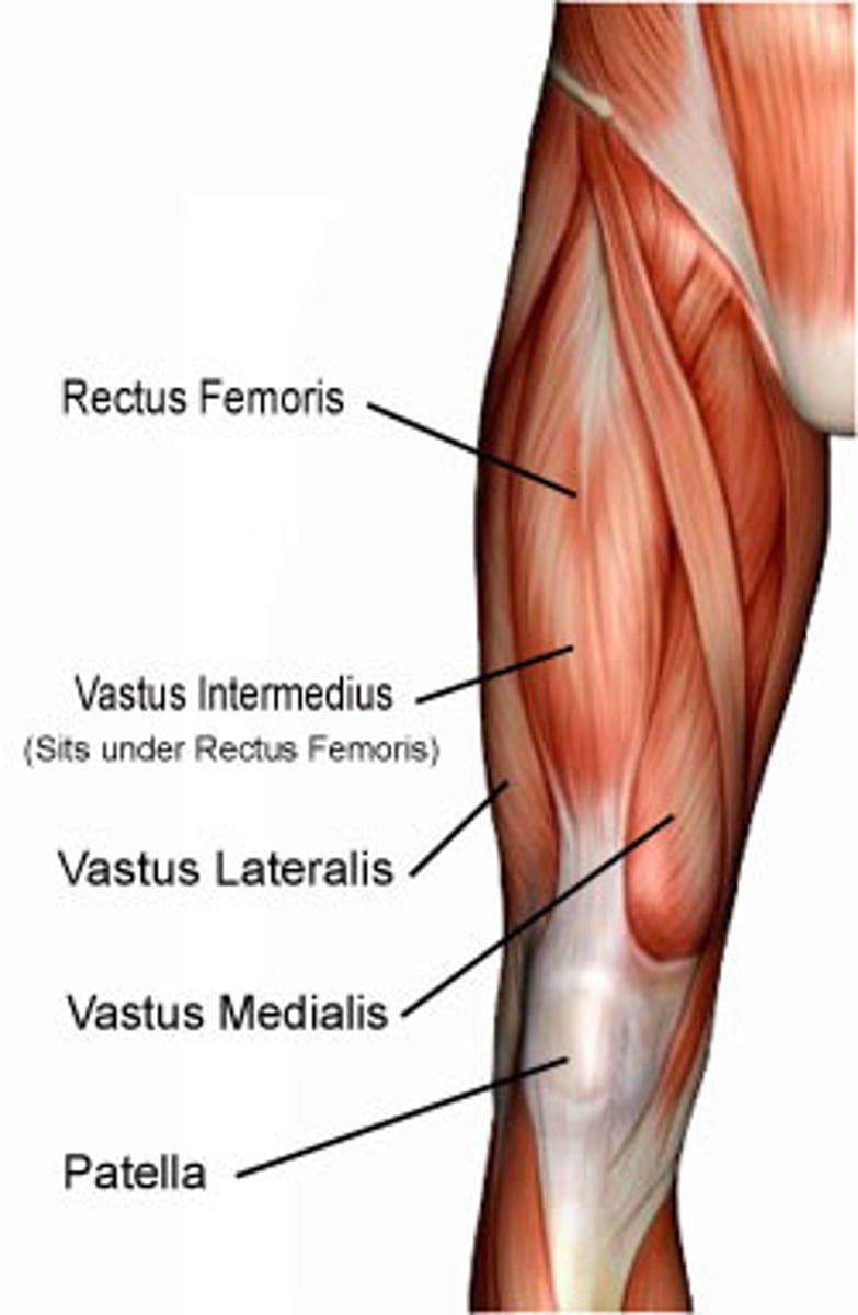
Rectus Femoris
Most superior muscle in quadriceps.
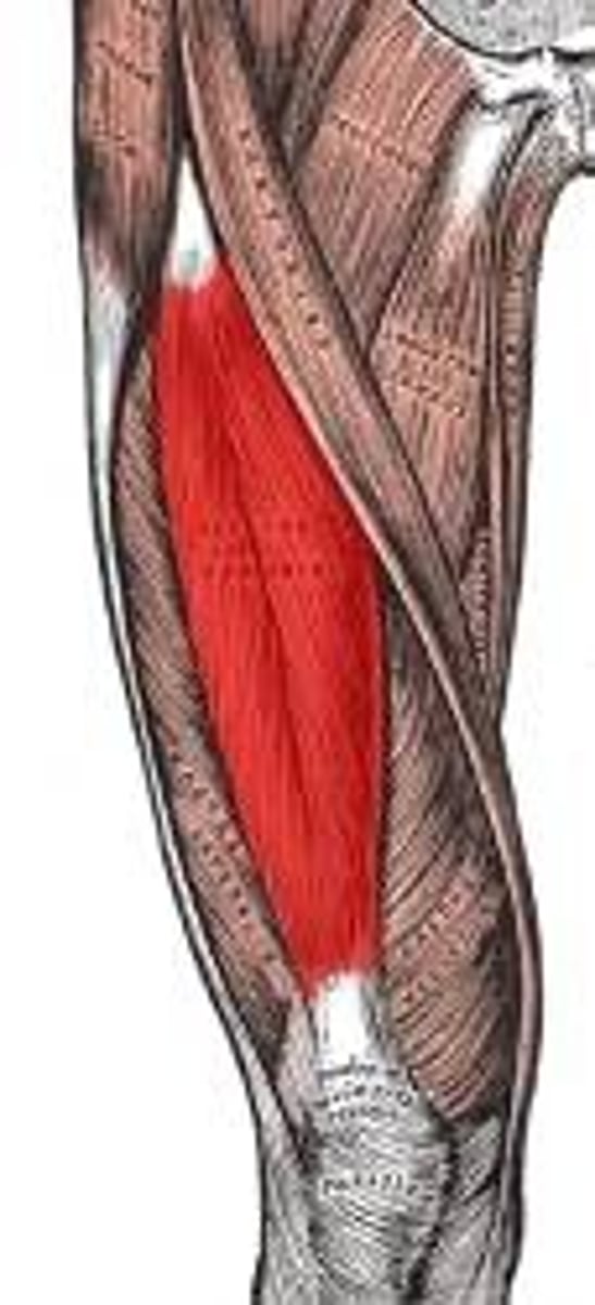
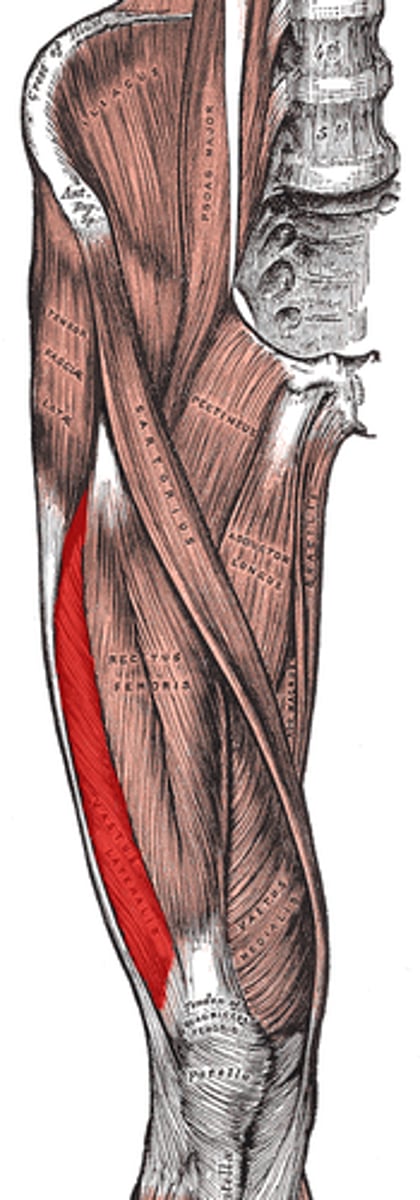
Vastus Lateralis
To the left and below the rectus femoris.
Vastus Medialis
Towards in the inwards of the midline on the thigh.
Vastus Intermedius
Intermediate-most muscle on the thigh.
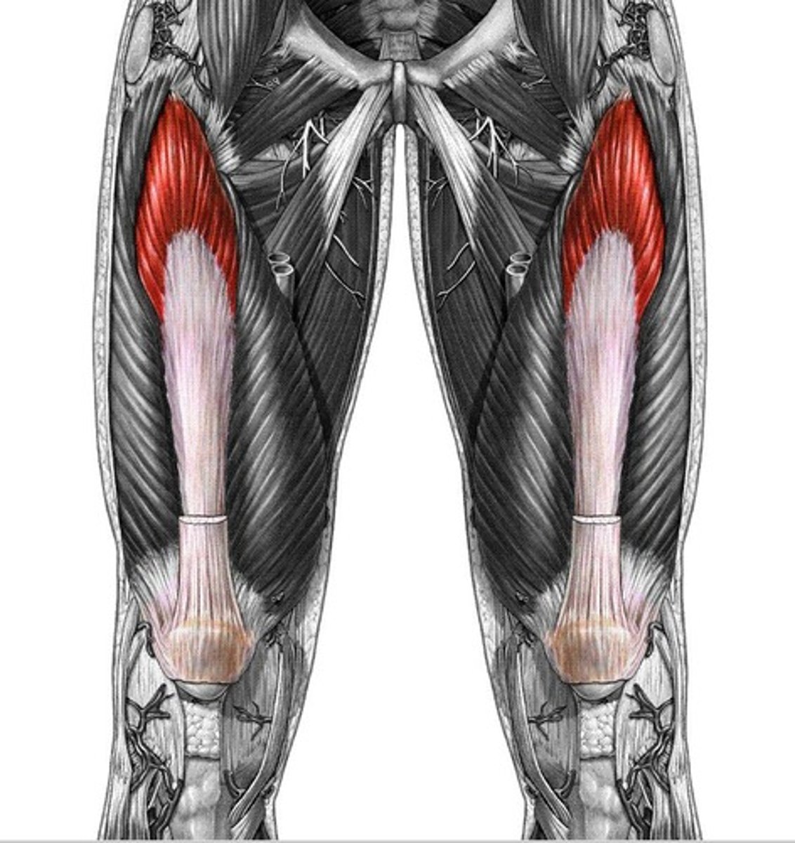
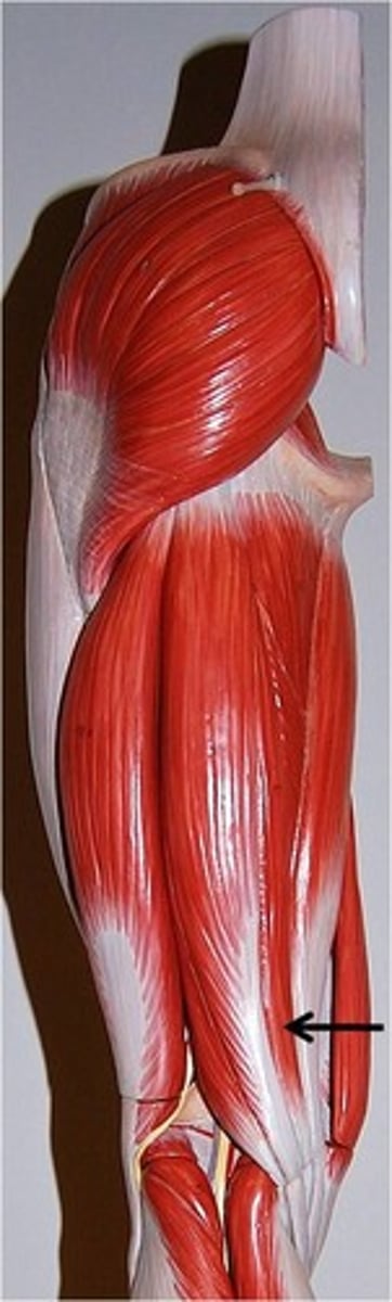
Hamstrings
Flexes knee.
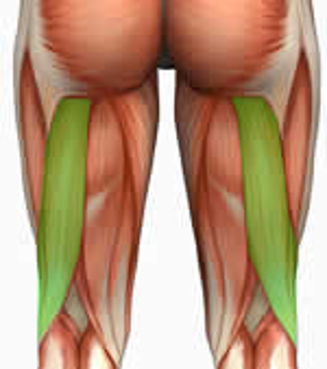
Biceps Femoris
Outside of posterior thigh.
Semimembranosus
Inside of posterior thigh.
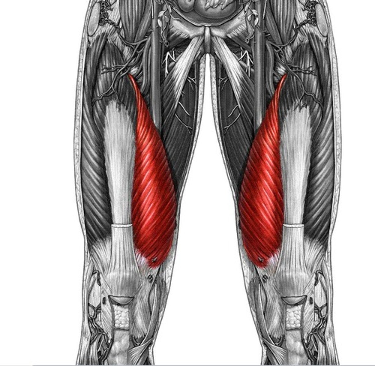
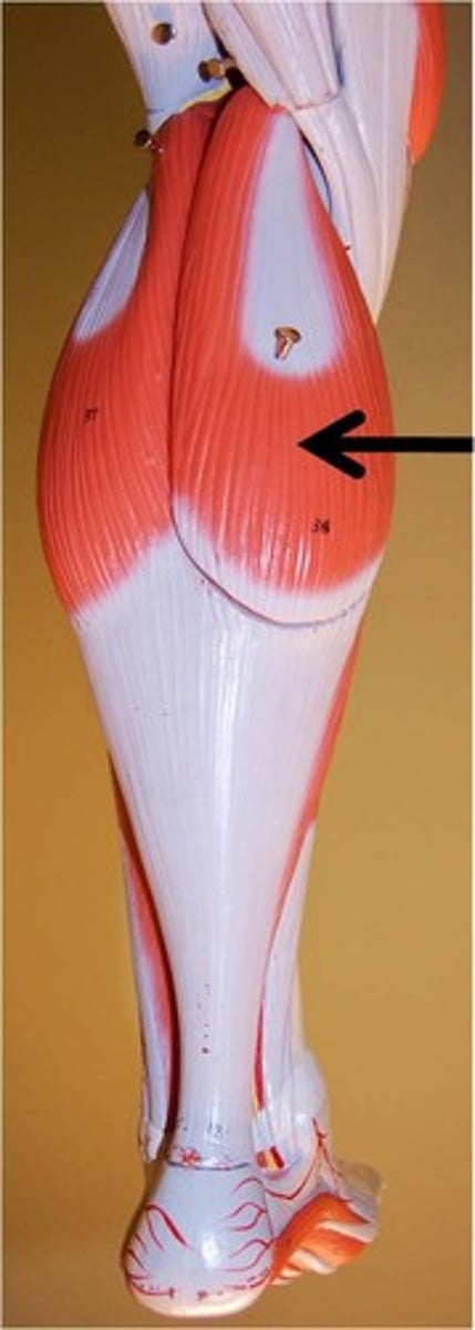
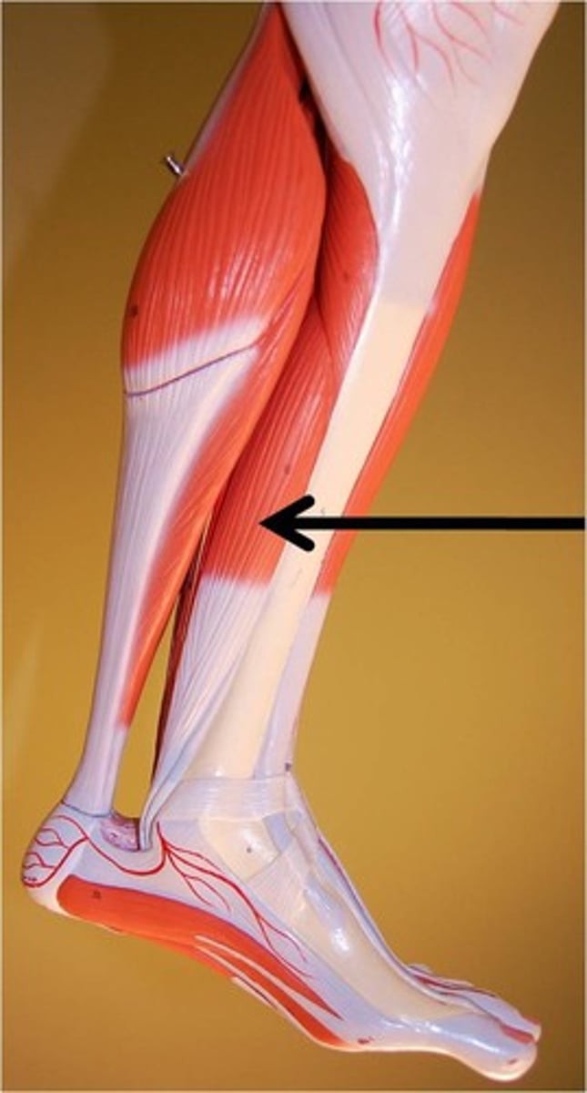
Femur (Origin)
Origin of Gastrocnemius
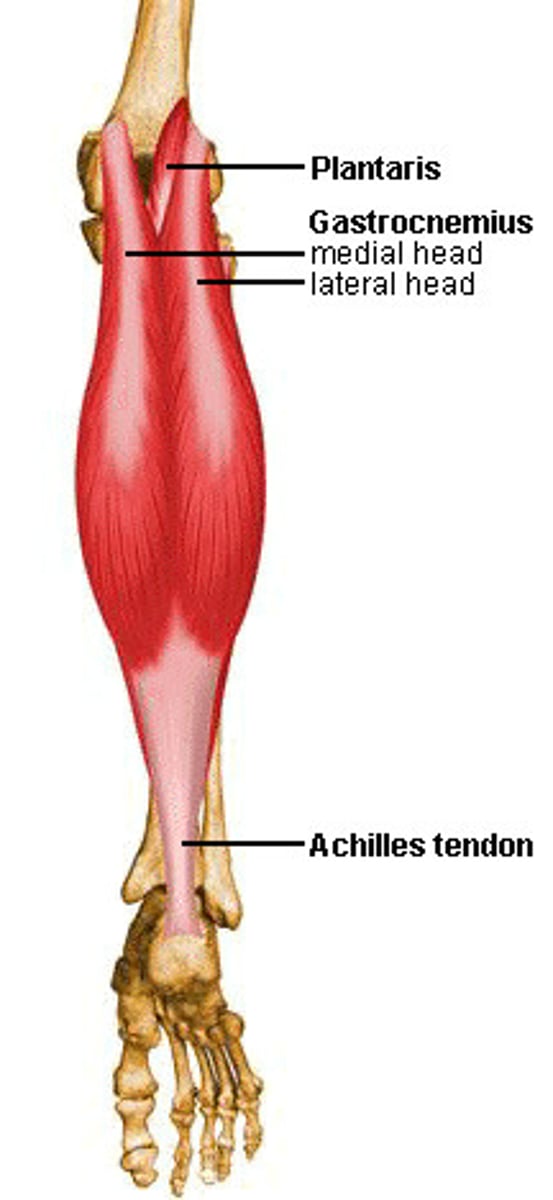
Calcaneus (Insertion)
Insertion of Gastrocnemius
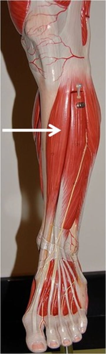
Gastrocnemius
Plantarflexes ankle; flexes knee.
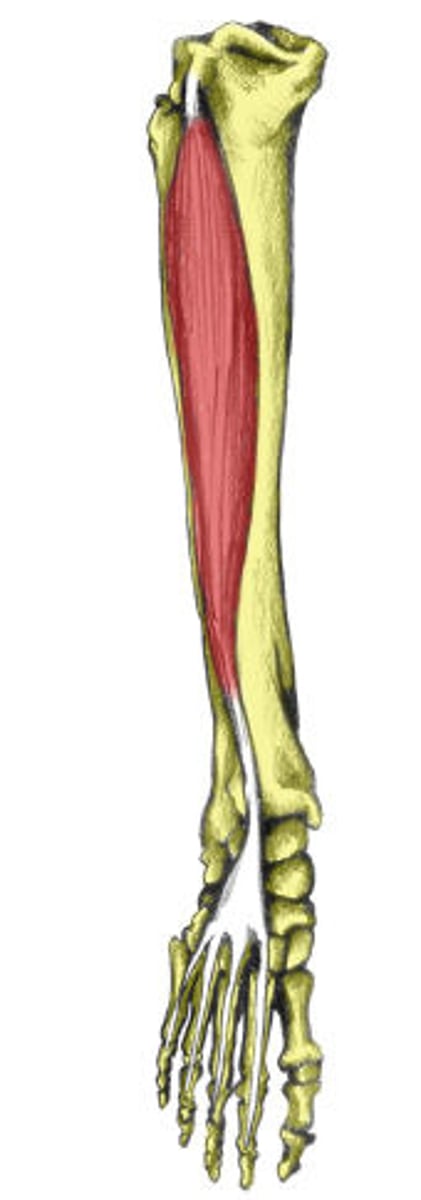
Posterior tibia. (Origin)
Origin of Flexor Digitorum Longus
Distal phalanges. (Insertion)
Insertion of Flexor Digitorum Longus
Flexor Digitorum Longus
Flexes toes 2-5; plantarflexes ankle; inverts foot.
Tibia/Fibula (Origin)
Origin of Extensor Digitorum Longus
Distal phalanges. (Insertion)
Insertion of Extensor Digitorum Longus
Extensor Digitorum Longus (EDL)
Extends toes and dorsiflexes foot.
Tibia (Origin)
Origin of Tibialis Anterior
1st Metatarsal (Insertion)
Insertion of Tibialis Anterior
Tibialis Anterior
Dorsiflexes and inverts foot.