URINARY SYSTEM
 Call Kai
 Learn
Learn Practice Test
 Spaced Repetition
 Match
Match Flashcards
 Knowt Play
1/68

Earn XP
Description and Tags
school notes
Name | Mastery | Learn | Test | Matching | Spaced | Call with Kai | Chat |
|---|
No analytics yet
Send a link to your students to track their progress
69 Terms
renal artery is _____ and ______ to the renal vein
posterior and superior
what is the layer of connective tissue encapsulating the kidneys and perinephric fat
gerota fascia
what is the process of urine formation
glomerular filtration
tubular reabsorption
tubular secretion
the left renal vein travels
anterior to the aorta
the right renal artery travels
posterior to the ivc
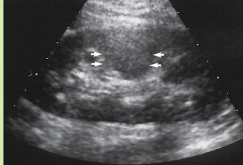
what is this
column of bertin
invaginations of the cortex between the medullae
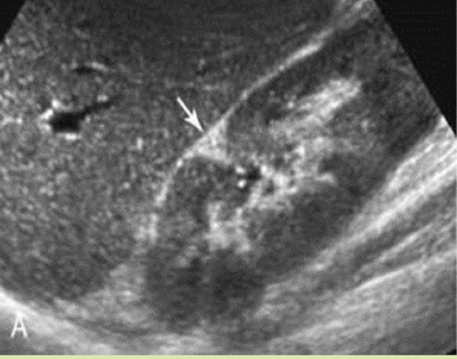
what is this
junctional parenchymal defect
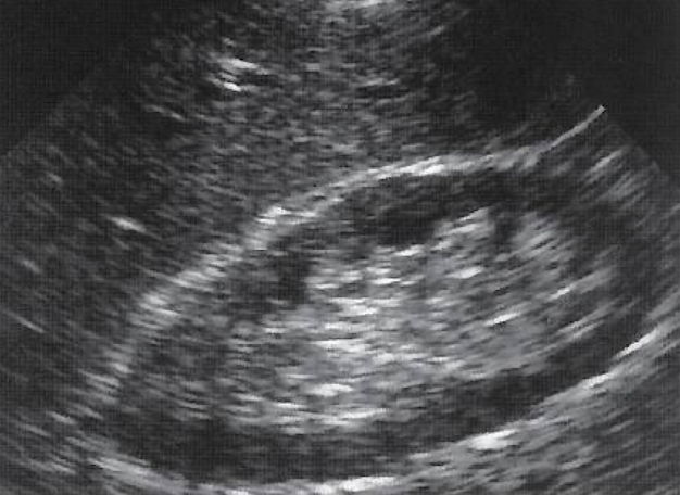
what is this
sinus lipomatosis'
deposition of a moderate amount of fat in the renal sinus
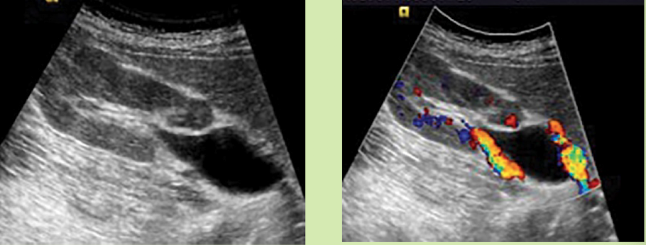
extrarenal pelvis
cystic area that extends beyond the renal borders
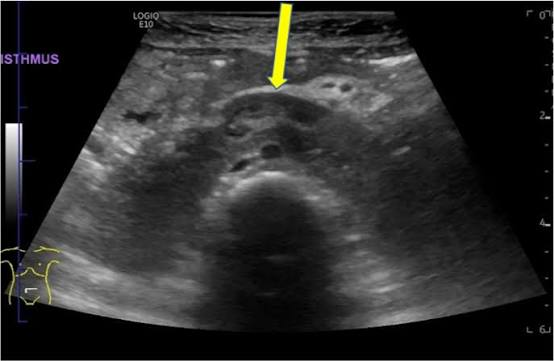
horseshoe kidney
lower poles of kidneys are fused
isthmus over aorta in trv plane
cross-fused ectopia
one kidney displaced across midline and the other fused to the other normally positioned kidney
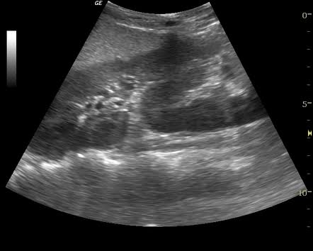
duplex kidney
complete duplication of collecting system
two distinct renal sinuses
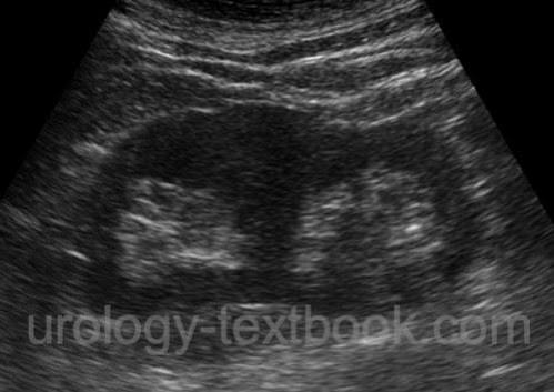
parapelvic cysts
cysts located in the renal sinus
do not communicate with collecting system
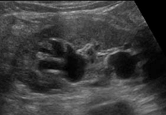
renal cysts associated with renal neoplasms
von Hippel-Lindau
several areas, abdominal cysts
Tuberous Sclerosis
multiple renal cysts/AMLs
mental retardation, seizures
Polycystic Kidney disease types
ARPKD (recessive)
infantile, rare
ADPKD (dominant)
adult, common
Autosomal Recessive polycystic kdiney disease (ARPKD)
dilation of renal collecting tubules causes renal failure
can be seen in utero (perinatal)
juvenile
hypertension
renal insufficiency
nephromegaly
periportal fibrosis
sono of ARPKD
symmetrically enlarged kidneys
microscopic kidneys
diffuse echogenicity
poor cortico-medullary differentiation
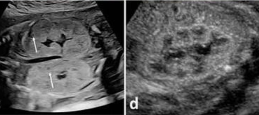
what are the s/s of autosomal dominant polycystic kidney disease (ADPKD)
pain
hypertension/headache
palpable mass
hematuria
UTI
renal insufficiency
what is the sono of ADPKD
bilateral
enlarged kidneys with multiple macroscopic asymmetrical cysts
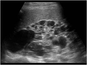
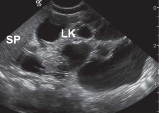
what is multicystic dysplastic kidney (MCDK)
common, nonhereditary
caused by early in utero urinary tract obstruction
bilateral in incompatible with life
large mass at birth
complications of MCDK
hypertension
hematuria
infection
flank pain
what is sono of MCKD
contralateral UPJ obstruction, nonfunctioning kidney, UNILATERAL
neonates/children
multicystic (bunch of grapes)
absence of renal parenchyma, renal sinus, atretic renal artery
adults
small atrophic/calcified kidney, echogenic
what is medullary sponge kidney (MSK)
occurs in medullary pyramids
dilation of distal collecting ducts, causing stasis of urine and stone formation.
may be unilateral or segmental
what are the s/s of medullary sponge kidney
asymptomatic. or
hematuria, infection, renal stones
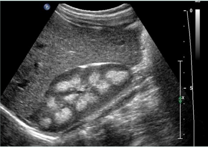
what is this
medullary sponge kidney
suspicious cysts look like
thick walls > 1cm
irregular walls
septations
calcifications
vascularity in septa/wall
what is the most common renal neoplasm
RCC
aka: Grawitz tumour, hypernephroma
what is the s/s of RCC
twice as common in men in their 60/70s
nonspecific s/s
hematuria, flank pain, palpable mass
what is sono of RCC
solid
most are isoechoic
the larger the more heterogenous
< 3cm they are usually hyperechoic (can look like AML)
hypo rim, intramural calcifications
invades IVC and renal veins
peripheral vascularity “basket sign”
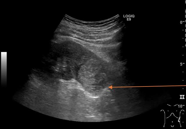
what comprises most of the malignancies that involve the renal pelvis, ureter, and bladder
TCC
what are the two types of TCC
Papillary
more common
Flat
smaller TCCs tend to be these
s/s of TCC
gross or macroscopia hematuria
flank pain
sono of TCC
hypo mass in collecting system
low vascularity
may invade adjacent renal parenchyma
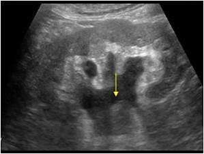
what is squamous cell carcinoma
rare, highly invasive tumour
hx of chronic irritation and gross hematuria
palpable kidney secondary to severe hydronephrosis
sono of squamous cell carcinoma
large mass is evident in renal pelvis
obstruction from kidney stones
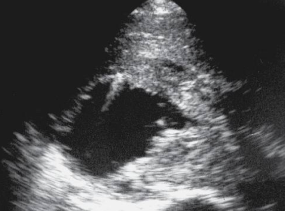
renal lymphoma
secondary is more common
bilateral invasion with multiple nodules
sono of renal lymphoma
enlarged, hypo kidneys
may simulate renal cyst without enhancement
very hypo, poorly defined margins
can be confused fro cyst
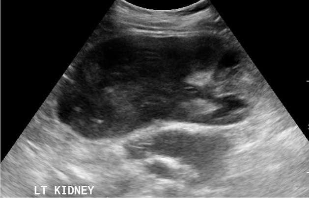
what is the m/c malignant renal tumour in pediatric pts
Wilms tumour (Nephroblastoma)
what is s/s of nephroblastoma (wilms)
palpable mass
non specific
pain, fever, malaise, weight loss, hematuria, hypertension
what is sono of wilms tumour
large, well circumscribed
homogenous
anechoic/cystic spaces
can invade LNs, renal veins, IVC
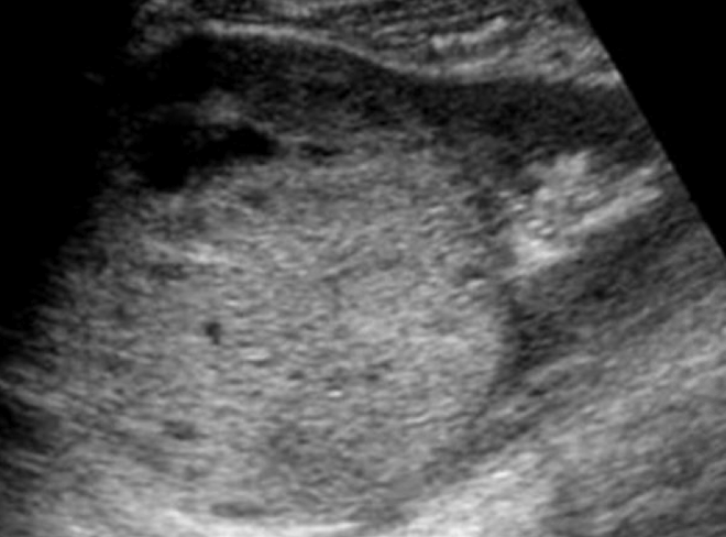
what is the most common benign renal tumour
angiomyolipoma
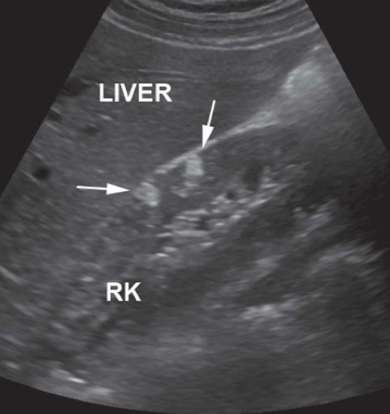
what are angiomyolipomas highly associated with
tuberous sclerosis
sono of renal adenomatous tumours
may be indistinguishable from RCC
solid
hyper to hypo
hypo vascular
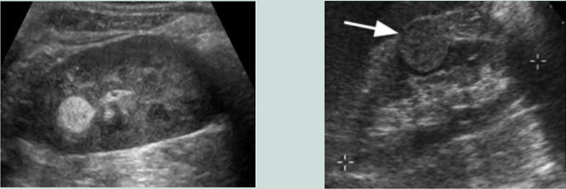
sono of oncocytoma
extremely difficult to differentiate from RCC
hypoechoic in 50% of cases
spoke wheel with central scar
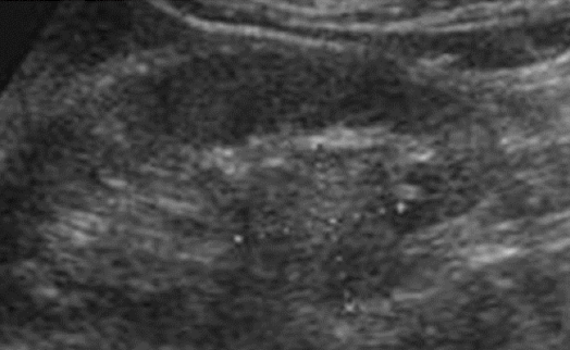
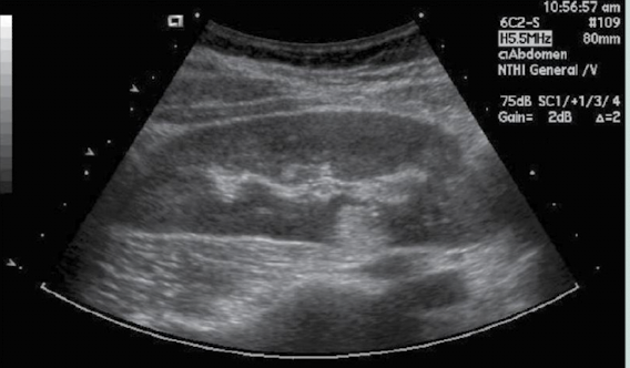
what is this
lipoma
what is acute glomerulonephritis
necrosis or proliferation occur in glomeruli
enlarged poorly functioning kidneys
suspected when echogenicity of renal parenchyma exceeds that of liver
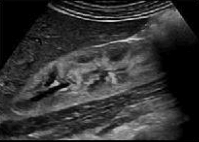
s/s of acute glomerulonephritis
nephrotic syndrome
massive protein uria
hypoalbuminea
hypertension
anemia
peripheral edema
what is acute interstitial nephritis associated with
infectious diseases
scarlet fever, diphtheria
sono of acute interstitial nephritis
enlarged mottles kidneys
increased renal cortical echogenicty
what is lupus nephritis
connective tissue disorder
affects women 20-40 more
s/s
hematuria, proteinuria, hypertension, RV thrombosis, renal insufficiency
sono of lupus nephritis
increased cortical echogenicity
renal atrophy
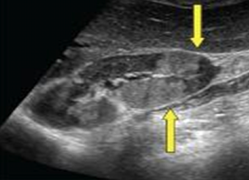
what does aids of kidney appear as
echogenic, enlarged kidneys
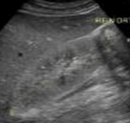
acute renal failure
can be caused by hypoperfusion of kidney
parenchymal disease, trhombosis, outflow obstruction
malignancy of bladder, prostate, uterus, ovaries, rectum
what is the most common renal disease to produce acute renal failure
acute tubular necrosis
what is the sono of acute tubular necrosis
bilaterally enlarged kidneys, hyperechoic pyramids
can appear as nephrocalcinosis
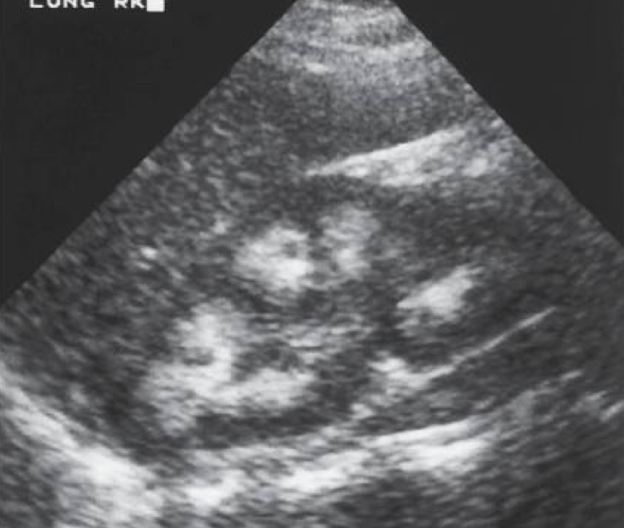
what is chronic renal disease
loss of renal fucntion as a result of disease
glomerulonephritis, chronic pyelonephritis, renal vascular disease, diabetes
sono of chronic renal disease
diffuse echogenic kidney with loss of normal anaotmy
nonspecific
Hydronephrosis common causes
UPJ obstruction (m/c)
UVJ/ distal urethral obstruction (by bladder)
duplication of the collecting system
what are s/s of hydronephrosis
renal insufficiency
low urine output
hypertension
sono of hydronephrosis
fluid-filled renal collecting system
thin parenchyma
hydroureter decrease
absent jets
highr RI > 0.7 suggests acute urinary obstruction
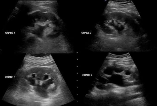
pyonephrosis
pus within collecting system
usually secondary to long standing ureteral obstruction from calculus
shows low level echoes with a fluid-debris level
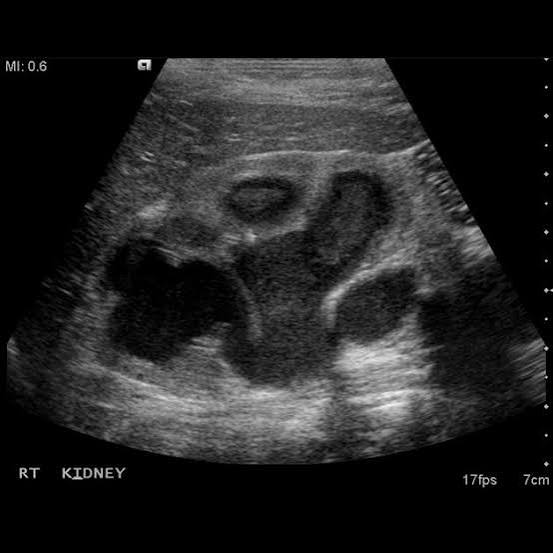
emphysematous pyelonephritis
when air is in parenchyma
may be caused by E.coli
diabetic pts
enlarged hypoechoic and inflamed kidneys
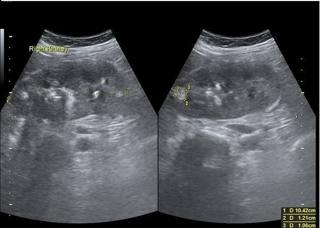
nephrocalcinosis
deposit of calcium in kidney
both kidneys affected
focal increases i echogenicity in renal pyramids
can be seen in medulla and cortex
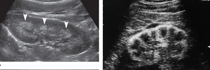
renal artery stenosis
> 180cm/s PSV
absence of early systolic peak
tardus parvus waveform
if a urolithiasis causes obstruction ______ will be seen
hydronephrosis
what is the m/c bladder tumour
TCC
s/s of bladder tcc
gross hematuria
dysuria
urinary frequency or urgency
what is this
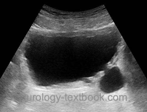
bladder diverticulum