Neurological System
 Call Kai
 Learn
Learn Practice Test
 Spaced Repetition
 Match
Match Flashcards
 Knowt Play
1/79

There's no tags or description
Looks like no tags are added yet.
Name | Mastery | Learn | Test | Matching | Spaced | Call with Kai | Chat |
|---|
No analytics yet
Send a link to your students to track their progress
80 Terms
Neuron components
Cell body
Dendrites
Axon
Neuron types
Sensory (afferent): To CNS
Motor (efferent): Away from CNS to body
Interneuron
Additional:
Supporting cells
Interneuron
Most abundant neuron type, provide connections between neurons and transmit signals between afferent and efferent neurons
Supporting cells
Neuroglia: provide support and nutrition, form myelin and support homeostasis
Membrane potential
Electrical charge difference between inside and outside of the cell
Action potential
A temporary, specific type of change in the broader membrane potential of a cell.
A rapid, temporary spike and reversal of the membrane potential when a cell is excited and fires a signal
Components of Action potential
Resting membrane potential: Na and K channels closed, inside of cell is more negative than outside
Depolarization phase: A stimulus hits the threshold, Na channels open and sodium rushes into the cell, making the inside positive
Repolarization phase: Na channels close and become inactive, K channels open, making potassium flow out of the cell turning the inside negative again.
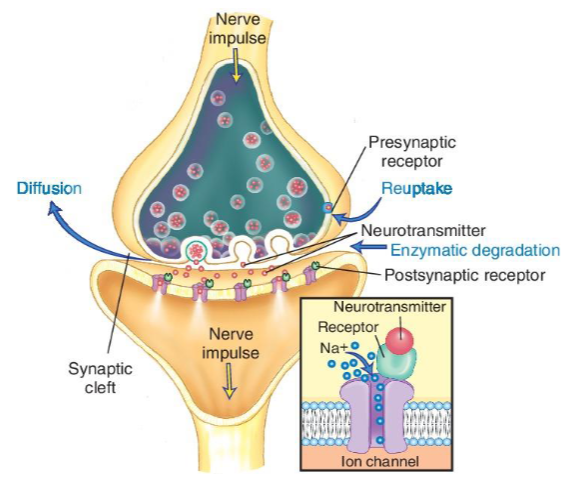
Flow of information across a synapse
Electrical
Chemical
Synapse structures: presynaptic terminal, synaptic cleft, postsynaptic membrane
Processes of Neural Injury
Types:
Chromatolyosis
Atrophy
Neuronophagia
Intraneuronal inclusions
Chromatolyosis
Neuron swells as a result of an injury
Atrophy
Neurons shrink
Neuronophagia
Dead neuron damages nearby cells, causing phagocytosis and inflammation
Intraneuronal inclusions
Structures formed in the nucleus or cytoplasm
Central nervous system organization
Spinal cord
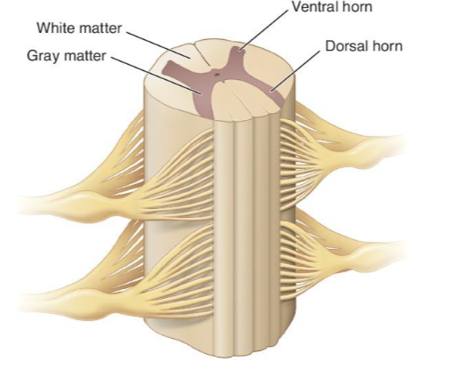
Ventral horns
Anterior extensions
Efferent motor neurons leaving cord
Dorsal horns
Posterior extensions
Sensory neurons receiving afferent impulses
Tissue composition
White matter: Axons and dendrites
Gray matter: Cell bodies
CNS Pyramidal and Extrapyramidal systems
Pyramidal
Extends from the sensorimotor areas of the brain to the motor neurons of the ventral horn of the spinal cord
Controls voluntary movement
Extrapyramidal
Basal ganglia
Fine-tunes and stabilizes movement
CNS protection
Cerebrovascular circulation: can control BP
Blood-brain barrier
CSF
Neural injury responses
Astrogliosis
Microglial nodules
Axonal degeneration
Axonal regeneration
Neuropathy
Demyelination
Astrogliosis
In CNS, astrocytes respond to tissue injury by forming a glial scar (examples of injury: contusions, wounds, tumors, hemorrhages)
Microglial nodules
Form when microglia is joined with astrocytes; help fight infection
Axonal degeneration
In PNS, necrosis resulting from injury to neuron cell body or axon. Inflammatory response leads to phagocytosis of cellular debris.
Axonal regeneration
May be possible if cell body and proximal axons are not damaged
Neuronopathy
Damage to cell body causes axonal degeneration
Demyelination
Caused by damage to oligodendrocytes (myelin segments that increase the speed of neuron impulse conduction)
Neuropathy
Nerve degeneration causing delayed nerve conduction and impaired sensory function
PNS organization
Somatic
Motor and sensory impulses between the CNS and periphery (peripheral nerves)
Cranial nerves
Spinal nerves
Dermatomes
Plexus formation
PNS Autonomic components
Preganglionic neurons
Autonomic ganglion
Postganglionic neurons
Activated Autonomic system = Automatic (Fight or Flight)
Sympathetic
Increased heart rate, BP, RR
Bronchodilation
Vascular smooth muscle constriction
Decreased urination and GI function
Activated Parasympathetic (Rest and Digest)
Heart rate and RR decrease
BP decreases
Smooth muscle relaxes to allow for blood flow
Bronchial constriction
Relaxation of anal and bladder sphincter
Neural Injury

Cerebral Palsy Pathophysiology
Group of neuromuscular disorders
Caused by event during the antenatal or postnatal period that damaged the upper motor neurons
Classification
Motor dysfunction type: Spastic, Athetoid/dyskinetic, Ataxic
Anatomic involvement: Hemiplegia, Diplegia, and Quadriplegia
Cerebral Palsy Clinical Manifestations
Variable severity
Limited fine motor skills
Lack of coordination and balance
Impaired cognitive function
Speech disorder
Seizure disorder
Cerebral Palsy Diagnostic Criteria
Diagnosis of exclusion
History and Physical examination
Neurologic exam
Testing motor skills and reflexes
Developmental milestones
Epilepsy
Seizure disorder; result of impaired chemical and electrical neurotransmission
Seizure types
Partial/Focal: are short lived
Partial seizures
Simple partial
Complex partial
Generalized: affect both sides of brain
tonic clonic
absence
myoclonic
atonic
epileptic spasm
Partial seizures
Start local and can spread to other side of the brain
Simple partial
Limited to originating hemisphere and can involve other motor or sensory brain components
Complex partial
involve both hemispheres and result in loss of consciousness and lack of memory
Generalized seizures
Caused by a more generalized electrical transmission
Absence seizures
Brief change in LOC, and eye/mouth movements. Can occur up to 100 times a day
Myoclonic seizures
Involuntary muscle movements of extremities not associated with LOC
Tonic clonic
Convulsive and associated with tonic (continuous muscle contraction) and clonic (alternating muscle contractions and relaxation) LOC and traumatic injury related to fall may result
Seizure Recovery (postictal state)
Extreme fatigue
Headache
Muscle pain and weakness
Status epilectus: continuous tonic-clonic seizures causing hypoxia
Multiple Sclerosis Pathophysiology
CNS demyelination also affects PNS
Etiology
Genetics
Environmental factors
Triggering event
Autoimmunity
Regional variation
Inflammatory T and B lymphocytes and IgG and IgM in CSF
MS Manifestation Types
Related to slowed nerve conduction
Clinical course variation:
Clinically isolated syndrome
Relapsing-remitting
Primary progressive: slow, chronic deterioration of neurologic function with plateaus
Secondary progressive: initially presenting with relapsing-remitting, followed by slow, chronic deterioration
MS Clinical Manifestations
Unilateral vision loss
Cognitive loss
Bowel and bladder dysfunction
Altered gait and balance
Spasticity (continuous muscle stiffness)
Pins and needles (Paresthesias)
Slurred speech
Fatigue
Pain
Pseudobulbar effect (uncontrollable laughing, crying)
MS Diagnostic Criteria
Complete medical history
Neurologic exam
MRI, CNS response to peripheral stimuli
Lumbar puncture to evaluate CSF
Evoked potential testing: time it takes brain to respond to stimuli
Hydrocephalus Pathophysiology
Increased ventricular accumulation of CSF
Ventricular enlargement
Increased ICP
Classifications
Noncommunicating
Communicating
Congenital
Acquired
Hydrocephalus causes
Intraventricular hemorrhage
Neural tube defect
Brain tumor
Meningitis
Traumatic injury
CSF flow obstruction
Impaired CSF absorption
Hydrocephalus Age dependent Clinical Manifestations
Infant
Enlarged head with bulging fontanels
Scalp vein distention
Difficulty feeding
High, shrill cry
Older children/adults
Impaired motor and cognitive function
Incontinence
Hydrocephalus Clinical Manifestations
Increased intracranial pressure
Increased BP
Altered HR
Headache
Vomiting
Decreased LOC
Papilledema
Hydrocephalus Diagnostic Criteria
Head circumference measurement
Transillumination
Imaging studies
Hydrocephalus treatment
Establishment and maintenance of normal CSF volumes and ICP
Ventriculoatrial shunt
Endoscopic third ventriculostomy
Incomplete Spinal Cord Transection Pathophysiology
Spinal cord injury altering neuronal transmission
Categories
Complete transection
Partial transection
Central cord syndrome
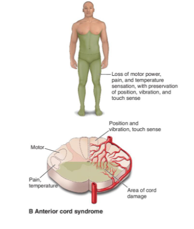
Anterior cord syndrome
Brown-Sequard syndrome
Incomplete Spinal Cord Transection Clinical Manifestations
Variable
Spinal segmental level
Type of injury
Degree of cord transection
Central cord syndrome

Anterior cord syndrome
Brown-Sequard syndrome

Incomplete Spinal Cord Transection Diagnostic Criteria
Physical examination
Cognitive function
Motor function
Sensory function
Imaging studies
Parkinson Disease Pathophysiology
Chronic, progressive neurologic condition
Leads to degeneration of pigmented dopaminergic neurons of the basal ganglia
Causes impaired transport of dopamine
Presence of Lewy bodies (protein)
Results in neuronal injury from oxidative damage being suspected
Parkinson Disease Clinical Manifestations
Tremor
Rigidity
Jerky movements
Flat affect
Bradykinesia (slowed movement)
Shuffling gait and stooped posture
Postural instability
Parkinson Disease Diagnostic Criteria
History and Physical exam
Hoehn and Yahr scale
Stage 1: mild, unliateral
Stage 2: bilateral, posture and gait affected
Stage 3: slowed movements, impaired balance
Stage 4: severe, limited walking, rigidity
Stage 5: cachectic, total disability
Stroke Pathophysiology
Any clinical event that leads to the impairment of cerebral circulation
Thrombotic, embolic, or hemorrhagic
Inflammation, ischemia, death of neurons
CVA or TIA
Stroke Clinical Manifestations
Focal brain injury
Dizziness, diplopia
Abrupt onset of hemiparesis
Ataxia, aphasia
Vision loss or field deficits
Sudden decreased in LOC
Stroke Diagnostic Criteria
History and Physical examination
Lab studies
CT scan
Alzheimer Disease Pathophysiology
Neurodegenerative disorder
Neurofibrillary tangles
Tau protein
Senile plaques
B amylod protein
Inflammation
Neuronal atrophy
Genetic and environmental factors

Alzheimer Disease Clinical Manifestations
7 stages
General
Global cognitive decline
Behavior changes
Eventual loss of function
Severity based on stage and progression
Initial symptoms
Memory loss
Confusion
Restlessness
Mood swings
Difficulty interpreting visual information
Alzeheimer Disease Diagnosis
History and physical exam
Neurologic evaluation
Mental status
Communication ability
Memory
Math ability
Balance
Imaging studies to rule out other conditions
Brain scan
MRI
CT scan
Definitive diagnosis at autopsy
Migraine Headache Pathophysiology
Recurrent headaches with aura
Moderate to severe intensity
Duration of 1-2 days
Cause: likely initiated by neurologic and biochemical events
Migraine Headache Clinical Manifestations
Pain
Unilateral
Pulsing and throbbing
Aura
Visual disturbances preceding headache may be associated
Prodromal symptoms
Mood changes
Food cravings
Constipation
Migraine Headaches Diagnostic Criteria
History and Physical examination
Imaging for acute, sudden onset
MRI
CT scan
Brain Cancer Pathophysiology
Metastasis much more common to brain than primary tumors
Primary tumors
Gliomas (in cerebrum)
Meningiomas
Pituitary adenomas
Acoustic schwannoma
Brain Cancer Clinical Manifestations
Loss of motor/sensory function
Cognitive or behavioral changes
Headache
Vomiting
Seizures
Brain Cancer Diagnostic Criteria
History and Physical examination
Imaging studies
Huntington Disease Pathophysiology
Progressive degenerative neurologic disorder with an autosomal dominant inheritance pattern
Defect on chromosome 4
Degeneration of the basal ganglia and cortical regions of the brain
Movement
Emotional
Cognitive impairments
Huntington Disease Clinical Manifestations
Present in all with inherited defective gene
Involuntary movements
Dyskinesia
Chorea (involuntary, rapid jerky movements)
Cognitive impairment
Emotional disturbance
Huntington Disease Diagnostic Criteria
Family and personal medical history
Genetic testing
Physical exam
Blood testing
Mental, cognitive, and emotional evaluation
CT, MRI, PET scans
Bacterial Meningitis
Inflammation of the meninges of the brain and spinal cord
Commonly caused by N. meningitidis
Respiratory droplet transmission; mechanism for entry into CNS unknown
Bacteria proliferate in CNS
Exudate damages and obstructs CNS structures, leading to reduced oxygen to the brain
Bacterial Meningitis Clinical Manifestations
Rapid and sever onset
Severe headache
Photophobia
Nuchal rigidity
Decreased alertness
Loss of consciousness
Changes in mental status
Vomiting
Seizures
Fever
Leukocytosis
Anorexia
Bacterial Meningitis Diagnostic Criteria
History and Physical examination
Kernig sign
Brudzinski sign
Blood cultures
CSF analysis and cultures

Meningitis types
