management of traumatic injury
 Call Kai
 Learn
Learn Practice Test
 Spaced Repetition
 Match
Match Flashcards
 Knowt Play
1/109

Earn XP
Description and Tags
lecture given 6/12/2026
Name | Mastery | Learn | Test | Matching | Spaced | Call with Kai | Chat |
|---|
No analytics yet
Send a link to your students to track their progress
110 Terms
dental trauma
an acute transmission of energy to the tooth and supporting tissues through a shock resulting in fracture and/or displacement of the tooth and/or separation or crushing of the supporting tissues (gingiva, PDL, bone)
what is the epidemiology of dental trauma?
peak ages 2-3 yr, 9-10 yr
incisors most affected
boys > girls
50% of children <15 yr, 60% deciduous teeth, 40% permanent
what is the etiology of dental trauma?
falls
traffic injuries
acts of violence
sports accidents
how should you begin an examination and diagnostic testing after trauma?
clean pts face and disinfect injured tissues
detailed history of the trauma
physical exam
radiographic exam
photographic registration
what questions should be asked after dental trauma?
when, where, how?
was there a loss of consciousness?
is there a change in occlusion?
is there an increased reaction to heat/cold?
medical history
what should be done during the physical exam after trauma?
pupillary reflex, blood pressure, pulse, bleeding (ear, nose)
head and neck hematomas, existence of an injury that would require the intervention of stitches
oral opening: amplitude, opening path, pain
soft tissue (throat, lips, cheeks, gums), in case of bleeding make a hemostasis with a sterilized gauze
in case of tooth loss look for foreign bodies, palpate all injuries and perform x rays
what should be done during the dental portion of a physical exam after trauma?
dental age of the child, search for the fractured/displaced tooth and classify the trauma
mobility, percussion, sensibility test
mobility test
to determie the extent of loosening of individual teeth and mobility of a group of teeth
0- no loosening
1- horizontal loosening <1mm
2- horizontal loosening >1mm
3- axial loosening
why is mobility testing important
distinguish between no mobility (normal physiologic) and no mobility (injured blocked tooth) after intrustion, lateral luxation, or ankylosis at the follow up visit
percussion test
to determine the health of the tooth and surrounding structures
if tenderness to percussion there is damage to PDL
if percussion not easily felt through the tooth and sounds like high metalic tone the tooth is blocked in the bone - lateral luxation/intrusion or ankylosis
pulpal sensibility test
to determine the nerve function and NOT to indicate the presence or absence of blood circulation within the pulpal space
cold test AND EPT
important for future comparison testing and follow up
t/f with pulp sensibility tests, there is a possibility of false negative readings
true- if the condution capability of the nerve endings is altered
this is NOT a sign of pulp necrosis but a sign of pulpal damange- many pulps will recover but it may take several months
why is radiographic examination done after trauma?
diagnosis, initial medical file, insurance
what are the 4 radiographs that should be taken after injury?
a direct 90 degree on the axis of the tooth
2 with different vertical/horizontal angulations
one occlusal film
what is the benefit of CBCT in dental trauma?
to determine the exact location, extent, and direction of fracture
should you take a CBCT after trauma just because?
no- there is a risk of hematological malignancies so use ALARA
x rays of soft tissue
check the presence of foreign substances or tooth fragments
reduce the intensity
when should the suturing of lip lesions be performed?
after the intra oral treatment has been completed
why is photographic registration important?
follow up examination
monitoring soft tissue healing
assessment of tooth discoloration
re-reuption of an intruded tooth
medico-legal documentation used in litigation cases
t/f no single element of the diagnostic process should be relied on to make even waht appears to be an uncomplicated diagnosis- before any treatment there should be at least 2 independent diagnostic test results
true
uncomplicated crown fractures
enamel infraction, enamel fracture, enamel-dentin fracture
complicated crown fractures
enamel-dentin-pulp fracture
enamel infraction
an incomplete fracture or crack of the enamel without loss of tooth structure
cracks visible by indirect illumination, + vitality test, normal mobility, - percission
x rays: 1 occlusal, 3 PA: straight on, mesial, distal, soft tissues in case of laceration
what is the treatment for enamal infraction?
abstention or polishing
no follow up is generally needed
6-8 weeks and 1 year recommended but not required
enamel fracture
a fracture confined to the enamel with loss of tooth fracture
+vitality test, normal mobility, - percussion
x rays: 1 occlusal, 3 PA: straight on, mesial, distal, soft tissues in case of laceration
what is the treatment for enamel fracture?
polishing, reconstruction
recall: 4 weeks, 6-8 weeks, 1 year (monitor vitality)
enamel- dentin fracture
a fracture confied to enamel and dentin without pulp exposure
+vitality test, normal mobility, - percussion
x rays: 1 occlusal, 3 PA: straight on, mesial, distal, soft tissues in case of laceration
what is the treatment for enamel-dentin fracture?
seal exposed dentin ASAP- minimize bacterial ingress, reduce patient discomfort
if remaining dentin thickness is <0.5mm (dentin is pink)- pulp capping
if remaining dentin thickness is >0.5mm- direct restorative treatment, possible to glue the fractured fragment if available
recall: 4 weeks, 6-8 weeks, 1 year (monitor vitality)
enamel-dentin-pulp fracture
a fracture confined to enamel and dentin, with pulp exposure
+vitality, normal mobility, - percussion
x rays: 1 occlusal, 3 PA: straight on, mesial, distal, soft tissues in case of laceration
recall: 4 weeks, 6-8 weeks, 6 months, yearly for at least 5 years
what is the treatment for enamel-dentin-pulp fracture?
depends on the stage of development of the tooth
immature- apexogenesis, apexification, regeneration
mature- non-surgical root canal treatment, vital pulp therapy (pulp capping/pulpotomy)
treatment also depends on the time between trauma and treatment, the concomitant periodontal injury, and the restorative treatment plan
immature teeth
immature permanent tooth has considerable capacity for healing after traumatic pulp exposure
every effort should be made to preserve the pulp to ensure continued root development
crown root fractures
the fracture includes enamel, dentin, and cementum with (complicated) or without pulp exposure (uncomplicated)
+vitality test, + mobility on the coronal fragment, +percussion
x rays: 1 occlusal, 3 PA: straight on, mesial, distal, soft tissues in case of laceration
CBCT: consider if needed to reveal the extension and direction of the fracture
what is the treatment for crown root fractures?
ensure restorability- periodontal challenge rather than an endodontic one
with or without gingivectomy or crown lengthening, orthodontic extrusion, surgical extrusion
without pulp exposure- cover the exposed dentin
with pulp exposure- immature tooth→vital pulp therapy, mature tooth→non surgical root canal treatment
recall: 1 week, 4 weeks, 6-8 weeks, 4 months, 6 months, 1 year, yearly for at least 5 years
root fractures
include cementum, dentin, and pulp
can be localized at the apical, middle, or cervical 1/3
unusual with permanent incisors with incomplete root develpment (more prone to avulsion)
tooth can appear elongated
coronal fragment may be mobile and is displaced sometimes
apical fragment is usually not displaced → apical blood circulation is not disrupted (apical necrosis is rare)
vitality may be initially - indicating transient pulpal damage, + percussion
x rays: 1 occlusal, 3 PA with different vertical/horizontal angulation (located fracture in the cervical 1/3), soft tissues in case of laceration
CBCT: if needed, location, extent, and direction of the fracture
why is it important to take different angulations of a root fracture?
the fracture can appear different on radiographs, need multiple angles to confirm number of fractures
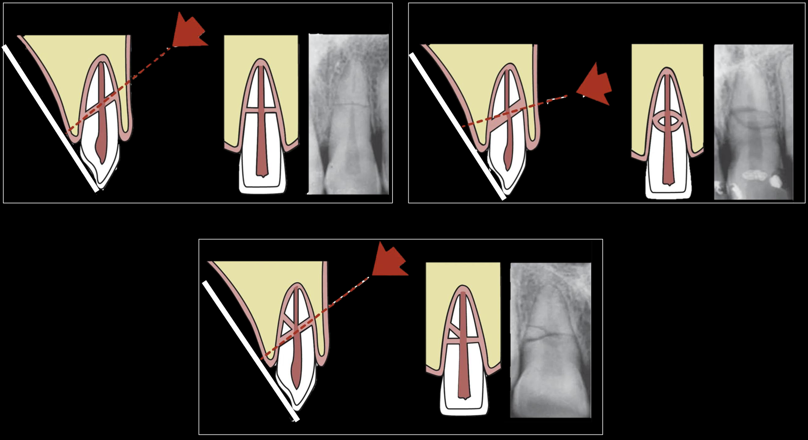
what is the treatment for root fracture
determine location of fracture
reposition coronal fragment ASAP, check with an x ray
stabilize the tooth with a flexible splint for 4 weeks (if the root fracture is near the cervical area, up to 4 months)
recall: 4 weeks, 6-8 weeks, 4 months, 6 months, 1 yr, yearly for at least 5 yrs
when indicated- NSRCT only to the coronal fragment, or surgical removal of the apical fragment
what are the responses to root fractures?
healing with calcified tissues
healing with interproximal connective tissue
healing with interproximal bone and connective tissue
no healing- interposition of granulated tissues
healing with calcified tissues
occurs after minimal or no displacement of the coronal fragment, most often in teeth with immature root formation
dentin (odontoblasts) + cementum (periodontium)
normal sensibility
normal mobility
fracture line discernible but fragments in close contact
pulp tissue of the coronal fragment: intact
successful healing!
healing with interproximal connective tissues
occurs after displacement of the coronal fragment
PDL cells
normal sensibility
increased mobility
fracture lines visible, fragments separated by a narrow radiolucent line
pulp space obliteration of coronal fragment is possible
successful healing!
healing with interproximal bone and connective tissue
occurs after root fracture prior to completed growth of the alveolar bone (young pts)
fragments separated by a distinct bony bridge
total pulp canal obliteration in both fragments is common
successful healing!
interposition of granulated tissues
occurs when bacteria gain access
granulation tissue is formed
negative sensibility
excessive mobility
a wide fracture line with a radiolucency (bone resorption)
necrosis of the coronal fragment
no healing!
supra-crestal root fracture in the coronal third
no PDL attachment to bone
pulp of both fragments will be susceptible to bacterial infection through the fracture line
healing is unlikely to occur
what should you do if your pt has a supra-crestal root fracture in the coronal third?
removal of the coronal fragment
if apical fragment restorable (gingivectomy, crown lengthening, orthodontic, or surgical extrusion) → RCT
if apical fragment not restorable → extraction
concussion
injury to the tooth supporting structures without mobility or tooth movement
+vitality, - mobility, + percussion
x ray: 3 PA different angulations to show normal tooth position in the socket
no treatment, release the traumatic occlusion
recall: 2 weeks, 4 weeks, 6-8 weeks, 3 mo, 6 mo, 1 yr, yearly for at least 5 yr
rare necrosis and root resorption
subluxation
injury to the tooth-supporting structures with mobility but without displacement of the tooth
+vitality, slightly + mobility, + percussion, bleeding from gingival suclus
x ray: 3 PA different angulations to show normal tooth position in the socket
no treatment (flexible splint if needed for 2wk), release from traumatic occlusion, pt under soft diet
recall: 2 wks, 4 wks, 6-8 wks, 3 mo, 6 mo, 1 yr, yearly for at least 5 yr
rare necrosis and root resorption
extrusive luxation
partial displacement of the tooth following the axis out of its socket but without leaving the socket
-vitality, ++ mobility, + percussion, tooth appears elongated
x ray: 1 occlusal and 3 PA different angulations- tooth appears dislocated with the apical part of the socket empty
what is the treatment for extrusive luxation?
repositioning and x ray, non rigid splint for 2 wks, release the traumatic occlusion, pt under soft diet
recall: 2 wks, 4 wks, 6-8 wks, 3 mo, 6 mo, 1 yr, yearly for at least 5 yr
if necrosis- immature teeth regeneration or apexification, mature teeth RCT
lateral luxation
lateral eccentric displacement of the tooth, usually associated with a fracture of the alveolar bone plate
-vitality, tooth in the bone immobile, + percussion with metallic sound, fracture of the alveolar bone may be palpable
x ray: 1 occlusal and 3 PA different angulations- tooth appears dislocated with the apical part of the socket empty
what is the treatment of lateral luxation?
tooth slightly pulled down to disengage the apex from the bone then repositioned (with anesthesia) and x ray
non rigid splint for 4 wks, release traumatic occlusion, pt under soft diet
recall: 2 wks, 4 wks, 6-8 wks, 3 mo, 6 mo, 1 yr, yearly for at least 5 yrs
if necrosis immature tooth regeneration or apexification, mature tooth RCT
intrusive luxation
displacement of the tooth deeper into the alveolar bone
it is a complex injury- compression and rupture of the PDL, the bone, and the neurovascular supply of the tooth
-vitality, locked tooth immoble, + percussion with metallic sound, infra occluded tooth, fracture of the alveolar bone may be palpable
x ray: 1 occlusal and 3 PA different angulation, tooth appears dislocated in an apical direction with partial or complete disapperance of PDL space
what is the treatment for intrusive luxation of an immature tooth?
if intrusion is <7mm- wait for re-eruption without intervention, if no movement initiate orthodontic repositioning within 4 wks
if intrusion is > 7mm- reposition orthodontically or surgically within 4 weeks
recall: 2 wks, 4 wks, 6-8 wks, 3 mo, 6 mo, 1 yr, yearly for at least 5 yrs
if necrosis- regeneration or apexification
what is the treatment for intrusive luxation of a mature tooth?
if intrusion <3mm, wait for re-eruption without intervention, if no movement position orthodontically or orthodontically after 8 wks
if intrusion >3mm but <7mm, reposition orthodontically or surgically within 3 wks
if intrusion >7mm, reposition surgically, flexible splint for 2 wks (4 wks if extensive), RCT (the pulp will likely become necrotic) → Ca(OH)2 recommended for 4 wks
recall: 2 wks, 4 wks, 6-8 wks, 3 mo, 6 mo, 1 yr, yearly for at least 5 yr (complications can occur due to the extensive PDL and pulpal injuries)
avulsion
the tooth is displaced completely out of its socket
young ages (7-9yr) most affected because there is no resistance due to the loosely structured PDL and low mineralized bone
no tooth!
x ray: to rule out intrusion or root fracture if the tooth is not found, to rule out root fracture of adjacent teeth
CBCT: if needed to rule out bone fracture
what is the first aid for avulsed teeth?
keep the pt calm, find the tooth and hold it by the crown (avoid touching the root)
if the tooth is dirty rinse it gently in milk, saline, or in the pts saliva → reposition within 15-20 min
if not possible to reposition → storage in a suitable storage medium
what are the suitable storage mediums for an avulsed tooth?
hanks balanced saline solution (HBSS)
milk
saliva
physiologic saline
water (rapid cell lysis and increased inflammation on replantation)
what conditions do you need to consider before replanting a permanent tooth?
no advanced periodontal disease
alveolar socket is reasonably intact to provide a seat for the avulsed tooth
the extra alveolar period (which affects the condition of the PDL cells)
should a deciduous tooth be replanted?
no! risk of damaging the permanent tooth germ
what is the goal of treatment of avulsion, and what does the prognosis depend on?
promote the survival of PDL, to minimize attachment damage and infection
the stage of development of the tooth, the extra-alveolar period and storage
how should you treat an avulsed mature tooth if it has already been replanted?
chlorhexidine/saline rinse
verify correct tooth position (clinically and w xray)
leave tooth in place (unless malposition)
flexible splint for 2 wks + suture lacerations if present
systemic antibiotics (based on pt risk factors) + tetanus shot
non surgical RCT 7-10 days
favorable prognosis
how should you treat an avulsed mature tooth if it been in a storage medium for < 60min?
rinse tooth with saline
local anesthesia (perferable without vasoconstrictor)
irrigate socket with saline
check the alveolar socket (reposition if fractured)
replant the tooth (do not use force) + xray
flexible splint for 2 wks + suture lacerations if present
systemic antibiotics (based on pt risk factors) + tetanus shot
non surgical RCT 7-10 days
favorable to guarded prognosis
how should you treat an avulsed mature tooth if it been in dry for > 60 min or long storage in non-physiologic media?
rinse tooth with saline
immerse the tooth in 2% sodium fluoride solution for 20 min → slow down osseous replacement (not an absolute recommendation)
local anesthesia (perferable without vasoconstrictor)
irrigate socket with saline
check the alveolar socket (reposition if fractured)
replant the tooth (do not use force) + xray
flexible splint for 2 wks + suture lacerations if present
systemic antibiotics (based on pt risk factors) + tetanus shot
non surgical RCT 7-10 days (Ca(OH)2 recomended 2-4 wks)
poor long term prognosis → high chance of ankylosis
decoronation in future to preserve the contour of the alveolar ridge
how should you treat an avulsed immature tooth if it has already been replanted?
chlorhexidine/saline rinse
verify position (clinically and w xray)
leave tooth in place (unless malposition)
flexible splint for 2 wks + suture lacerations if present
systemic antibiotics (based on pt risk factors) + tetanus shot
monitor for root formation- regeneration if tooth gets necrotic, apexification if regeneration fails
favorable prognosis
how should you treat an avulsed immature tooth if it been in a storage medium for < 60min?
rinse tooth with saline
local anesthesia
irrigate socket with saline
check the alveolar socket
replant the tooth (do not use force) + xray
flexible splint for 2 wks + suture lacerations if present
systemic antibiotics (based on pt risk factors) + tetanus shot
monitor for root formation- regeneration if tooth gets necrotic, apexification if regeneration fails
favorable to guarded prognosis
how should you treat an avulsed immature tooth if it been in dry for > 60 min or long storage in non-physiologic media?
rinse tooth with saline
immerse the tooth in 2% sodium fluoride solution for 20 min → slow down osseous replacement (not an absolute recommendation)
local anesthesia
irrigate socket with saline
check the alveolar socket
replant the tooth (do not use force) + xray
flexible splint for 2 wks + suture lacerations if present
systemic antibiotics (based on pt risk factors) + tetanus shot
monitor for root formation- regeneration if tooth gets necrotic, apexification if regeneration fails
poor long term prognosis → high chance of ankylosis
decoronation in future to preserve the contour of the alveolar ridge
what are patient instructions for an avulsed tooth?
systemic antibiotics- pencillin class for 7 days, azithromycin if allergy to penicillin (IF NEEDED, use AAE guidlines for pt immune status, level of contamination, presence of severe gingival injuries)
avoid sports
soft diet for 2 wks
0.12% CHX mouth rinse for 2 wks (to reduce bacterial load)
recall: 2 wks, 4 wks, 3 mo, 6 mo, 1 yr, yearly for 5 yr
alveolar fracture
the bone segment containing the involved teeth is fractured and mobile, it usually involves more than 2 teeth
complex injury- damage to supporting bone, pulp, PDL, and gingiva
mobility may be segmental (more than 1 tooth involved)
percussion dull sound
occlusal interference
displacement of an alveolar segment
vitality of involved teeth may be initially -
x ray: 1 occlusal + 3PA different angulations- fracture line move up or down along the root surface according to the xray beam angulation
with root fracture- fracture position on the root surface is not altered with the x ray beam angulation
pano- for extension and direction of fracture
CBCT- full set of labio-lingual information about the fracture
what is the treatment for alveolar fracture?
reposition the fragment ASAP
stabilize it with a flexible splint for 4 wks
suture gingival and soft tissue lacerations if present
recall: 4 wks, 6-8 wks, 4 mo, 6 mo, 1 yr, yearly for at least 5 yr
necrosis frequent, root resorption rare
for an alveolar fracture, if the fracture line does not involve root apices…
less chance of necrosis
for an alveolar fracture, if the fracture line does involve root apices…
higher chance of necrosis
when can you get ortho after dental trauma?
suggestion of waiting 3 mo after minor injuries, 6-12 mo for more severe injuries
what are the types of pulpal responses?
pulp healing, pulp necrosis, pulp canal obliteration, internal root resorption
what are the types of periodontal response: external root resorption?
repair-related resorption (surface resorption), infection related resorption, replacement resorption/ankylosis
what does the pulp response depend on?
the type and degree of displacement, the time until repositioning of the tooth, the stage of root development, the bacterial contamination of the affected tissues
what is the prognosis for concussion and subluxation injuries?
best prognosis
what is the prognosis for extrusion, lateral luxation, intrusion, and avulsion?
increasingly greater risk of pulp necrosis and infection
pulp healing
revascularization starts to occur in an apico-coronal direction at 4 days after injury at a growth rate of 0.5mm/day
vitality + (after 2-3 mo)
narrowing of the pulp canal
root maturation
pulp necrosis
discoloration of the crown
-vitality
persistent tenderness to percussion
periapical radiolucency on x ray
if 2 or more signs → RCT
pulp canal obliteration
when revascularization and re-innervation of the ischemic pulp succeed → hard tissue deposition along the pulp canal walls
the exact mechanism is unknown, usually occurs within 1 yr after trauma, more common in open apices (>0.7mm)
frequent after extrusive or lateral luxation injuries and rigidly splinted teeth
presents challenge to NSRCT
internal root resorption
the inflammed pulp is the tissue involved in resorbing the root structure
on x ray- a fairly uniform radiolucent enlargement of the pulp canal
RCT with Ca(OH)2
external root resorption
most commonly seen after intrusive luxation
severe lateral luxation
subluxation, concussion (rare cases)
repair related resorption (surface resorption)
response to mild injury to PDL or cementum- concusssion to subluxation
limited, localized PDL and cementoblast damage
no pulp necrosis/infection involved
odontoclast activity
cementoblast spontaneous repair (new cementum and PDL)
on x ray- not usually seen due to their small size
self limiting and reversible (healing response)
infection related resorption (inflammatory response)
response to moderate injury to the PDL and cementum
damage to PDL and cementum and apical neurovascular
pulp necrosis and infection through dentinal tubules to root surface
granulation tissues adjacent to exposed root surface
odontoclastic activity stimulation
continuous loss of cementum and dentin
on x ray: root resorption and radiolucency
once infection is removed → new repair cementum and PDL expected
ankylosis related resorption (replacement resorption)
response to intensive injury to PDL and cementum (intrusive luxation or extended avulsion)
extensive damage: PDL to cementum (failure of regeneration)
ankylosis- direct bone to root contact
bone remodeling- osteoclast (resorb)/odontoblast (replace)
progressive bone replacement of the resorbed root surface
irreversible and not affected by RCT
splinting
indicated in all cases after repositioning (luxation, avulsion, root or alveolar bone fracture)
a flexible splint may optimize pulp and PDL healing
orthodontic and composite wire- the most used (buccal side, away from marginal gingiva)
wire composite splints- 0.4mm (physiologic stabilization)
avoid rigid and extended duration splint (can lead to ankylosis)
which injuries should be splinted for 2 wks?
subluxation, extrusive luxation, avulsion
which injury should be splinted for 2-4 wks?
intrusive luxation
which injuries should be splinted for 4 wks?
lateral luxation, root fracture (apical and middle third), alveolar bone fracture
which injury should be splinted for 4 mo?
root fracture (cervical third)
why is radiographic examination important for primary dentition injuries?
to see the degree of development of the primary tooth and its permanent successor
to determine if the primary tooth has invaded the follicle of the developing permanent tooth
when the primary root apex is away from the follicle…
forshortened image
developing teeth symmetric
when the primary root apex is forced into the follicle…
elongated image
developing teeth asymmetric
what is the treatment for primary dentition injuries?
whatever the type of injury is, the main goal is to optimize periodontal and pulpal healing in the primary dentition to minimize the disturbance to the developing permanent tooth germ, that can affect both mineralization and malformation
depends on: the relatively short period primary teeth are in function in the child’s mouth, the close proximity of the root of the primary tooth to its developing permanent successor, the difficulty of gaining the child’s compliance
what is the treatment for a crown fracture in primary dentition?
restorative and pulpotomy if needed and if possible
what is the treatment for a crown-root fracture in primary dentition?
extraction
what is the treatment for a root fracture in primary dentition?
if minimal displacement of the coronal fragment→ no treatment
if coronal fragment displaced → extract it, keep apical fragment (physiological resorption)
what is the treatment for concussion and subluxation in primary dentition?
observation, pulp healing is frequent
permanent tooth: rare complications
what is the treatment for extrusive luxation in primary dentition?
reposition or extraction, pulp healing depends on the stage of root development
permanent tooth: moderate complications
what is the treatment for lateral luxation in primary dentition?
if minimal displacement → no treatment, occlusion check
if severe displacement → extraction
pulp healing depends on the stage of root development
permanent tooth: moderate complications
what is the treatment for intrusion in primary dentition?
if intrusion to labial bone plate → no treatment
if intrusion into the developing germ (rare cases) → extraction
pulp necrosis
permanent tooth: frequent complications
what is the treatment for avulsion in primary dentition?
x ray to ensure that the missing tooth is not intruded
permanent tooth: frequent complications