Brain bee part 2
1/112
There's no tags or description
Looks like no tags are added yet.
Name | Mastery | Learn | Test | Matching | Spaced |
|---|
No study sessions yet.
113 Terms
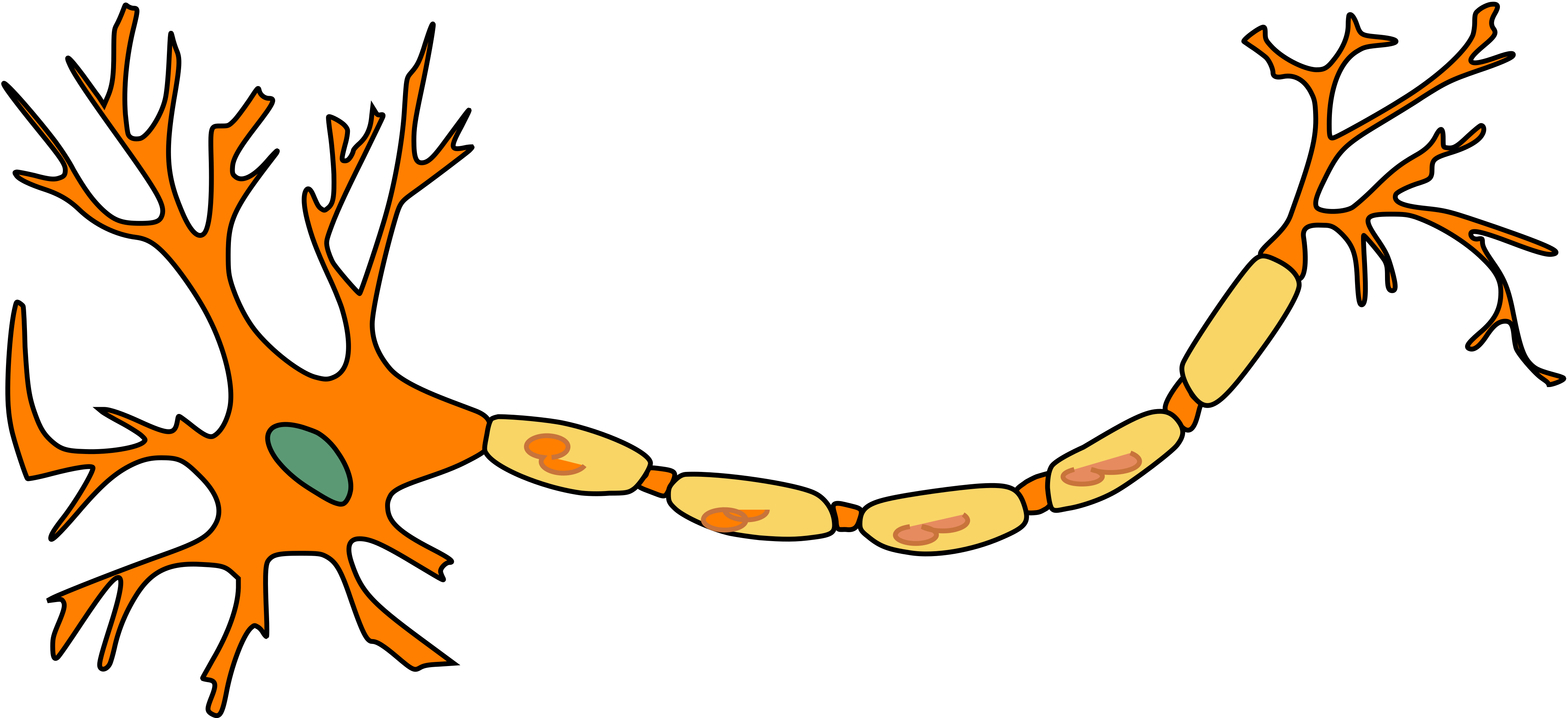
Draw all the components of a neuron from the start of receiving neurotransmitter to the
end, moving to another neuron. Label the following on your drawing: dendrites, soma,
nucleus, axon, myelin, nodes of Ranvier, axon hillock, and axon terminal.
What is the purpose of the myelin sheath? Where is it located on a neuron? What are the sections called where there’s no myelin? Why are those sections important? Why do signals degrade when an axon is unmyelinated?
Myelin acts as in insulator on the axon, it surrounds most of the axon of a neuron. It speeds up the electrical signal transmitted down the axon.
The sections of no myelin are called Nodes of Ranvier.
Nodes of Ranvier are important because they contain ion transporters to keep the electrical gradient to continue to propagate the signal down the axon.
When an axon is unmyelinated, the axon is not well insulated enough to keep all the ions in the axon to maintain the electrical gradient, so an action potential may fizzle out.
There are sensory and motor neurons. Explain to someone the difference between them in terms of to and from the CNS. What two types comprise the motor neurons (explain a bit on each)?
Sensory neurons:
Afferent-relay information from inside/outside environment and carry it to the CNS (brain and spinal cord).
They can also detect a variety of stimuli-ranges from photons of light (visual), to chemicals in the air (olfactory), to CO2 lvls in the bloodstream (chemoreceptors).
Have a large variety of shapes and structures
Motor neurons:
Efferent, they carry from the CNS to the body to carry out movement of muscles.
2 types:
Somatic
Control movement of skeletal muscles-voluntary
Autonomic.
Controls smooth muscles, cardiac muscles and glands automatically-no thought needed
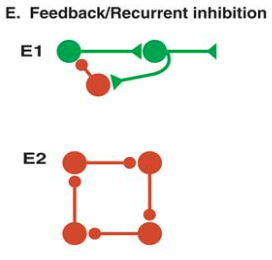
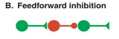
Explain what an interneuron is and what it does in feedforward and feedback inhibition.
(Tip: It might help to draw this one out!)
An interneuron is a short neuron that acts as a messenger to relay information sent from a pre-synaptic neuron to a post-synaptic neuron. It relays information between sensory or motor neurons and the CNS.
In feedforward inhibition, the presynaptic neuron excites an interneuron, which then elicits an inhibitory post-synaptic potential in the next neuron in line.-this is a way of shutting down of limiting excitation in a downstream neuron in a neural circuit
In feedback inhibition, the presynaptic neuron excites a postsynaptic neuron which excites an interneuron, but that interneuron then starts an inhibitory postsynaptic potential back in the same presynaptic neuron. Generally, it dampens excitation thru a neural circuit
What are 2 examples of feedback inhibition. What does the term recurrent inhibition apply to? What’s the difference btween feedback inhibition and recurrent inhibition?
The Renshaw cell in the spinal cord. The axon of a spinal motor neuron branches. One main branch innervates and goes to the muscle and the other branch (axon collateral) makes an excitatory synaptic connection with an inhibitory interneuron called the Renshaw cell.
The interneuron then inhibits the motor neuron that activated it, thereby closing the loop-can prevent overexcitation of the motor neuron and prevent straining of the muscle, etc.
Another example of feedback inhibition is found in the hippocampus. CA3-type pyramidal cells make excitatory connections to basket cells and the basket cells feedback to inhibit the CA3 cells.
Note: .this would be feedback inhibiton in a microcircuit
The term recurrent inhibition is applied to simple feedback inhibition circuits such as the Renshaw circuit in the spinal cord and the basket cell circuit in the hippocampus
Recurrent inhibition is more specific while feedback inhibition is more general and broad a category-when a neuron inhibits itself indirectly through an interneuron-can stop overexcitation of a single neuron, while recurrent inhibition is a specific case of feedback inhibition-A neuron sends an axon collateral (which is basically a branch of an axon that extends off the main axon + allows signals to be sent to multiple targets simultaneously) to an inhibitory interneuron (Renshaw cell), which inhibits that same neuron.

Feedforward excitation
Allows one neuron to relay info to its neighbor
Long chains of these can be used to propagate info thru NS

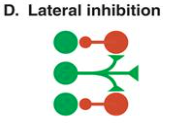
Lateral inhibiton
A presynaptic cell excites inhibitory interneurons and they inhibit neighboring cells in the network
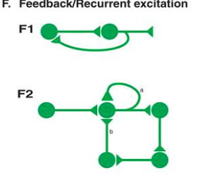
Feedback/recurrent excitation
A presynaptic neuron excites a postsynaptic neuron and that postsynaptic neuron excites the presynaptic neuron
This type of circuit can act as a switch like function-cause once the presynaptic cell is activated, that activation could allow it to stay on (cause of the cycle of excitation) even after the og stimulus stops

Autapse and recurrent excitation (types of recurrent/feedback excitation)
Autapse: A neuron excites itself directly-releases neurotransmitters that bind to tis own receptors
Recurrent: Presynaptic neuron excites postsynaptic neuron which eventually loops back to excite the og neuron again
Give an example of a feedforward excitation and feedforward inhibition microcircuit
Feedforward excitation:
A circuit that mediates simple reflex behaviors-eg. would be the knee reflex
When rubber tapper hits the knee, it stretches the muscle-initiates action potential in sensory neurons in the muscle that are sensitive to stretch
This action potential goes to the synaptic terminal of the sensory fenuron, which will release neurotransmitter-leads to excitation of the motor neuron
This will then cause an action potential in the motor neuron-which then spreads out the peripheral nerve? to go to the synapse at the muscle-will then cause the release of neurotransmitter and an action potential (AC) in the muscle
This action potential will cause contraction in the muscle and extension of the limb
Now for the feedforward inhibition part:
Similar to feedforward excitation, will allow the AC in the sensory neuron to go to the synaptic terminal-causes a release of neurotransmitter and excites the postsynaptic interneuron in between the neurons
This excitation causes the initiation of an AC and release of neurotransmitter from the presynaptic terminal of the interneuron
BUUT-this elands to an IPSP in the postsynaptic flexor motor neuron-which will lead to feedforward inhibition-will help decrease the probability of the flexor motor neuron becoming active and producing an inappropriate flexion of the leg-makes sure u don’t over flex and injure urself-regulates excitatory neuron
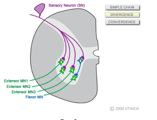
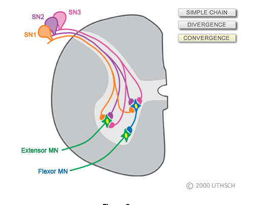
Convergence and divergence and stretch reflex
Th proper function of the circuit of the stretch reflex also relies on convergence and divergence-a single sensory neuron has multiple branches that diverge and make synaptic connections with many individual motor neurons.
Therefore, when the muscle contracts as a result of the neurologist’s tapper, it does so because multiple muscle fibers are activated simultaneously by multiple motor neurons.
Also, when the muscle is stretched, not one, but multiple sensory neuron are activated and these sensory neurons all project into the spinal cord where they converge on to individual extensor motor neurons
So, the stretch reflex is due to the combined effects of the activation of multiple sensory neurons and extensor motor neurons.
Lateral inhibition and edge enhancement
Helps with edge enhancement in the retina-makes edges of things better/easier to see (important so u can see the edge of a cliff even if its dark outside for eg.)
It does this by reducing light intensity received by the second neuron (just any postsynaptic neuron)
Without lateral inhibiton, the intensity of light would be the exact same everywhere-but with it, the intensity is reduced
Edge enhancement can also cause an illusion called Mach bands-the thing in the diagram
This happens because horizontal cells inhibit adjacent neurons at some level (cause they block signaling pathways when activated)
Endogenous bursting behavior in neurons
Some neurons naturally fire in rhythmic bursts due to intrinsic ion channel dynamics (not external feedback), with silent periods in between each burst
Feedback inhibiton can help regulate these bursting patterns but the bursting itself is an intrinsic property of the neurons
Its basically just neurons firing spontaneously without any external stimulus
This bursting is, as mentioned before, intrinsic to the neuron tho-meaning its not dependent on a network-instead controlled by Ca2+ dynamics- Ca2+ enters the cell-causes depol. and firing. As it accumulates, it inhibits further influx, stopping the burst. Ca²⁺ levels then drop due to buffers and pumps, removing inhibition and restarting the cycle.-This is a form of feedback inhibition at the cellular level, where excitation triggers inhibition, leading to rhythmic activity.
Circadian rhythms
Are regulated by feedback inhibition at the molecular level, not just in neural circuits.
The vertebrae circadian rhythm is controlled by neurons in the suprachiasmatic nucleus (SCN)-located above the optic nerve-, which influence hormones (melatonin, cortisol) and body temperature.
The key gene involved is per (period gene), first discovered in fruit flies but also found in vertebrates.
The per gene produces per mRNA, which makes PER protein.
PER protein enters the nucleus and inhibits further per gene expression (feedback inhibition).
Over 24 hours, PER protein degrades, removing inhibition and restarting the cycle.
This repeating cycle forms the basis of circadian rhythms, influencing many body functions.-Circadian rhythms are controlled by a molecular feedback loop where gene activation leads to self-inhibition, then resets—just like a rhythmic neuron but at a much slower, daily pace.
How does the whole per cycle repeat itself if it inhibits itself?
The PER protein is degraded over a 24 hour period. So, as the PER protein is degraded, the inhibition or repression is removed (disinhibition) allowing this gene to start making messenger RNA and protein all over again. So once this cycle begins, it is repeated over and over again at a 24 hour period.
Recurrent Inhibition in ring circuits
May help explain how animals generate different walking patterns (gaits) like walking, trotting, bounding, and galloping.
Ring circuits are a network of four inhibitory neurons, each controlling a limb.-Each neuron has endogenous bursting activity (fires rhythmically on its own). The neurons are inhibitorily connected in a loop, ensuring they fire in a coordinated sequence. Small adjustments to neuron properties can shift the phase relationships (the timing of limb movements relative to each other in a cycle-eg. in phase=2 legs move together), allowing different gaits (gaits are basically diff types of limb movement-walking, trotting, etc.) to emerge from the same circuit.
Feedback/Recurrent Excitation in Learning & Memory
Plays a big role in learning and memory, particularly in the hippocampus (CA3 region)-strengthens neural connections through synaptic plasticity and allows for auto-associative memory storage.
How It works:
Neurons excite each other in a loop, forming a recurrent excitatory circuit
Hebbian Learning Rule: “Neurons that fire together, wire together.”-If two neurons are active at the same time, the connection between them strengthens.
Convergence & Divergence: Each neuron receives input from multiple others and sends output to multiple others.
Memory Storage: The pattern of connectivity changes based on activity—storing information across the network, not in a single synapse.
This process helps with pattern completion, meaning if part of a memory is triggered, the network can reconstruct the full memory.
What do convergence and divergence do?
Convergence:
Allows a neuron to receive input from many neurons in a network
Divergence:
allows one neuron to communicate with a bunch of other neurons in a network
What is the difference between reticular theory and the Neuron Doctrine? Which one is most supported by evidence?
The Reticular theory suggests that parts of the nervous system are all one very large, connected network
The Neuron doctrine suggests that the nervous system is comprised of individual units that are separated from each other physically.
Currently, the neuron doctrine is the theory most supported by evidence.
Who were the reticular and neuron doctrine theories supported by and why?
Reticular was supported by Golgi because of his staining experiment results-the neurons looked connected like a net
Neural doctrine was supported by Cajal because when he redid Golgi’s staining experiment, he got a diff result that showed that the NS was more a series of individual units that were physically separated-not all connected like Reticular theory states
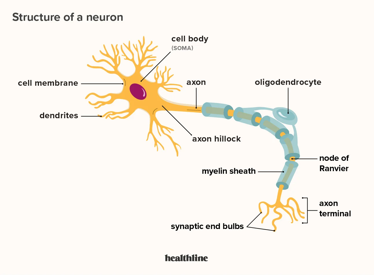
What are dendrites? What is their function?
Dendrites are branch-like extensions that protrude (stick out) from the cell body (soma). On the dendrite are spines-tiny protrusions of the cell membrane where chemical signals from other cells are received.-where info first enters neurons
Dendritic plasticity
Tiny changes to the morphology of dendritic spines may represent a single complex memory that u from-thought to underlie the reason we can learn new facts or maintain memories for long periods of time
However, a neuron doesn’t require a spine to receive info for plasticity to take place-input site can be anywhere along the dendrite
Cell body (soma)
Info coming from dendrites goes thru here
Diff sizes for diff neurons-Betz cells are largest
Contain organelles needed for producing proteins, etc.
Axon
Main output extension of neuron
Neurons only have one axon extending from the cell body, but it can branch off multiple times after exiting the soma-this branching allows the neuron to communicate with many other neurons at the same time (instead of just open)
They are thinner than dendrites-several axons can bundle and travel together-called nerves
Cellular arithmetic
Just the action of the axon hillock weighing all incoming signals-excitatory, inhibitory, modulatory, etc., and deciding whether it should move on or not
What is the axon hillock?
The axon hillock is the section between the soma and the axon, the output extension of a neuron.
This area acts as an integration center where all the signals coming into a cell are added up, to decide if an action potential will be sent to continue the signal to the next neuron.
What part of the axon releases neurotransmitters?
At the end of an axon is the axon terminal, or terminal bouton, which houses the active zone.
The active zone contains a variety of proteins essential to neurotransmitter release in order to transmit a signal to the next neuron.
Anterograde + retrograde transport
Retrograde transport: When substances are moved towards the cell body
Anterograde transport: When substances are moved away rom the cell body
What is a neurofilament?
Organelles found along the length of an axon to maintain axon structure.
What is the difference between an electrical and a chemical synapse?
An electrical synapse is between two neurons with distinct cell membranes, but that share cytoplasm. This allows the ion signaling to continue through the shared cytoplasm.
A chemical synapse occurs between two neurons that don’t share cytoplasm and are physically separated, which requires the use of neurotransmitter release by the first neuron and uptake by the second using vesicles
What are the different types of glial cells?
Astrocyte
Oligodendrocyte
Schwann cell
Microglia
Ependymal cell
Endfeet
Protrusions at the end of astrocytes and release biological compounds that allow endothelial cells to stay healthy as they function in maintaining the BBB
Wrapped around endothelial cells that surround blood vessels
Astrocytes
Maintain BBB by wrapping endfeet around endothelial which line the blood vessels, to help maintain the blood-brain barrier (BBB).
Decrease strength of glutamate signals at the synapse-Have proteins on their cell surface that can transport molecules of things like the neurotransmitter glutamate inside the astrocyte-able to decrease strength of glutamate signal
Produce trophic factors to signal which neurons/synapses should continue to live-helper molecular signals (chemical messengers) do this-anyway this helps guide neurons as they reach out and form synapses when needed
Star shapes (astro)
Have a protein called glial fibrillary acidic protein (GFAP)-often used as a marker for differentiating astrocytes from other cells
Can affect the extracellular concens of ions like potassium, which can have an influence on cellular excitability
Oligodendrocyte
Myelinate up to 50 axon segments in the CNS per 1 oligodendrocyte
Only in CNS
Add a layer of myelin around axons of nearby CNS neurons-therefore increases conduction speed of neurons as they send signals
Produce myelin (duh)
Able to produce almost 3x their weight in membrane per day once they start myelinating
Have highest metabolic rate of any type of cell in the brain
Schwann cell
Myelinate a single axon segment in the PNS-function similarly to oligodendrocytes-but oligs do more haha
Clear out old myelin in event of axonal injury
Produce signaling molecules to guide damaged axons to correct targets
Can only be found in PNS
To regenerate injured axons, Schwann cells quickly go to the injured area. Since the loose leftover myelin from the injury can actually make it harder for the nerve to heal now, Schwann cells will get rid of these leftover pieces-allow the nerve to regenerate more effectively
Microglia
Immune cells of the CNS-identify and destroy pathogens, dead/dying cells, protein clumps, react to injury, etc. that enter our brain
More like immune cells rather than neural cells haha
They travel thru the brain and spinal cord
Make up 10-15% of all cells in the brain
After injury to the CNS, microglia quickly react to it-marker Iba1 is used to identify when microglia are reacting to an injury
Ependymal cell
Part of the choroid plexus (network of blood vessels and cells that form a boundary between the blood and CSF) responsible for producing CSF
Along inside of ventricles
Are arranged in a column against cilia-extend into ventricles, to the central canal and lung down and inside the spinal cord
Explain the difference between an excitatory post-synaptic potential and an inhibitory post-synaptic potential. How do they relate to summation?
An excitatory post synaptic potential is a positive graded potential (meaning it is not yet an all or nothing action potential- it has a magnitude) that brings the membrane potential closer to the threshold to start an action potential, making an action potential more likely. (same as EPP)
An inhibitory post-synaptic potential is a negative graded potential that brings the membrane potential further from the threshold, making an action potential less likely.
The sum of all the excitatory and inhibitory post synaptic potentials is taken at the axon hillock, and if this sum reaches the threshold, an action potential is fired.
What is meant by the terms convergence and divergence?
Convergence is when a neuron receives input from many neurons, so all their signals converge on that neuron
Divergence is when a neuron sends signals down to multiple neurons, so the signal diverges away from that neuron.
What is the difference between feedforward and feedback control?
Feedforward control refers to how a presynaptic neuron affects the next neuron (postsynaptic), while feedback control refers to how downstream neurons connect back to affect the presynaptic neuron.
Vesicles (include where, what its made of, and what it contains)
In the axon terminals
Small spheres made up of CM and proteins
Contain molecules used for chemical communication between neurons-when an AC travels down the axon and reaches the terminal the CM changes in electric charge-causes vesicles to fuse with the inner membrane of the neuron-allows contents of the vesicles to be released outside the cell
Remember when we discussed Nodes of Ranvier. Now we can add more context as to their importance in a neuron. The voltage-gated sodium (Na+) channels are located here. Explain how sodium enters the cell through these channels and how the signal is passed down the axon.
When the depolarization of the membrane potential is large enough, an action potential is triggered.
Firstly, sodium ions are moved down their concentration gradient (move from outside the membrane to inside) which are in the Nodes of Ranvier-The sodium ions are less likely to move in the opposite direction (inside to outside) because the outside of the membrane has a higher concentration of sodium ions.
Since the previous section of the membrane is in the absolute refractory period, no action potential can travel backward. The action potential “bounces” from node to node, this is known as Saltatory conduction. The rest of the axon that is wrapped in myelin increases the speed the action potentials moves down the axon to the axon terminal.
Saltatory conduction
Refers to the jumping of a signal from node to node.
Changes in electrical charge are detected in intervals.
Its just the way action potentials travel down a myelinated axon, and it makes nerve signals much faster and more efficient.
Its just the process of the myelin sheath insulating the axon, preventing ion leakage. The Nodes of Ranvier (gaps in myelin) have voltage-gated ion channels. The action potential "jumps" from node to node instead of traveling the whole axon (where there is myelin cause it blocks the ion channels).
This makes nerve signals much faster and more energy-efficient.
In simple terms, it is the rapid method by which nerve impulses move down a myelinated axon with excitation occurring only at nodes of Ranvier.
What is an action potential? What is a graded potential? What is difference?
An action potential is a change in the membrane potential.
It’s an all-or-nothing response because only a large change in the membrane potential will push the signal down the axon.
A graded potential is a small, temporary change in Vm of a neuron that is sub-threshold-note: an EPP is a type of graded potential
Unlike action potentials, they don’t always reach threshold (not all or nothing) and trigger an action potential
They can also add together-thru spatial or temporal summation
If enough graded potentials add up at the axon hillock and allow it to reach threshold, a full action ptoential is triggered
If the don’t however, reach enough amplitude at the axon hillock, they just fade away
A graded potential isn't always the same size too (varies in amplitude). Instead, it can be stronger or weaker depending on how much stimulus is applied.
For example:
A small stimulus → small graded potential (low amplitude).
A stronger stimulus → bigger graded potential (higher amplitude).
It’s like tapping vs. slamming a piano key—the harder you press, the louder the sound. Similarly, the stronger the stimulus, the bigger the graded potential!
Spatial and temporal summation
Spatial:
When multiple presynaptic neurons release neurotransmitters at different locations on the postsynaptic neuron at the same time.-it is two small graded potentials from2 adjacent inputs being triggered
If enough inputs combine, they can push the membrane potential to the threshold and trigger an action potential.
Temporal:
When one presynaptic neuron fires multiple times in rapid succession.-multiple graded potentials for the same input occur close together in time
The repeated signals build on each other, increasing the membrane potential until it reaches the threshold.
What happens at the neuron level during the depolarization phase of an action potential?
Depolarization occurs when voltage-gated sodium channels open.
There is an influx of sodium (Na+) ions into the cell because the concentration of sodium is greater outside the cell than inside (follows the chemical gradient).
What happens at the neuron level during the repolarization phase of an action potential?
Repolarization occurs when voltage-gated potassium channels open.
There is an efflux of potassium (K+) ions out of the cell because the concentration of potassium is greater inside the cell than outside.
Explain the term net flow. When does it occur?
Net flow is the movement of ions across the cell membrane.
This occurs when the electrical and chemical gradients favor one ion.
What does selectively permeable mean? What part of the cell has this feature?
Selectively permeable means that only specific molecules can pass through.
The cell membrane has this feature. Only small molecules such as water and gases can pass through easily, but other larger molecules, such as glucose can’t.
What helps larger molecules pass through the cell membrane?
Transmembrane proteins. These are big protein complexes that span the membrane. They have a pore in the middle (essentially a tunnel that allows molecules and ions to pass across the CM-cause they cant get in normally-allows less permeable molecules to pass thru. Its the middle protion of the transmembrane portein).
They can distinguish ions based on chemical properties-eg. some are selective for Na+ -this prevents the passage for other ions.
These are also called ion channels + work passively-don’t use cellular energy to move ions-just provide easy passage
If you break down the term, trans- means across, and membrane just means membrane. It spans the whole cell membrane.
What are features that allow for distinction between ions in channels?
Pore size
Shape of the channel
eg. Would exclude larger molecules if its a smaller shape, etc.
Electrical charge
Prevents certain ions from passing thru based on charge-eg. sodium channels have negatively charged glutamate in their pores-causes things like negatively charge chlorine ions to be repelled while attracting positively charged sodium ions
Hydration shell
Every ion comes with a hydration shell around it-water molecules surrounding them
Io channels are designed to fit specific ions based on size and charge (the 2 above points)
To enter the channel, an ion needs to shed its hydration shell-lose some water molecules-so even if the ion is smaller, like Na+, but has a big hydration shell, it might not be able to lose enough water molecules in order to fit thru the channel-makes sure that only ions that can properly shed their water fit thru. But if the ion can’t lose enough water or isn’t stabilized correctly, it is kept out
What does electroactive mean? Are cell membranes electroactive?
Yes cell membranes are electroactive-means that they are sensitive to electric charges
Neurons can change the electrical potential of their membranes
What is the absolute refractory period?
The absolute refractory period is the window of time during right after an action potential is fired.
This time doesn’t allow for another action potential to be fired.
This is because the voltage gated Na+ channels are inactivated-prevents the generation of another action potential until the membrane potential returns towards the resting state and the channels reset. Also sodium ions can’t enter the cell anymore
How does an action potential fire during the relative refractory period?
An action potential can fire during the relative refractory period only if there’s a greater depolarization than the first.
If the depolarization is below threshold (-45 mV), then no action potential is triggered.
What are the 4 major classes of ion channels?
Leak channels
Voltage-gated ion channels
Ligand-gated ion channels
The variety of channels used by the sensory systems
Leak channels
Persistently open-act like revolving doors that never lock
Neurons usually have several-eg. K+, Cl-, etc.
Since ions are at an equilibrium, they contribute to properties of neurons t rest-go back and fort across the CM
Voltage-gated ion channels
Sensitive to electrical potential of surrounding membrane
For example voltage-gated Na+ channels will stay close dat negative resting potentials and only open when positive
Channels used by the sensory systems
Open and close in response to unique stimuli on what they’re able to sense
Eg. Some open and close when u move physically-like a stretch
We have these in our ears, skin, etc.
Electrochemcal gradient
Just what the electrical and chemical gradients are collectively called
To predict the forces acting on an ion, u need to know the charge of the ion and the relative concentrations of the ions across the membrane
Dynamic equilibrium
Ok for example for K+, it’s positive which means it goes inside the cell (electrical gradient. However, there is a high concentration of K+ inside the cell-therefore the chemical gradient pushes the K+ out of the cell.
Since these 2 forces oppose each other perfectly, it causes dynamic equilibrium (dynamic because there is a constant movement of ions in and out of the cell yet equilibrium because there is no net movement of charge-charge moving in as balanced by charge moving out
So it’s just the constant movement of ions going in (Vm) and out- therefore equilibrium because there is no net movement of charge. The membrane voltage can change, but not the ion concentration.
Equilibrium potential (E) (also add how it affects the membrane potential in phases like depolarization and repolarization of the action potential)
The exact value of the Vm when an ion is in dynamic equilibrium is called the equilibrium potential-when force pushing in is same as force pushing out. Each specific ion has its own equilibrium potential, and the overall membrane potential of the cell is a result of the combined effects of equilibrium potentials of all ions present weighed by permeability-therefore, the resting potential wont be the exact same value as the equilibrium potential for K+ because the cell is also permeable to thinks like Na+-might make he resting ptoential more positive
Note: During smth like the depolarization phase, the Vm will shift towards the equilibrium potential of Na+ (which is pretty positive). This happens because of the whole permeability thing said above-as the permeability for a given ion increases, it shifts to balance the Vm more towards the equilibrium potential of that specific ion. therefore, when channels of a specific ion open, permeability increases which moves the Vm towards the E (equilibrium potential) of Na
Likewise, during the repolarization phase, the Vm will shift more towards the K+ equilibrium potential, which is pretty negative (because the K+ channels r open)
What is a change in Vm made in an action potential made by?
The movement of ions thru voltage-gated ion channels
Steps to an action potential
Depolarization from incoming neurons
U usually need multiple EPSPs to reach threshold (add either by spatial or temporal summation)
Anyway how does this depol. occur? Well a PSP (postsynaptic potential) is involved. -the release of excitatory neurotransmitters will cause small depolarizations called EPSPs (excitatory postsynaptic potentials) while the release of inhibitory ones will cause small hyperpolarizations (IPSPs-Inhibitory postsynaptic potentials)
PSPs are characterized by a quick change in Vm going up followed by a decreasing phase where Vm resets to og resting potential (the decay period)-adding a second EPSP in this time could cause summation btw
Opening of voltage-gated Na+ channels
At rest, the voltage-gated ion channels are almost all closed, but when Vm depolarizes, some channels open
Due to the electrochemical gradient, we can determine that once they do, Na+ will move inside the cell (cause both in favor of this)
This will cause more depol. to occur (cause Na+ is positively charged)
Opening of voltage-gated K+ channels
K+ channels will then open when the cell starts to depolarize-allows K+ to move across the membrane
When the cell depolarizes to the point where it becomes positive, K+ will be pushed out of the cell (cause now + and high concentration of K+ in cell).
This will cause the interior of the cell to become more negative-cause K+ is positive and now its outside the cell
Cells at this pint will become more negative than the resting potential
Inactivation of voltage-gated Na+ channels
Voltage-gated Na+ channels not only have a pore, but also an inactivation gate-block flow of Na+
When the cell membrane gets positive, the inactivation gate will close-prevents further movement of Na+ ions
This process is very fast
Deactivation of voltage-gated K+ channels
This is the last step to the action ptoential
The main current flow will now be outward-cause K+ now being driven out of the voltage-gated K+ channels due to electrochemical gradient, making the cell negative again
This deactivation process, however, is much slower than Na+’s
But once they do deactivate, then hyperpolarization current stops-causes the membrane potential to gradually return to the equilibrium resting potential
PSP
These are small deviations in voltage caused by ion movement that the presynaptic neurons cause when they release neurotransmitters onto dendrites via postsynaptic ligand-gated ion channels.
What are the 3 parts we can divide an action potential into (based on te steps)
Depolarization
This would be when the Na+ enters the cell, making the Vm more positive
At this step, voltage-gated potassium channels also start to open
This would be the upward deflection in the action potential
The membrane potential becomes more positive (from -70mV to +40mV)
Repolarization
This would be when the Na+ channels have almost all been inactivated
K+ will also be going out of the cell thru the potassium channels, making the Vm more negative-(from +40mV to -70mV)
This would be the rapid decline/deflection downwards
Afterhyperpolarization
It would be when the gradual deactivation of the voltage-gated potassium channels occurs-making the membrane potential more negative than the resting potential
By the end of this deactivation in the K+ channels, there would be the slow return back to the resting membrane ptoential because the hyperpolarization current would stop (form -70 mV to -80mV and back to -70 mV)
add pic form notes here
Movement of an action potnential
For an AC yo travel down from the axon hillock to the axon terminal, the Vm change must physically move down the axon
This can occur because the Na+ that enter the channels are not restricted to the cytoplasmic volumes beneath the ion channels-they will instead diffuse to an area of lower concentration (which happens to be lower and lower down the axon)
Then they move down to the next section of the membrane, the membrane will depolarize and the process of the AC occurs again and keeps repeating until it reaches the axon terminal
Why does an action potential only move in one direction?
2 reasons:
The chemical gradient
Na+ will move down their concentration gradient (from higher concen., to lower)
The Na+ ions that enter thru voltage-gated Na+ channels don’t go in reverse/opposite direction because the area will have a high concentration of Na+
The previous patch of the membrane will at that point be in the absolute refractory period, which makes it impossible for an action ptoential to travel backwards
What is the relative refractory period?
Occurs after the absolute refractory period.
During this period, an action potential is hard to fire compared to the resting condition.-cause only some of the sodium channels have reset from their inactive states and since many of the K+ changes are still open, the neuron will have a more negative charge/potential cause K+ is still moving in the neuron. The increased movement of K+ ions also hinders depol. This happens because potassium movement pulls Vm closer to the E (equilibrium potential) of K+-which is usually more negative
This period lasts as long as the afterhyperpolarization (slow increase in membrane potential).-therefore, it is a gradient-the sooner after the repolarization phase of the action potential, the more difficult it will be to reach threshold for the second action potential (cause more negative therefore we need more positive to reach threshold)
Draw a diagram of a pre-synaptic neuron releasing the neurotransmitter, acetylcholine, onto a post-synaptic neuron.
Label the following: pre-synaptic neuron, post-synaptic neuron, synaptic vesicle, receptors ,synaptic cleft, and acetylcholine.
add pic here
Explain what myasthenia gravis is in terms at the neuronal level.
Myasthenia gravis is severe muscle weakness due to the lack of acetylcholine receptors in the muscle cell.
Muscular weakness occurs because the endplate potential is smaller than normal.
Since the endplate potential cannot reach threshold, there is no action potential in the muscle, and no muscle contraction.
What are the 2 types of synapses? What is a synapse?
Synapse:
Not actually a a part of the structure of a neuron-just the site of close proximity between 2 communicating neurons
The 2 types are electrical and chemical synapses
Electrical synapse
Refers to the entire connection/space between neurons
The synapse allows the cells to share a cytoplasm with the adjacent cell so molecules like ATP, ions, etc. can move between the cells
This would be what Golgi thought all of the nervous systems would be like
Neurons connected by an electrical synapse are usually closer together than those that are connected by a chemical synapse
Each cell in an electrically coupled network can receive multiple inputs at the same time from any of the cells
Note: Each neuron connected by an electrical synapse is still tis own neuron tho
What was the likely reason that electrical synapses evolved?
Because of the need for speed-can pass signals fast and can therefore be useful for things like reflexes (eg. the escape reflex to escape predators)
Because of this whole survival reflex thing, electrical synapses are often found in less complex organisms-because their reflexes are more critical for survival
What is a connexon/hemichannel? What is formed by two connexons?
A physical channel between 2 neurons that allows the cytoplasm of two neurons to connect. Its like a physical bridge between the 2 cells
Each hemichannel is made of 6 transmembrane proteins called connexins.
A gap junction is formed by two connexons, this is the structure that connects neurons electrically.
Can chemical synapses pass information in either direction?
No, only electrical synapses pass information bidirectionally (from both sides/neurons), ions and signaling molecules can move either direction through the connexon.
Chemical synapses have space between the two neurons, not a tunnel. Neurotransmitters are released from the pre-synaptic neuron to the post-synaptic neuron.-and probs won’t go in the opposite direction
Chemical synapse. What organisms are they usually found in?
These synapses are like actually space between the neurons-not connected by smth like a bridge like electrical synapses are
Therefore, to communicate, they release signaling molecules (usually neurotransmitters) from the presynaptic to the postsynaptic neuron
After being released, neurotransmitters will diffuse across the synapse, where they can affect nearby neurons once the chemical binds to its corresponding receptor
They ca also pass different signals depending on neurotransmitter and receptor-eg. some signals are excitatory and allow positive ions to enter the neuron which cause a depol. while other might be inhibitory
Chemical synapses are usually found in more complex organisms like humans. This is because some are more complex and can do things like modify cellular excitability over time, which allow for fine-tuning of neural networks-which gives the NS a larger range of possibilities. Therefore, these signals are necessary for more complex behaviors found in more complex organisms (like cognition, etc.
What is the space called between a motor neuron and muscle tissue? Explain what happens in this space
Neuromuscular junction (NMJ).-its is a specific type of chemical synapse
The motor neuron releases synaptic vesicles that contain the neurotransmitter called acetylcholine (ACh).
Once this signaling molecule is released, the receptors on the muscle detect it and cause the muscle to contract.
What are the two types of vesicles? What is the purpose of each?
Small vesicles
Store neurotransmitters such as GABA, dopamine, glutamate, etc. These vesicles are usually found in the axon terminals.
Found exclusively in axon terminals
Large dense-core vesicles
Much larger than small vesicles, so they store larger structures than neurotransmitters. For example peptides such as dynorphin or enkephalin.
These large-core dense vesicles can be found in the cell body (soma), along axons, and the axon terminal.
The loading process of neurotransmitters requires energy. Where does this energy come from and what type of port is this called?
Vesicles can be filled/loaded with neurotransmitters thru the action of giant transmembrane proteins called vesicular transporters
These vesicular transporters function to take molecules of neurotransmitters from the intracellular space of the axon terminal and pump them into the vesicles
The way this works/the way they do this is Neurotransmitters enter the vesicle via the energy produced from the movement of H+ protons of the vesicular transporters. (cause the movement of H+ from high to low concentration produces energy)
The transporters use the high intravesicular H+ concentration to move molecules of neurotransmitters from the axon terminal into the vesicle.
Since the H+ ions move from vesicle to the axon terminal and the neurotransmitter moves inside the vesicle, this vesicular transporter is called an antiport.-because the H+ ions are moving opposite of the neurotransmitters -alr so basically the H+ moves into the axon terminal (because there is a high concentration of H+ in the vesicles due to the transmembrane enzyme vesicular-ATP-ase or V-ATP-ase. These proteins used molecular energy in ATP to concentrate H+ in the intravesicular space (for each ATP molecule used, one proton (H+) was pumped into the vesicles and a low concen. in the axon terminal) this movement from area rea of ig-to low concen. releases energy. The neurotransmitters will then use this energy to move form the axon terminal into the vesicles.
What three locations of the axon terminal can synaptic vesicles be found on?
Readily releasable pool (RRP) vesicles are located close to the cell membrane at the axon terminal. These vesicles are “docked” meaning that their coat protein (think of it as an identification marker) interacts with the proteins inside the cell membrane.
Once the depolarization of an action potential reaches the axon terminal, these vesicles are the first to fuse with the cell membrane.
Recycling pool are the vesicles that are always in the process of being refilled with neurotransmitter. They are farther from the cell membrane and the protein machinery is not primed for release, therefore it requires more stimulation to release its contents of the vesicle.
Reserve pool vesicles are the furthest away from the cell membrane. The stimulus required for the contents of the vesicle to be released is very intense. These vesicles may not be released under physiological conditions.
Explain the purpose of the proteins embedded in the cell membranes of the vesicles or the neuronal membrane.
V-SNAREs are proteins expressed on the vesicle. (v for vesicle). Synaptobrevin and synaptotagmin are 2 specific v-SNARE proteins involved during synaptic release
T-SNAREs are proteins expressed on the cytoplasmic side of the axon terminal. They are the “target” for the vesicles is the cytoplasm. (the t in t-SNARE). Syntaxin and synaptosomal nerve-associated protein 25 (or SNAP-25) are t-SNAREs that function during vesicular fusion
The v-SNARE proteins interact with t-SNARE proteins for vesicular fusion and the release of neurotransmitter. They are important because the release of neurotransmitters must be tightly regulated in order to not deplete their entire stock of neurotransmitter, which would cause signals to constantly go off. And this process of regulation is dependent of these proteins.
When does a neurotransmitter release from one neuron to the next? What is involved in fusing of vesicles? What is a voltage-gated calcium channel (VGCCs) used for?
An elevation of Ca2+ in the intracellular space is the signal that causes neurotransmitter release.
The V-SNARE synaptotagmin protein is required for vesicular fusion with the cell membrane. This protein detects high level of Ca2+ in the axon terminal.-it gives the “go ahead” signal that causes neurotransmitters to release
VGCCs remain closed until the surrounding membrane depolarizes.-like Na+ channels. Therefore, as the change in ptoential moves down the axon (action potential), it causes a depol. which allows the enter of Ca2+ in the VGCCs. Therefore This causes a physical change in the conformation to allow Ca2+ ions to selectively pass.
After this, the Ca2+ at the terminal will bind with synaptotagmin. The v-SNARE and t-SNARE will then interact with each other in the response of Ca2+-which will form a molecular structure called a SNARE complex.
As they twist tighter together, the vesicular membrane will approach the inside of the cell membrane-causes vesicular fusion.
How are vesicles capable of fusing?
Vesicles can undergo full fusion; the vesicular membrane becomes completely integrated with the cell membrane. All neurotransmitters release into the synapse (total exocytosis)
Vesicles can also fuse by kiss-and-run; the vesicle partially connects with the interior of cell membrane (quick smooch). Few neurotransmitters are released into the synapse. Its an incomplete fusion-no one talked just ran
Receptors
Proteins that can send a signal to change the function or activity of a neuron
Most that function in neurotransmission are large transmembrane proteins
On the extracellular surface, there is a series of amino acid residue-called the active site (or orthosteric site). This is shaped to allow molecules of neurotransmitter to bind to the receptor
There are 2 categories
Ionotropic
Metabotropic
Which receptor, ionotropic or metabotropic, is slower?
The metabotropic receptor is the slower out of the two because it has a complex protein attached to the receptor that signals more pathways.
The ionotropic receptor is faster because ions flow through the channel once a neurotransmitter binds to the receptor.
Use the terms recycling, mobilization, release, docking, and fusion to explain exocytosis during the synaptic vesicle cycling.
Exocytosis is explained by vesicles releasing neurotransmitters into the synaptic cleft.
The process of vesicles migrating to the edge of the axon terminal is called mobilization.
Docking happens next, this is the vesicle touching the cell membrane.
Fusion is the vesicle combining with the membrane. The neurotransmitters are released into the synaptic cleft and the vesicle is recycled to form new synaptic vesicles.
Ion channels
A specific type of transmembrane protein
a passive channel because it doesn’t use cellular energy to move the ions through the membrane.
Channels are selective in the ions based on chemical properties.
Electrical gradient
Electrical forces acting on charged molecules
Pull opposite charges together and push like charges away
Chemical gradient
Natural process of a high concentration of a substance diffusing to a lower concentrat89n and will settle evenly over time
Voltage-gated Sodium (Na+) channel
Sodium (Na+) enters the cell because the
concentration of sodium ions on the outside
is greater than on the inside. Once these
channels open, there’s an influx of sodium.
This causes the depolarization phase. During
an action potential.
Voltage-gated Potassium (K+) channel
Potassium (K+) leaves the cell because the concentration of potassium on the inside is greater than the outside. Once these channels open, there’s an efflux of potassium. This causes the repolarization phase during an action potential.
Equilibrium potential (Ex)/ Reversal potential
Exact value of Vm, for a specific ion. This
differs for each ion. Matched ion movement
into the cell and out of the cell.
Nernst equation
Used to predict the direction ions will move when an ion channel opens.-thru the equilibrium potential calculations
Nernst potential
Ion direction reverses once the cell membrane crosses from the equilibrium potential (Ex)
If the membrane potential is more positive than the equilibrium potential (Ex), the ion will move in one direction. If the membrane potential is more negative than Ex, the ion will move in the opposite direction. When the membrane potential equals Ex, there is no net movement of the ion
It represents the change in membrane voltage that would be required to reverse the direction of ion flow for a particular ion.
Conduction velocity
Myelin wrapped around the axon of a neuron increases the speed at which action potentials travel down the axon. Myelin physically blocks leaking potassium channels in the cell membrane-Positive charges are unable to exit the cell, causing the signal to move rapidly (cause trynna get to places of less concen. faster
Its basically just the speed at which an action potential propagates along a neuron. In simple terms, it’s how fast an electrical signal travels down the axon.
Note: Myelinated axons do still need an influx of Na+ for signal to travel (need Na+ to cause an AC so yk)
Action potential threshold
The average neuron has a threshold of –55
mV. If the depolarization exceeds this value,
the neuron will fire and action potential.
Charcot-Marie Tooth (CMT)
Rare genetic disorder that damages select parts of the PNS.
Influences/damages motor and sensory nerves-results in muscle weakness and difficulty with walking for motor and abnormal sensations like tingling or pain in extremities (a limb of body)
These symptoms are characteristic of signal transduction failure resulting from deficits in myelin
A connexin protein called Cx32 is heavily expressed in Schwann cells (the glia that produce myelin in the PNS). Mutations in the gene that codes for Cx32 is associated with the X-linked form of CMT + knocking out the gene in experimental mice can cause similar symptoms as in CMT
Ionotropic receptor (also called a ligand gated ion channel)
Channel that requires neurotransmitter to bind to receptor on the active site (extracellular side of protein) to open
They are transmembrane proteins with large pores so ions can pass thru-but need the neurotransmitters to bind in order to open
Also called ligand-gated ion channels (because the chemicals that are needed to open the channel are called ligands)-neurotransmitters are a type of ligand btw
Basically, these receps physically change shape when bonded with a molecule of neurotransmitter-results in ion flow across the membrane
Metabotropic receptor (or G protein-coupled receptors-GPCRs)
The slower channel as it requires G proteins to send the signal onwards.
It causes cells to change metabolism so that it leads to either excitation or inhibiton
Ions don’t pass thru these receptors-they instead use G proteins-induce a change in neuronal excitability thru the action of 2nd messenger signaling molecules
Receptors are transmembrane proteins
Can also be called GPCRs because they are physically linked to G proteins on the inner surface of the CM
Basically, the binding of a neurotransmitter changes the excitability of neurons
Acetylcholine (ACh)-include what its made by, what is it used for mainly, does it act at inotropic or metabotropic receptors?
One of the many neurotransmitters used in neuronal communication
Made by chAT (choline acetyltransferase)
The first neurotransmitter discovered
Able to act at ionotropic and metabotropic receptors and activity at both is essential for normal function
ACh is the main neurotransmitter that the NS uses to comm with muscles at the NMJ-since ACh is released by motor neurons-activates nAChRs on muscle cells and causes constriction or flexion
In the CNS, ACh can help with things like attention and learning
The ionotropic receptors:
Nicotine ACh receptors (nAChRs)-can be activated with nicotine and AC. These receptors are ligand-gated sodium channels-therefore excitatory
Metabotropic receptors:
Called muscarinic ACh receptors (mACRs). They are activated by muscarine and can be coupled with Gs or Gi -can be either excitatory or inhibitory
mAChRs are usually located t the heart-activationc auses a decreased heart rate