Positioning - upper extremities
1/239
There's no tags or description
Looks like no tags are added yet.
Name | Mastery | Learn | Test | Matching | Spaced |
|---|
No study sessions yet.
240 Terms
What two carpal bones on the lateral side of the wrist should be clearly demonstrated in the image of the PA oblique projection of the wrist?
- scaphoid
- trapezium
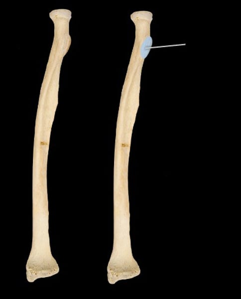
radial neck
Name the structure.
radial tuberosity of radius
Name the structure.
the body of the radius
name the structure
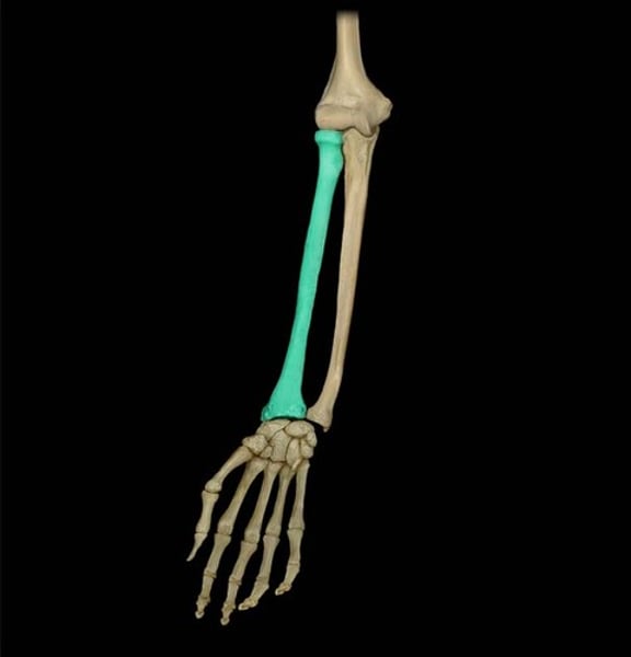
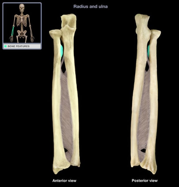
the radius distally includes:
1) the radial styloid process
radial styloid process
Name the structure.
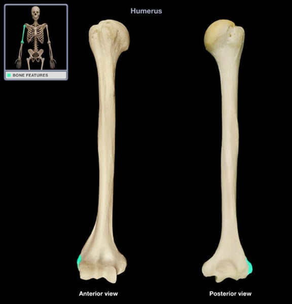
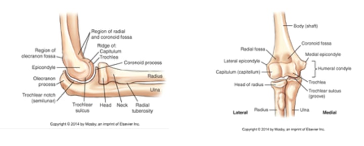
the distal humerus includes:
- (2) condyles
- (2) epicondyles
- trochlea - the medial aspect
- capitulum - lateral aspect
- coronoid fossa - anterior
- olecranon fossa - posterior
AP Projection: Forearm
-14 x 17
-lengthwise
-done on table top w/ 40" SID
-use a small focal spot
-in a supinated position
-patient is seated at 90 degrees
-you can fully extend the limb, the entire limb needs to be within the same space
-long axis of IR is parallel with the forearm
-the epicondyles should be equidistant
CR: perpendicular to the mid shaft
the radial head is located proximally and the ulnar head is located distally:
TRUE or FALSE
TRUE
good lateral position: forearm
3 important lateral forearm positioning points
1) elbow flexed at 90 degrees
2) hand and wrist in true lateral position
3) humerus should be resting on the table top
*if the above three points are met, a good lateral position of the forearm will most always result*
lateral projection: forearm (lateromedial)
-flex elbow 90 degrees on medial side [thumb is up]
-depress shoulder
-both joints shown
-CR: to midpoint of forearm
-Shows: bones of forearm, elbow joint, and proximal row of carpals
basic routine: elbow
-Ap - supine
- (2) AP obliques
*medial rotation
*lateral rotation
-lateromedial - lateral position
optional images of the elbow:
- (2) AP's - partial fexion
- axiolateral - greenspan & norman method (coyle method) vs. radial head series
AP projection of the elbow
- 10 x 12
- lengthwise
- sfs
- 40 " SID
- supinated position
- fully extend the limb, the entire limb in the same plane, epicondyles need to be parallel
- elbow is centered
- CR: perpendicular to the joint
Lateromedial Projection: Elbow
- 10 x 12
- lengthwise
- sfs
- 40 " SID
- done in the lateral position
- pt. seated at 90 degrees
- flex elbow 90 degrees & depress the shoulder
- hand and wrist are lateral
- center to the space
- CR: perpendicular to the elbow joint
AP oblique projection: elbow (medial)
- 10 x 12
- lengthwise
- sfs
- 40 " SID
- medial (rotation) oblique position
- pt. seated at 90 degrees
- fully extend the limb, medially rotate
- anterior surface is 45 degrees
- elbow is centered
- CR: perpendicular to joint
AP oblique projection: elbow (lateral)
- 10 x 12
- lengthwise
- sfs
- 40 " SID
- lateral oblique position
- need to fully extend the limb, and laterally rotate
- the posterior surface needs to be 45 degrees
- elbow needs to be centered
- CR: is perpendicular to the joint
AP projection: elbow (for a partially flexed elbow)
- 10 x 12
- lengthwise
- sfs
- 40 " SID
- a partial flexion position - proximal forearm
- pt. needs to be seated at 90 degrees
- limb needs to be partial flexed
- the forearm needs to be within the same plane
- epicondyles are parallel
- CR: perpendicular to proximal forearm
axiolateral projection: elbow (greenspan & norman method) when demonstrating the radial head fx
- done in lateral position
- pt. seated at 90 degrees
- flex the elbow at 90 degrees & depress the shoulder
- hand and wrist need to be pronated
- center to the space
-CR: elbow joint, needs to be 45 degrees medially
axiolateral projection: elbow (greenspan & norman method) when demonstrating the coronoid process
- done in a lateral position
- pt. seated at 90 degrees & depress the shoulder
- hand and wrist need to be pronated
- center to the space
- CR: to the elbow joint, 45 degrees distally
lateral epicondyle of humerus
Name the structure.
Which structure articulates with the trochlea?
proximal ulna
radial tuberosity of radius
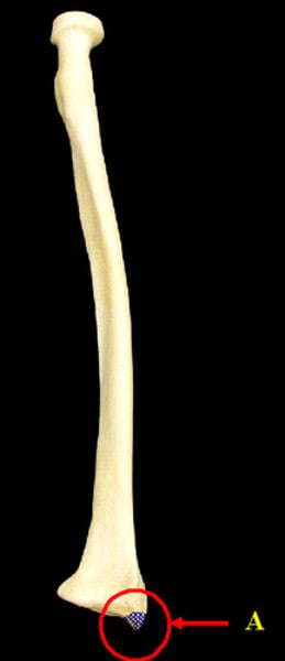
medial prominence just below the head of the radius; site of attachment of the biceps brachii
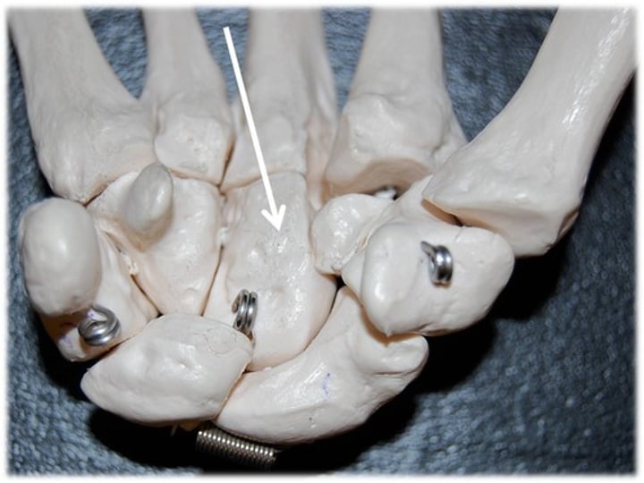
Scaphoid Series PA and PA Axial Rafert-Long method wrist
- 10 x 12
- lengthwise
- seat the pt. at the end of the radiographic table, with the arm and forearm resting on the table
- position the wrist on the IR for a PA projection
- without moving the forearm, turn the hand outward until the wrist is in extreme ulnar deviation
- CR: is perpendicular and with multiple cephalad angles, and should directly enter the scaphoid
- there must be no rotation of the wrist
- scaphoid with adjacent articular areas open
- maximum ulnar deviation
- bony trabecular detail and surrounding the soft tissues
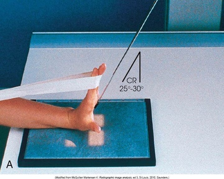
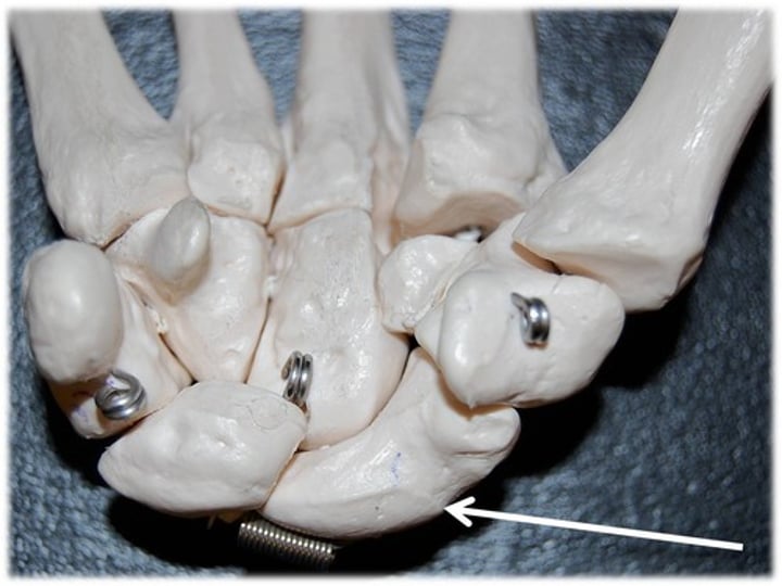
Carpal Canal Tangential (Gaynor-Hart Method)
SID: 40 in
Position: Hyperextend wrist, long axis of hand as vertical as possible
Pull fingers back with band
CR: 1 in distal to base of third metacarpal at 25 - 30 degree angle
posterior fat pads in elbow:
- covers the largest area and lives within the olecranon fossa of the posterior humerus
supinator fat pad of the elbow:
- is positioned anterior to and parallel with the anterior aspect of the proximal radius
What two bony landmarks are palpated for positioning of the elbow?
humeral epicondyle
humeral epicondyle
name the structure
for the AP projection of a forearm, how should the elbow be positioned?
needs to be fully extended
If the hand is pronated for the AP projection of the forearm, the image will demonstrate the:
radius and ulna crossing over each other
For the lateral projection of the forearm, the elbow should be flexed:
90 degrees
for the AP projection of the elbow, why should the hand be positioned with the palm facing up?
to prevent rotation of the bones of the forearm
for the lateral projection of the elbow, how should the hand be adjusted?
lateral with the thumb side up
How should the humeral epicondyles appear in the image of the lateral projection of the elbow?
superimposed
How much medial rotation of the elbow is needed to position it for AP oblique projections?
45 degrees
Which AP oblique projection positioning movement (medial rotation or lateral rotation) requires the hand to be pronated?
medial rotation
for the AP distal humerus projection (partially flexed elbow), what part of the upper extremity should be parallel and in contact with the IR?
the distal humerus
in the AP distal humerus projection (partially flexed elbow) image, what part of the upper extremity will appear greatly foreshortened in the image?
the proximal radius and ulna
For the AP proximal forearm projection (partially flexed elbow), what part of the upper extremity should be parallel and in contact with the IR?
radius and ulna
in the AP proximal forearm projection (partially flexed elbow) image, what part of the upper extremity will appear greatly foreshortened in the image?
the distal humerus
what position is the hand in for the axiolateral projection (Coyle method) of the elbow?
hand should be pronated for the axiolateral projection (Coyle method) of the elbow
what specific anatomy is best demonstrated on the axiolateral projection (Coyle method) of the elbow when the central ray is directed 45 degrees towards the shoulder?
an open elbow joint between the radial head and the capitulum
where is the centering point for the central ray for the AP projection of the thumb?
the first metacarpophalangeal joint
which projection of the thumb requires the patient to rotate the hand into extreme internal rotation?
AP projection
where is the centering point for the central ray for the PA projection of the third digit of the hand?
the proximal interphalangeal joint (PIP) to the third digit
explain why the hand should be rotated into extreme internal rotation until the lateral surface of the index finger is in contact with the IR, rather than positioning that finger with its medial surface toward the IR, for the lateral projection of the index finger
to minimize the OID
Name the four bones that should be completely seen in the image of the AP projection of the thumb:
- distal phalanx
- proximal phalanx
- the first metacarpal
- trapezium carpal
describe how and where the central ray should be directed for the PA projection of the hand:
needs to be perpendicular to the third metacarpophalangeal joint
What surface of the hand should be in contact with the IR for the PA projection of the hand?
anterior (palmar) surface
From the prone position, how many degrees should a hand be rotated for the PA oblique projection of that hand? For the lateral projection?
- 45 degrees
- 90 degrees
For the best demonstration of all digits how should the thumb and index finger be positioned with respect to the IR for the PA oblique projection of the hand?
needs to be elevated from the IR and parallel with the plane of the IR
for the PA projection of the wrist, why should the hand be slightly arched by flexing the fingers?
to place the anterior surface of the wrist in contact with the IR
describe how and where the central ray should be directed for the PA projection of the wrist:
perpendicular to the midcarpal area
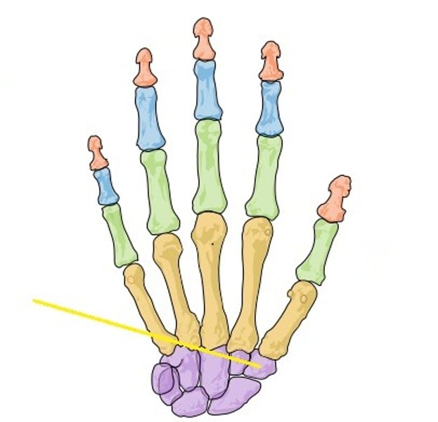
In addition to the eight carpal bones, what other bones should be seen in the image of the PA projection of the wrist?
- distal radius
- distal ulna
- proximal metacarpals
how many degrees from the prone position should the wrist be rotated for the PA oblique projection of the wrist?
45 degrees
which projection of the wrist requires the superimposition of the radial and ulnar styloid processes?
lateral
which surface of the wrist should be in contact with the IR for the lateral projection of the wrist?
ulnar (medial)
in which projection of the wrist should the metacarpals appear superimposed in the image?
lateral
for the PA oblique projection of the wrist, which side of the wrist should be elevated from the IR?
lateral (radial) side
how should the hand be positioned for the lateral projection of the forearm?
- true lateral
- thumb side up
PA oblique projection of the hand:
name the projection.
another name for triquetrum:
triangular or cuneiform
which of the following articulates with the bases of the metacarpal bones?
carpals
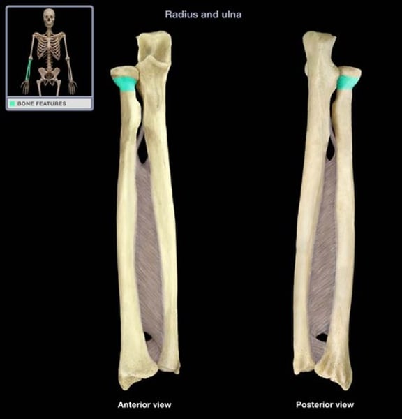
Which two structures articulate to form the proximal radioulnar joint?
the head of the radius and the radial notch of the ulna
How may bones are in the human body?
206 bones
how many bones in the axial skeleton?
80 bones
how many bones in the appendicular skeleton?
126 bones
Osteology
Study of bones
compact bone
dense, hard layers of bone tissue that lie underneath the periosteum
spongy bone
Layer of bone tissue having many small spaces and found just inside the layer of compact bone.
trabeculae
supporting bundles of bony fibers in cancellous (spongy) bone
medullary cavity
cavity within the shaft of the long bones filled with bone marrow
Periosteum
A dense fibrous membrane covering the surface of bones (except at their extremities) and serving as an attachment for tendons and muscles.
Endosteum
membranous lining of the hollow cavity of the bone
Ossification
process of bone formation
intermembranous ossification
bones develop from fibrous membranes in the embryo; creates the flat bones, such as the skull, clavicles, mandible, and sternum
primary ossification
begins before birth and forms long central shaft in long bones
secondary ossification
-occurs after birth when a separate bone begins to develop at both ends of each long bone
-near the age of 21
Long bones are:
-longer than they are wide
-typically known for being the limbs
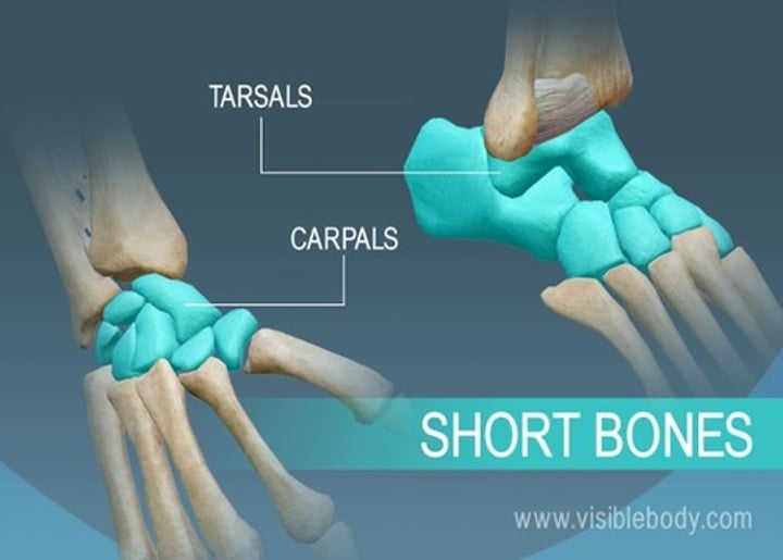
Short bones are:
cube shaped; carpals and tarsals
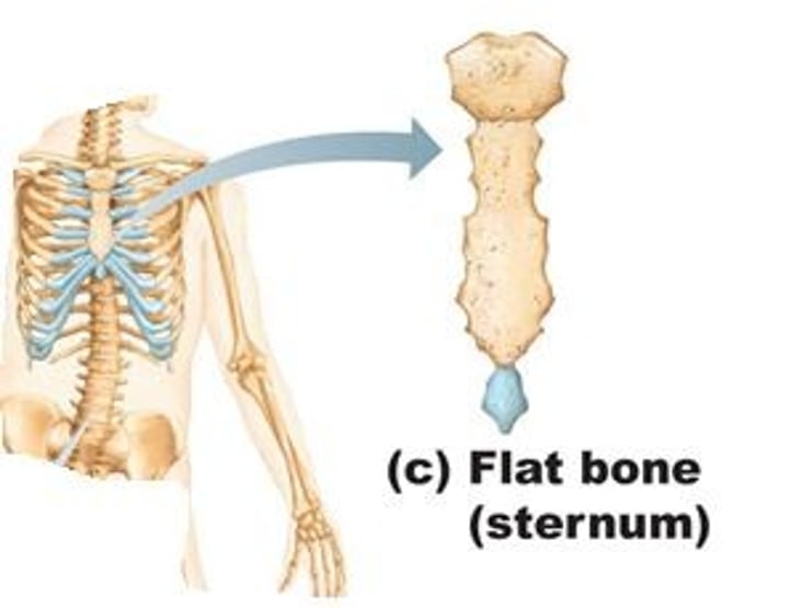
flat bones:
These bones are thin, flat, and curved. They form the ribs, breastbone, and skull.
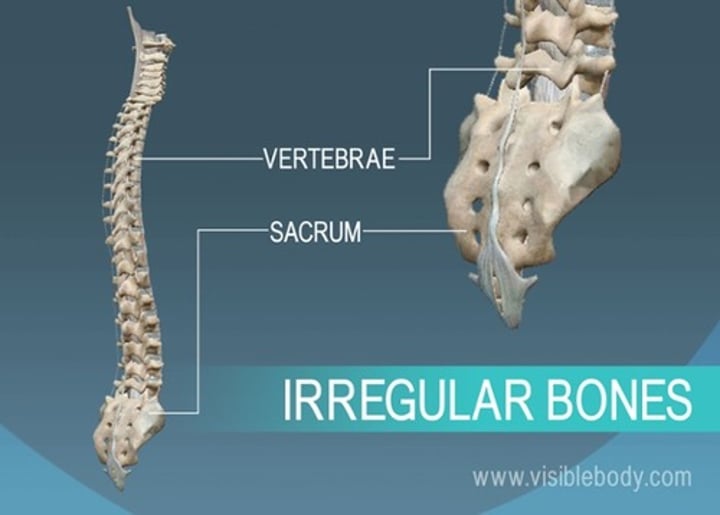
Irregular bones include
-peculiar shapes
-vertebrae, pelvic bones, and certain facial bones
Sesamoid bones are:
small and flat
develop inside tendons near joints of kneecap, hands, and feet
arthritis
inflammation of a joint
Arthology is the study of
joints
small to medium dry plaster cast
Increase mAs 50%-60% or +5-7 kV
Large or wet plaster cast
Increase mAs 100% or +8-10 kVp
Fiberglass cast
increase mAs 25-30% or +3-4 kVp
Distal phalanx
Name this specific bone.

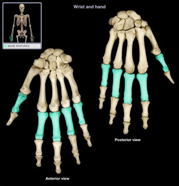
middle phalanx
Name this specific bone.
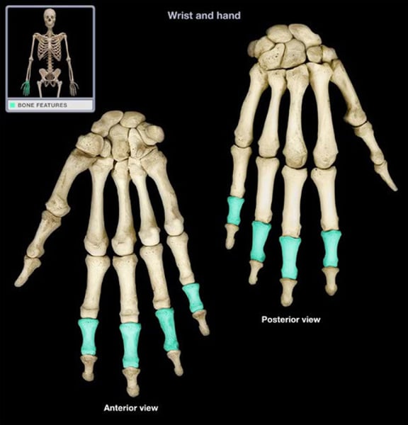
Proximal phalanx
Name this specific bone.
Hamate
Name the bone.
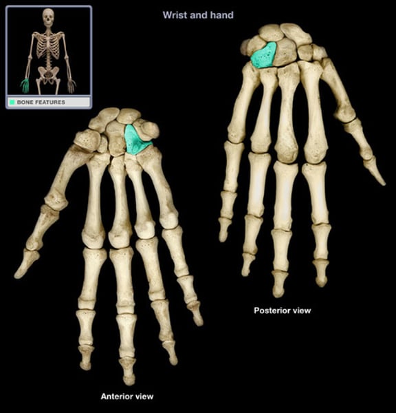
Capitate
Name the bone.
Trapezoid (Hand Bone)
Name the bone.
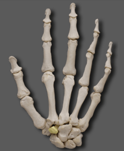
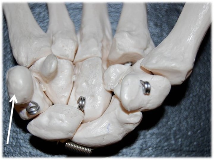
Trapezium
Name the bone.
Scaphoid
Name the bone.
Lunate
Name the bone.
Triquetrum
Name the bone.

Pisiform
Name the bone.