8. Nervous tissue
1/127
There's no tags or description
Looks like no tags are added yet.
Name | Mastery | Learn | Test | Matching | Spaced |
|---|
No study sessions yet.
128 Terms
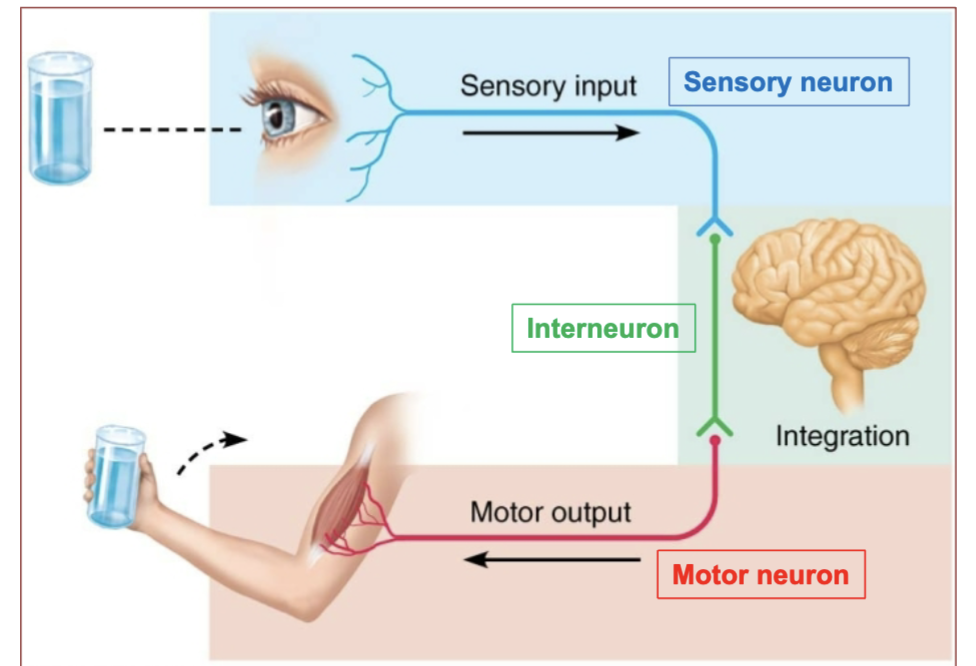
3 Functions of the nervous system
Sensory Input
Integration
Motor Output

Sensory Input 3 jobs
(Functions of the nervous system)
Gathering information.
Monitoring changes in and outside body.
Sense organs receive information and send stimuli to brain and spinal cord
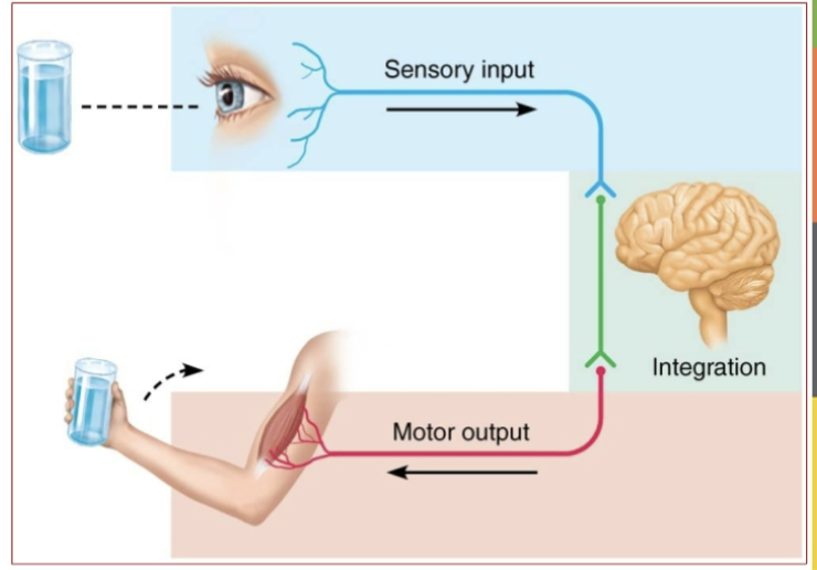
Integration - where does it occur & what does it do?
(a functions of the nervous system)
Occurs in our brain and it processes and interprets sensory input to determine appropriate response.

Motor Output definition/job
(Functions of the nervous system)
The response to stimuli.
→ It will send commands to activate a muscles or organ in
response to the input

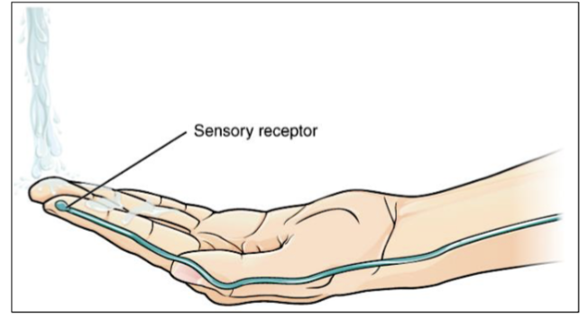
Example of a sensory input
Receptors in the skin that sense the temperature of the water
What does the Central nervous system (CNS) Consist of
Brain & Spinal Cord
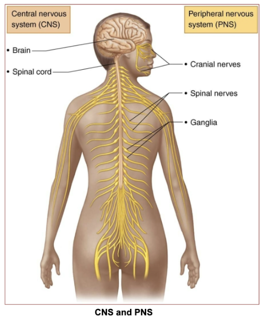
What does the Peripheral nervous system (PNS) consist of
Cranial nerves & spinal nerves. (& ganglia)

What does Somatic mean in the nervous system
Somatic refers to the part of the nervous system that controls voluntary movements of skeletal muscles.
→ Signals from skin, muscles, joints. Sends response to skeletal muscles.
What does Visceral mean in the Nervous system?
Visceral refers to organs.
signals from viscera (internal organs) send response to cardiac muscle, smooth muscle and glands.
Includes: Heart, lungs, digestive system, glands.

Which Nervous System, functions as the body’s main control center & and what does it include?
CNS or PNS?
the CNS functions as the body's main control center, it includes the brain and spinal cord
What 2 systems make up the Nervous system + what do they contain and there jobs?
Central Nervous System (CNS)
Contains: Brain and spinal cord
Job: Processes and integrates information, acting as the control center of the body.
Peripheral Nervous System (PNS)
Contains: Nerves that connect the CNS to the rest of the body
Job: Carries messages to and from the CNS. Moves the limbs, organs, and muscles.
Job of the CNS ?
Processes/collects & integrates information and decides how the body should respond
(includes: Brain & spinal cord)
Job of the PNS?
The PNS carries messages to and from the CNS. Moves the limbs, organs, and muscles.
(includes: All the nerves that extend from the brain and spinal cord to the rest of the body.)
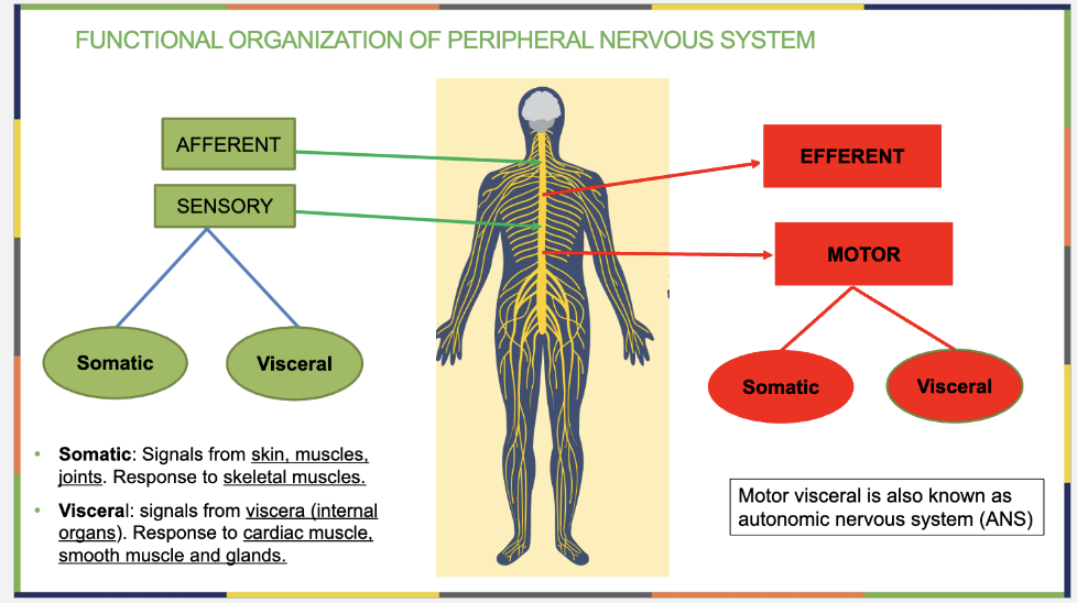
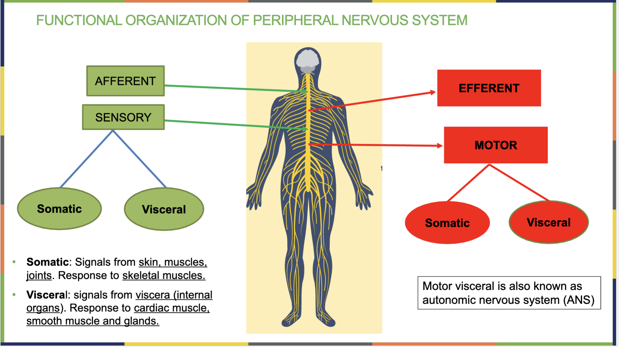
What does Afferent mean?
Signals going INTO the brain/spinal cord (CNS) (e.g., touch, pain, temperature).

What does Efferent mean?
Efferent → Exit
Sends signals FROM the brain/spinal cord TO muscles/organs (e.g., movement, gland activity)
to cause movement or responses.
Motor Neuron : ______ Sends motor response away from CNS.
Afferent, Efferent, Integration
Efferent

Sensory Neuron: ______ Detects Sensory stimuli & Conducts them to CNS.
Afferent, Efferent, Integration
Afferent

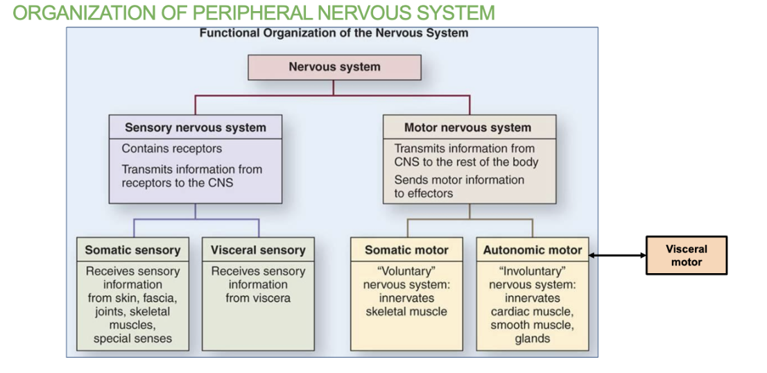
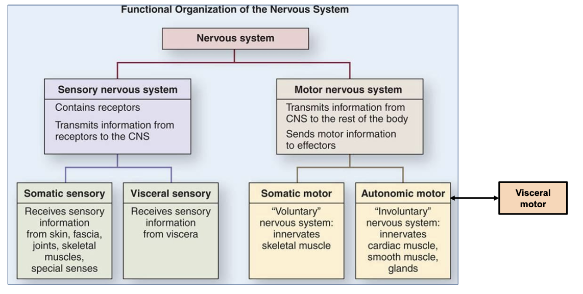
What are the 2 main divisions that make up the PNS?
Sensory (afferent) division & Motor (Efferent) Division
(these each have 2 divisions of their own)
What are the 2 divisions under the Sensory (afferent) division of the PNS?
Somatic & Visceral Sensory
Somatic Sensory: Brings information from the skin, muscles, and joints (e.g., touch, pain, temperature).
Visceral Sensory: Brings information from internal organs (e.g., stomach pain, heart rate).
(This division carries sensory signals from the body to the CNS to be processed)
What are the 2 divisions under the Motor (efferent) division of the PNS?
Somatic & Visceral Motor
Somatic Motor (Voluntary): Controls skeletal muscles (e.g., moving your arms, walking).
Visceral Motor (Involuntary) = Autonomic Nervous System (ANS): Controls organs, smooth muscle, and glands (e.g., heart rate, digestion).
(This division carries signals from the CNS to muscles and glands to make the body respond)

Interneuron : _______ Inside the CNS. Connects Incoming sensory input with outgoing motor.
Afferent, Efferent, Integration
Integration
Blood vessels constricting to raise my blood pressure is an example of …
Afferent/Efferent, Sensory/Motor , Somatic/visceral(autonomic)
Efferent → Motor → Visceral (autonomic)
Muscles in Gallbladder contracting to secret bile is an example of ….
Afferent/Efferent, Sensory/Motor , Somatic/visceral(autonomic)
Efferent → Motor → Visceral (autonomic)
Biceps contracting while pumping weights is an example of….
Afferent/Efferent, Sensory/Motor , Somatic/visceral(autonomic)
Efferent → Motor → Somatic
_______ : Integration Inside the CNS. Connects Incoming sensory input with outgoing motor.
(neuron name)
Interneuron
______ Neuron: Afferent Detects Sensory stimuli & Conducts them to CNS.
(neuron name)
Sensory Neuron

_____ Neuron : Efferent Sends motor response away from CNS.
Neuron name
Motor Neuron

Autonomic Nervous System (ANS) Definition/Controls
Definition: The part of the nervous system that controls involuntary bodily functions.
Controls: Internal organs, blood vessels, and smooth muscles.
(also known as the Motor visceral, it’s under the Motor (efferent) division of the PNS)
Motor visceral is also known as the ______ ______ system because it controls involuntary movements of internal organs, glands, and smooth muscles.
autonomic nervous system (ANS)
_______ visceral is also known as the autonomic nervous system (ANS) because it controls involuntary movements of internal organs, glands, and smooth muscles.
Motor visceral
2 cells of the nervous system
Neurons → Actual nerve cells that transmit electrical signals
Neuroglia → Support system for the neurons
Neurons definition/job
Neurons are the actual nerve cells that transmit electrical signals
Neuroglia (glial cells) definition
They are the support system for the neurons
There are about 50 Neuroglia (glue cells)for each neuron that help support and nourish it.
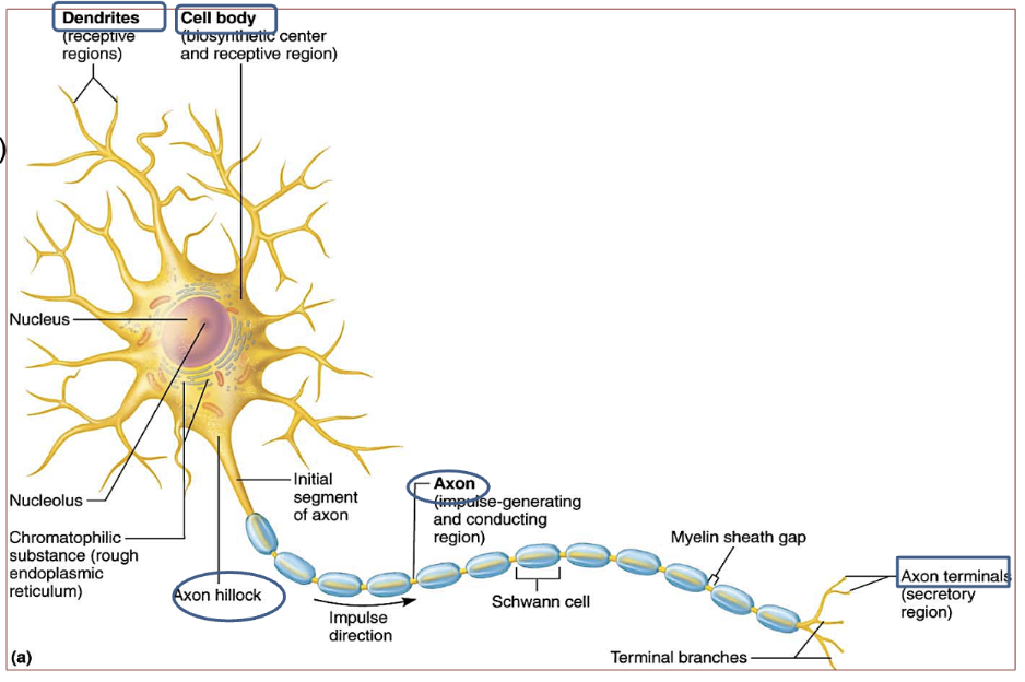
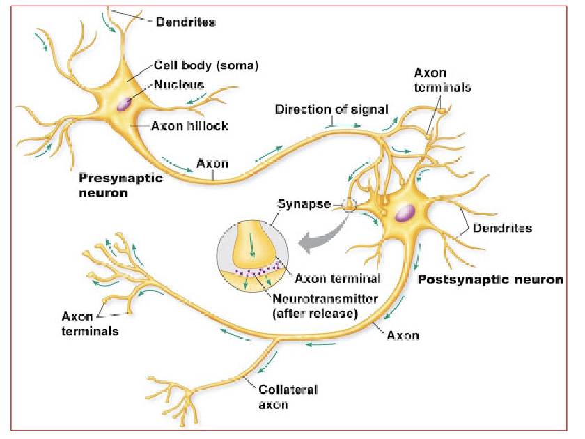
4 Neuron Structures
Soma (cell body)
Dendrites
Axons : Axon Hillock (trigger zone) & Axon
Axon terminal
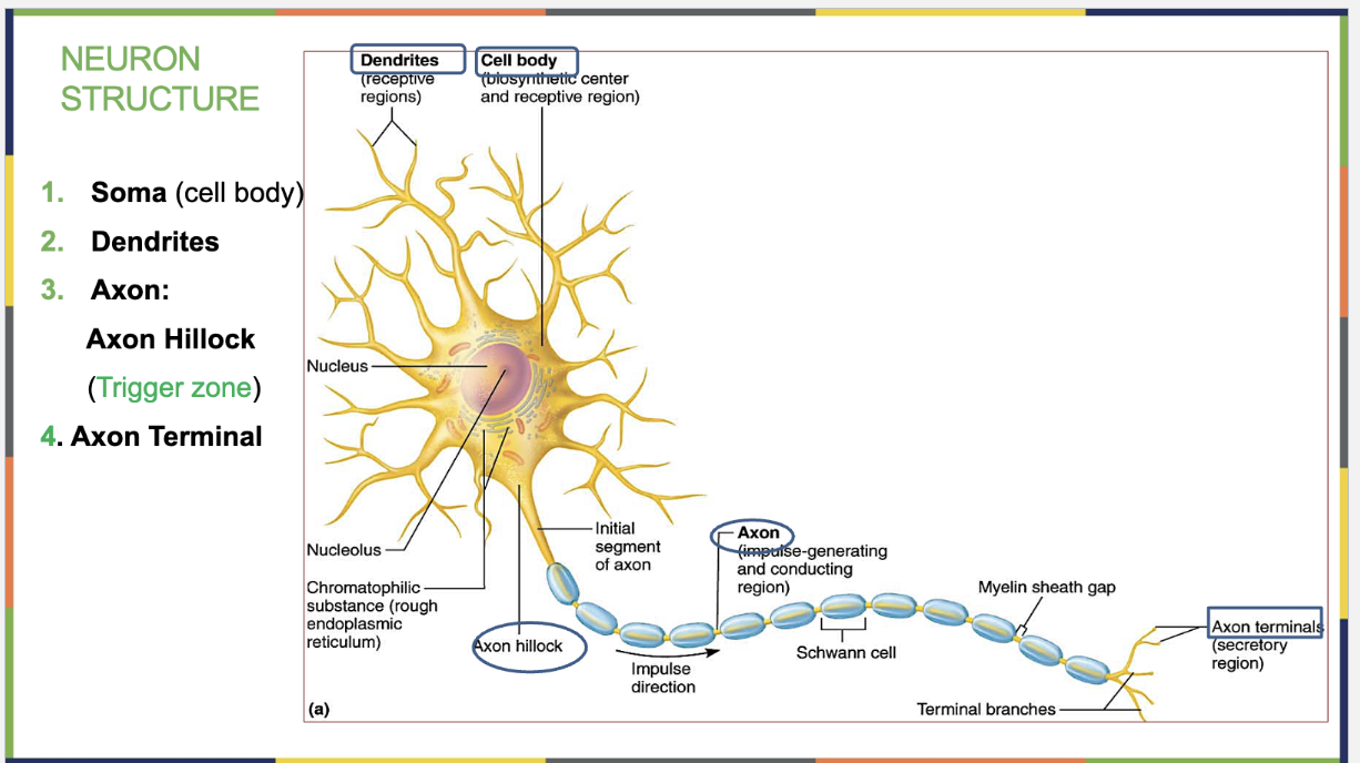
Function of the Soma (cell body) of a neuron (& what it contains)
Metabolic functions occur in the soma
Contains organelles except centrioles → No Cell Division
RER is called Nissl bodies.

What’s the RER of the Soma (cell body) of a neuron called?
Nissl bodies
In which structure of the Neuron doe metabolic functions occur?
At the Soma (cell body) of the neuron
Function of the Dendrites of a neuron
Receptive: receives stimuli from other neurons
→ They convey stimuli to the soma by graded potentials.

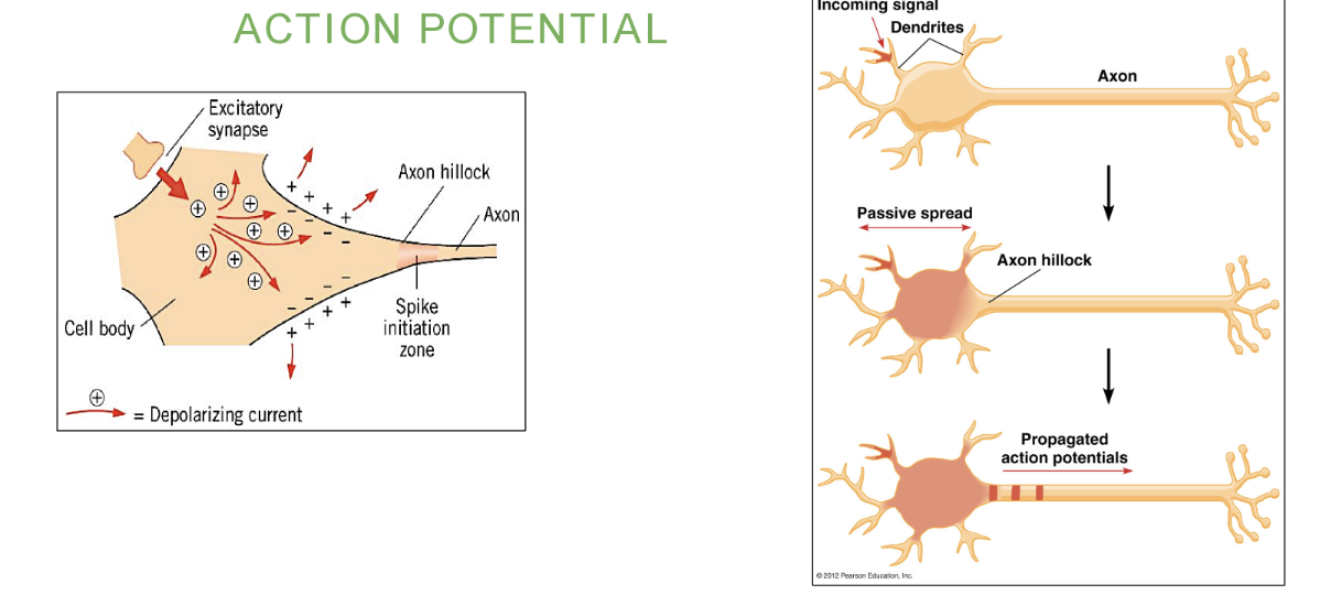
Function of the Axons (hillock (trigger zone) & axon) of a neuron
Axon Hillock (trigger zone) – The starting point of the axon; nerve impulse (action potential) is generate here & it’s sent down the axon.
Axon – Conducting area. Transmits electrical signals away from the neuron’s cell body to other neurons, muscles, or glands by sending it to the axon terminal. The terminal contains the synaptic vesicles with the ACH.

Function of the Axon Terminal of a neuron
Its the Secretion area.
The terminal branches of the axon terminal have the synaptic knobs containing synaptic vesicles of neurotransmitters (ACH).

What organelle do neurons (nerve cells) not have & what can they not perform?
Centrioles & they CANNOT perform cell division (mitosis).
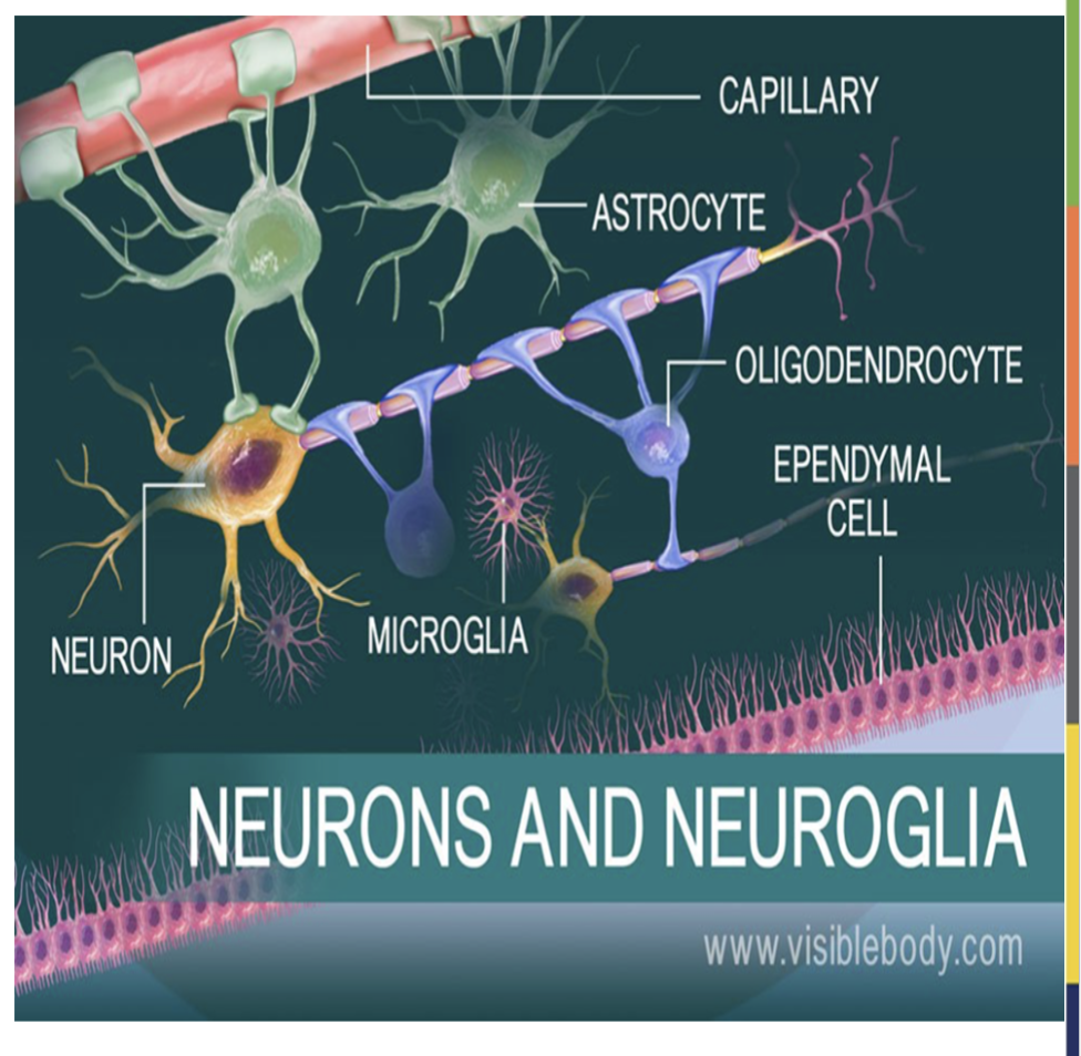
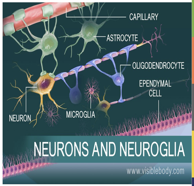
4 Neuroglia (Glial Cells) in CNS & their jobs.
Astrocytes - brings nutrition to neurons. Attached to blood vessels & also form
blood brain barrier
Oligodendrocytes - Secrete myelin around axons of neurons in CNS
Microglia - Tiny Phagocytic immune cells. Destroy microorganisms and remove debris.
Ependymal cells - Secrete and circulate cerebrospinal fluid (CSF). Line cavities in the brain and spinal cord that contain cerebrospinal fluid.
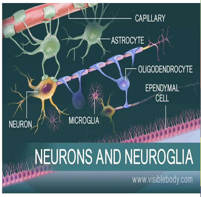
Job of the Astrocytes (Glial Cell) & where are they, CNS or PNS?
They bring nutrition to neurons & are attached to blood vessels (Form blood brain barrier)
→ In CNS

Job of the Oligodendrocytes (Glial Cell) & where are they, CNS or PNS?
They Secrete myelin around axons of neurons in CNS.

Job of Microglia (Glial Cell) & where are they, CNS or PNS?
They’re tiny Phagocytic immune cells. Destroy microorganisms and remove debris in CNS.

Job of Ependymal cells (Glial Cell) & where are they, CNS or PNS?
Ependymal cells line cavities in the brain and spinal cord that contain cerebrospinal fluid & they Secrete and circulate cerebrospinal fluid (CSF)
→ in CNS

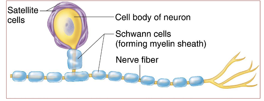
2 Neuroglial Cells of PNS & their jobs?
Schwann cells : Secrete myelin around axons of neurons in PNS.
Satellite cells : Surround soma of neurons in the ganglia for protection and electrical insulation.

Job of Schwann cells (Glial cell) & where are they, CNS or PNS?
They Secrete myelin around axons of neurons in PNS.
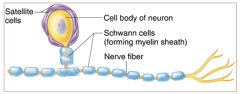
Job of Satellite cells (Glial cells) & where are they CNS or PNS?
They surround the soma of neurons in the ganglia for protection and electrical insulation.
They’re in the PNS.

Which 2 Neuroglia Cells Secrete Myelin & where are they?
Oligodendrocytes : In CNS → Secrete myelin around axons of
neurons in CNS
Schwann cells : In PNS → Secrete myelin around axons of neurons in PNS
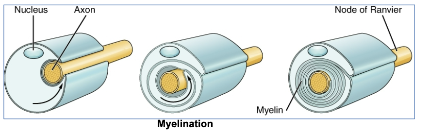
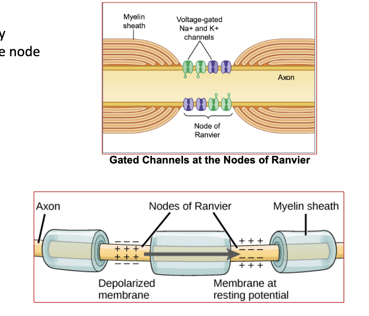
What’s Myelin & what’s it’s job?
Myelin is a fatty, insulating layer that wraps around axons, formed by Oligodendrocytes (CNS) and Schwann cells (PNS).
→ It Speeds up nerve impulses by allowing signals to "jump" between gaps (nodes of Ranvier).
Protects by insulating the axon to prevent signal loss.
(Myelinating glia wrap several layers of myelin around the cell membrane of a segment of the axon)
Damage to which neuroglia cell of the CNS would affect the Secretion of cerebrospinal fluid?
The Ependymal Cells
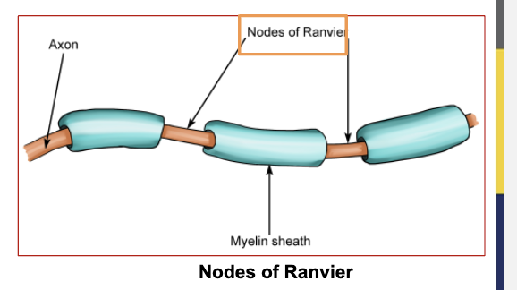
Areas not covered with myelin are called _________?
Nodes of Ranvier

What are “Nodes of Ranvier”?
Areas not covered with myelin.
Schwann cells insulate _______ nerves.
Peripheral nerves
Oligodendrocytes insulate ______ nerves.
Central
What is White Matter & what is it made up of?
The areas that appear white in the brain or spinal cord, and are occupied by dense collections of myelinated axons.

What is Gray matter & what is it made up of?
Area with no myelin.
Made up of mostly cell bodies, dendrites, neuroglia, and unmyelinated axons.
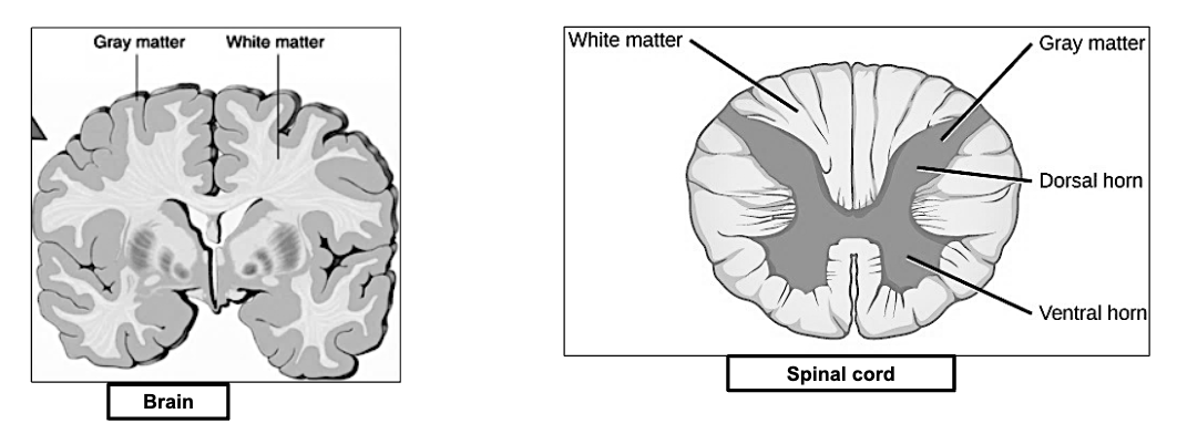
Which area is made up of Myelin in the brain and/or spinal cord, white or gray matter?
White

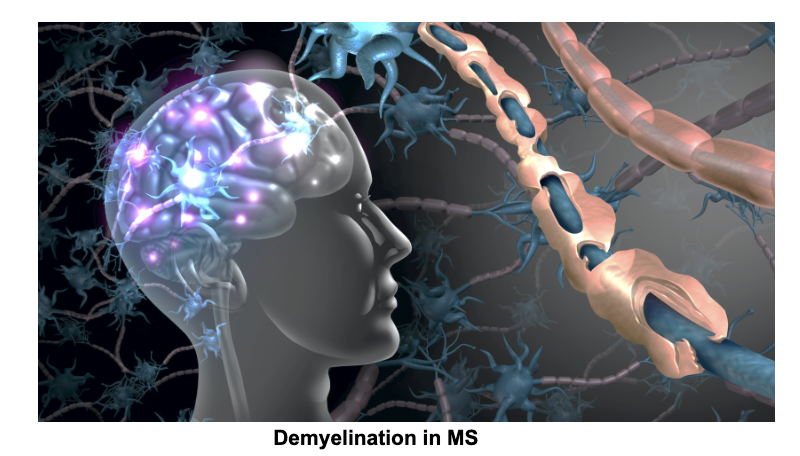
What is Multiple Sclerosis (MS) ?
The loss of myelination in CNS.
It will disrupt nerve conduction and result in multiple sensory and motor symptoms.
( Ex : can move but not feel or feel but not move)
The Resting Membrane potential is _____ mV.
-70 mV
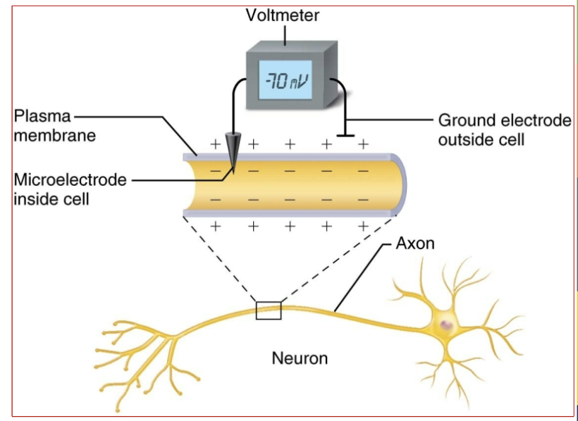
Inside the membrane (neuron) , it is more ______ charged.
negatively
-70mV

When the membrane (neuron) is resting it is also referred as _______.
Polarized.
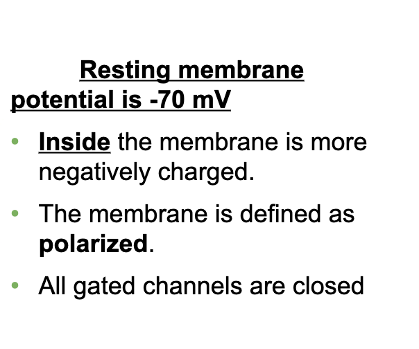
All gated channels are _____ when the membrane (neuron) is resting.
opened/closed
Closed

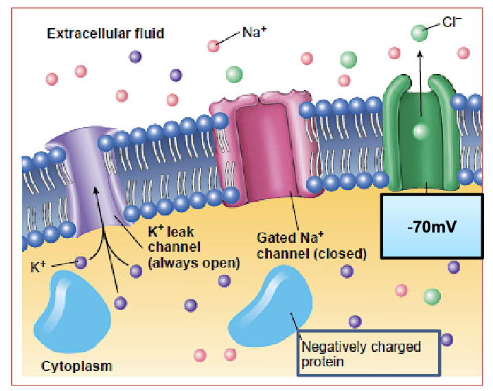
3 Factors Generating Resting Membrane Potential
(3 factors keeping a neuron at rest with a negative charge inside)
Difference in Concentration & Leak Channels (ionic gradient)
ECF: Higher Na⁺ concentration
ICF: Higher K⁺ concentration
LEAK CHANNELS → K⁺ (potassium) leaks OUT
Na⁺ (sodium) leaks IN, but there are fewer Na⁺ leak channels.
SO → More positive charge leaves than enters → inside stays negative.
(all gated channels CLOSED)
Fixed Negative Charged Protein in ICF
ICF has negative proteins that can’t move out, keeping the inside more negative.
Resting Potential: -70mV.
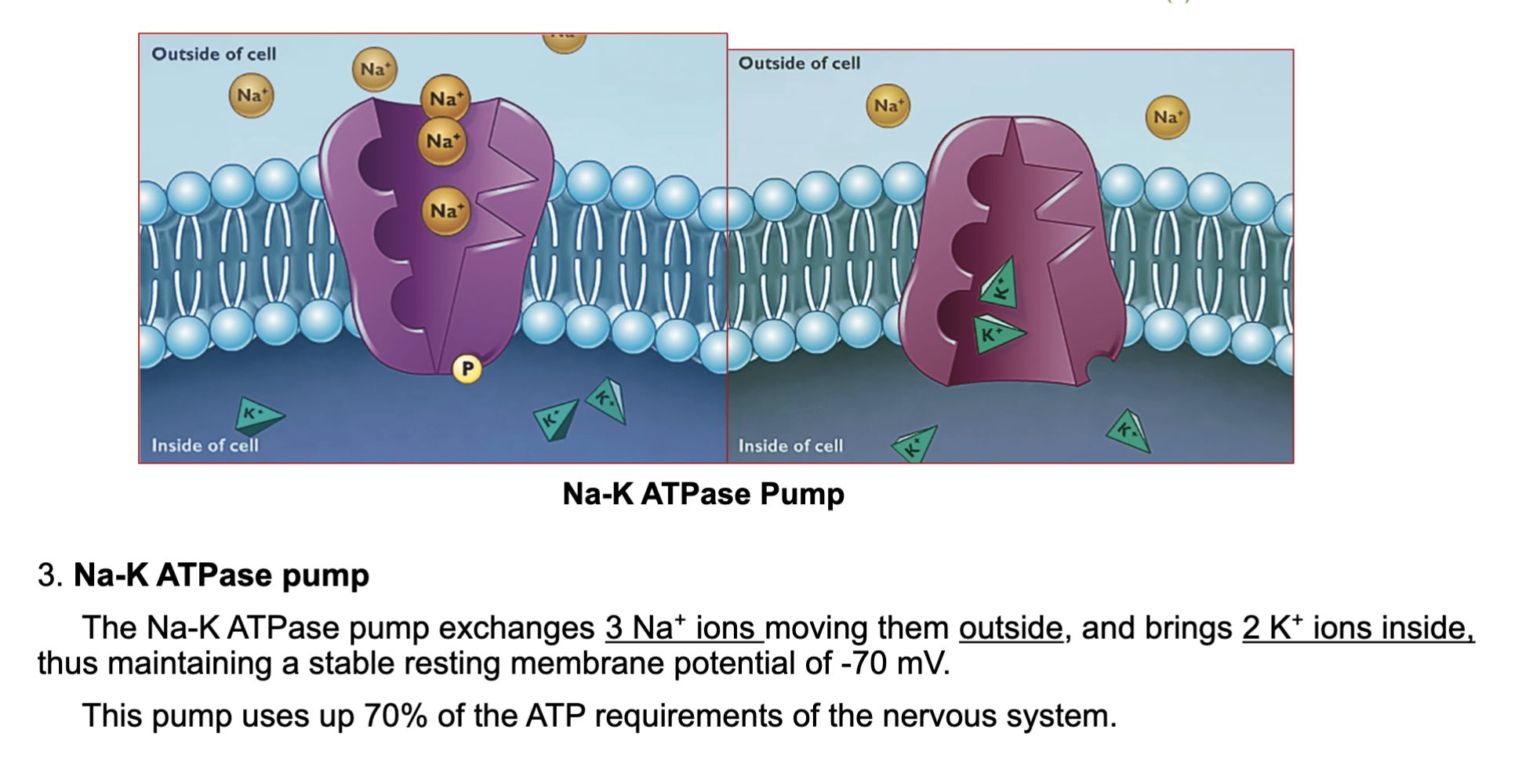
Sodium/Potassium Pump
Pumps out 3 Sodiums and Pumps in 2 Potassiums
Maintains resting membrane potential at -70mV.
This pump Uses 70% of ATP in the nervous system.
What uses 70% of the ATP reuirements of the nerbous system?
the Sodium/Potassium Pump that pumps out 3 Na⁺ out and 2 K⁺ in, thus maintains resting membrane potential at -70mV.
2 Types of Nerve potentials
Graded potential - Stays at dendrites
Action Potential - Come from axon hillock, all the way down through the axon to the terminals.
What are the 2 types of Graded Potentials?
Weak & Strong Graded potenials
What are graded potentials?
Localized stimuli that travel short distances
mainly at the dendrites.

What happens with a weak stimulus in graded potentials?
Opens a few Na⁺ channels, resulting in a short, localized current that will not travel far.
dies at dendrites
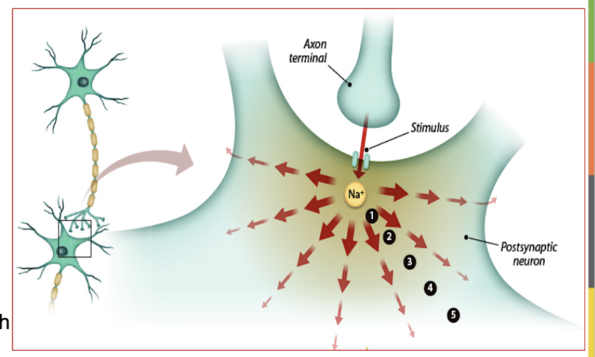
What happens with a strong stimulus in graded potentials?
Opens more Na⁺ channels, creating a stronger current that may reach the trigger zone.
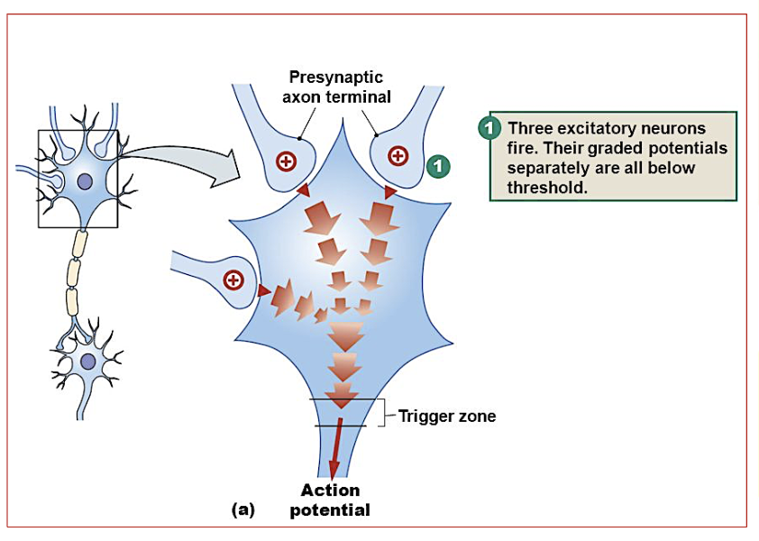
How does stimulus strength affect graded potentials?
It determines how many Na⁺ channels open, and if strong enough it can travel down to the axon hillock (trigger zone) and become an action potential
(needs to be at least -55mv)

What happens if the electrical impulse from a graded potential reaches the trigger zone?
It will result in an action potential. (-55 mV needed at trigger zone to start action potential)
This travels all the way down the axon to the terminals.

Action potential is generated at the _____ Zone in the ____ Hillock, when graded potentials are strong enough.
Trigger , Axon
Action potential is generated at the Trigger Zone in the Axon Hillock, when graded potentials are strong enough.
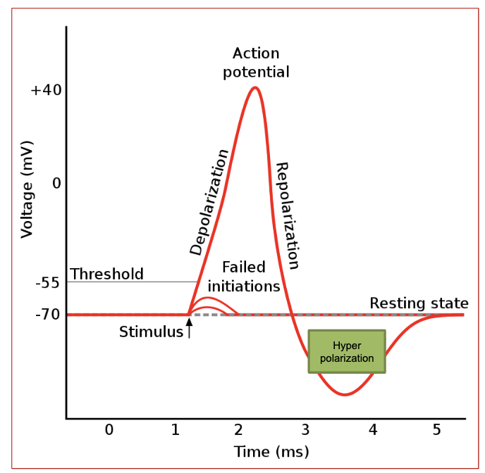
3 Phases of an Action Potential (AND describe each)
Depolarization - Na⁺ channels open, Na⁺ rushes in. Membrane potential rises to +30mV.
Repolarization - Na⁺ channels close, K⁺ channels open. K⁺ exits, restoring negativity inside membrane.
Hyperpolarization - K⁺ channels stay open longer, causing voltage to drop below -70mV.
→ Na-K pump restores resting potential (-70mV).

Which channels are open & closed during Depolarization ?
(1st phase of an action potential)
Sodium/Na⁺ channels are open, so sodium rushes in.
(potassium channels are closed)

Which channels are open & closed during Repolarization ?
(2nd phase of an action potential)
Na⁺ channels close, & Potassium/K⁺ channels open.
K⁺ exits, bringing the voltage back down.
(sodium channels are closed)
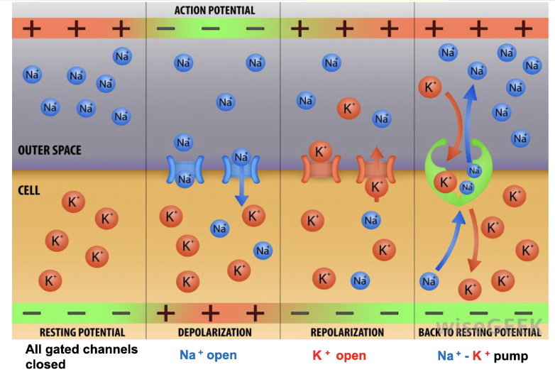
How are the gated channels during RMP?
→ All gated channels are closed.
Leak channels are open, allowing some K⁺ to exit and a little Na⁺ to enter.
(The Na-K pump maintains balance by moving 3 Na⁺ out and 2 K⁺ in, keeping RMP at -70mV.)

To what mV must local potential change to, to start an action potential? This is called the threshold potential.
It will cause a change in voltage and carry the signal to the nerve terminal.
-55 mV

What is the resting membrane potential (RMP)?
-70 mV

What is the threshold potential?
-55mV; the voltage needed at the trigger zone to start an action potential.
What happens during depolarization?
Sodium/Na⁺ channels open, Na⁺ enters, and voltage rises to +30mV.

What happens during Repolarization?
Na⁺ channels close, K⁺ channels open, K⁺ exits, and voltage starts becoming negative again.

What happens during Hyperpolarization?
K⁺ channels stay open longer, making the voltage more negative than -70mV.
(the potassium channels are a little too slow at closing up)

What restores the resting membrane potential after hyperpolarization?
The sodium-potassium pump returns the membrane to -70mV.

What happens when the membrane reaches -55mV?
This is called the threshold potential and it will reach the Axon hillock (trigger zone) and send Action Potential down the Axon.

How does -55mV lead to depolarization?
This is the voltage that the local potentials must change to, in order to trigger the action potential down the axon.
-55mv is called the threshold potential

What is the result of depolarization?
The membrane potential increases quickly, leading to an action potential.

What returns the membrane back to resting state?
The Sodium/Potassium (Na+/K+) pump returns the membrane back to resting state!
An action potential starts when the membrane reaches ___mV (threshold potential) and fully activates at __mV during depolarization.
-55mv, +30mV

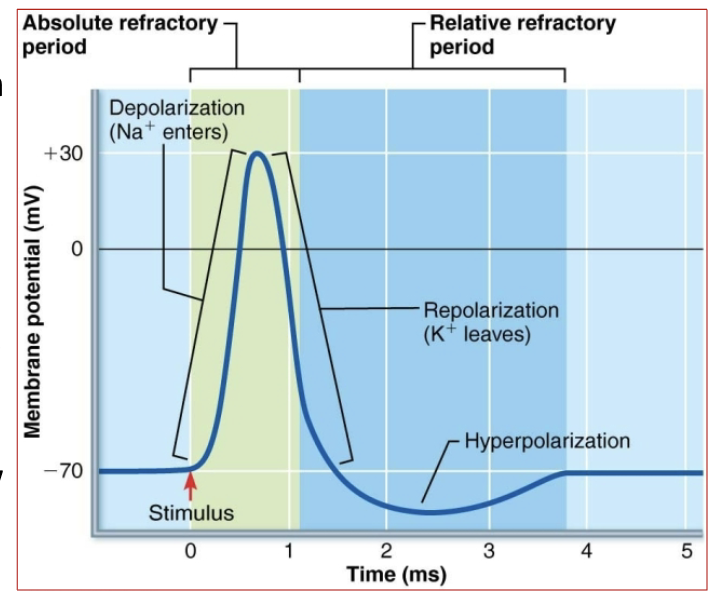
What is the absolute refractory period?
the time during and immediately after an action potential (during depolarization) when the neuron CANNOT fire another action potential—no matter how strong the stimulus. its unresponsive.
→ This Ensures one-way AP (action potential) transmission and prevents exhaustion.
WHY? - because Na⁺ channels are either already open or inactivated, so another action potential cannot start until they reset.
Why is the absolute refractory period important? (3 reasons)
It ensures AP(action potential) moves in one direction,
prevents overlapping signals,
and allows neurotransmitter replenishment.

What is the relative refractory period?
the phase after repolarization when the neuron CAN fire another action potential, but only with a much stronger stimulus.
WHY? : because K⁺ channels are still open, so the neuron is in hyperpolarization (more negative than -70mV).
Since it’s further from the threshold (-55mV), a stronger stimulus is needed to trigger another action potential.

What 2 things affects axon conduction velocity?
Axon Diameter & Myelination
Axon diameter : Larger diameter of neuron = more surface area = faster.
Myelination : Myelinated axons= faster
unmyelinated axons = slower.
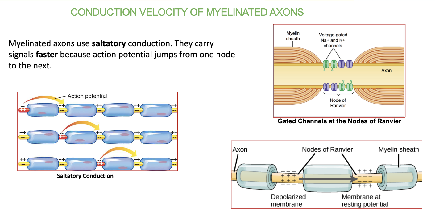
How do myelinated axons conduct signals?
Saltatory conduction – AP jumps between Nodes of Ranvier, speeding up transmission.
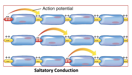
What is Saltatory conduction?
the process where the action potential jumps from one Node of Ranvier to the next in a myelinated axon, making signal transmission faster and more efficient.

What happens at the Nodes of Ranvier w the channels?
The Nodes of Ranvier are gaps in the myelin sheath where voltage-gated Na⁺ and K⁺ channels are located.
→ (the gap that the AP jumps becomes positive because of the channels)
When the action potential reaches a node, these channels open, allowing ion exchange to regenerate the signal and keep it moving quickly down the axon.
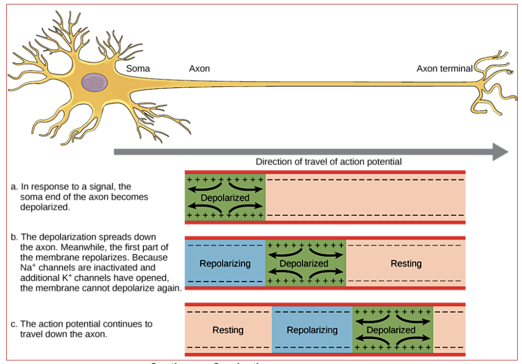
Conduction velocity in unmyelinated axons is _______ and _____.
Continuous & Slower
What is a synapse?
A connection between two neurons that allows signal transmission.
(or neuron w a muscle/gland)
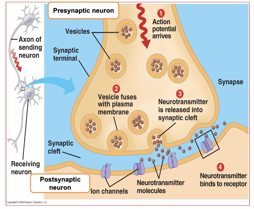
What does the presynaptic neuron contain?
Synaptic vesicles that store neurotransmitters (e.g., Acetylcholine).
(this is also the Synaptic Knob)
Also contains the voltage gated channels
