Micro2 (microscopically slide)
 Call Kai
 Learn
Learn Practice Test
 Spaced Repetition
 Match
Match Flashcards
 Knowt Play
1/22

There's no tags or description
Looks like no tags are added yet.
Name | Mastery | Learn | Test | Matching | Spaced | Call with Kai | Chat |
|---|
No analytics yet
Send a link to your students to track their progress
23 Terms
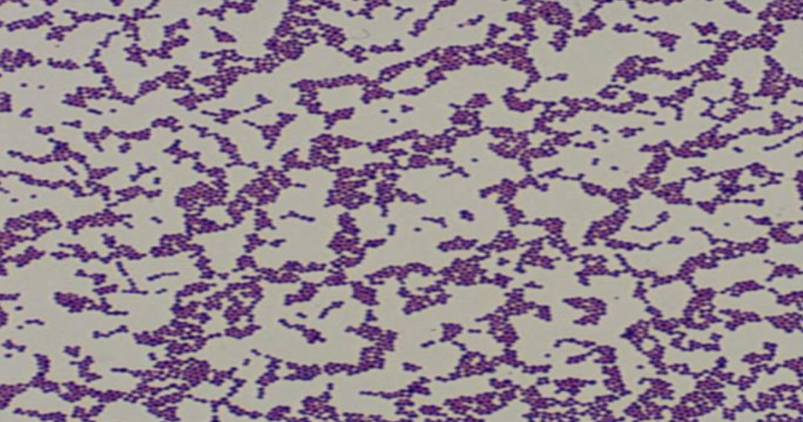
Specimen (microscopically slide):
• Name of organism:
• Stain:
• Sample from: culture or pus or in tissue
• Morphology
• Name of organism:
Staphylococci
• Stain:
Gram stain
• Sample from:
culture
• Morphology:
Gram-positive cocci arranged in grape-like clusters

Specimen (microscopically slide):
• Name of organism:
• Stain:
• Sample from: culture or pus or in tissue
• Morphology
• Name of organism:
Capnocytophaga
• Stain:
Gram stain
• Sample from:
culture
• Morphology:
Gram-negative, long thin fusiform bacilli
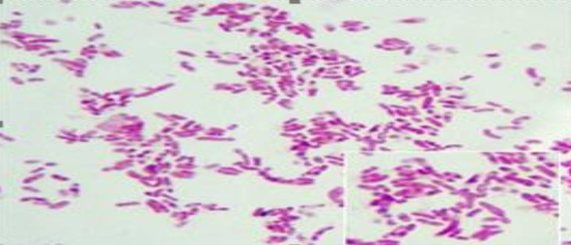
Specimen (microscopically slide):
• Name of organism:
• Stain:
• Sample from: culture or pus or in tissue
• Morphology
• Name of organism:
Bacteroides fragilis
• Stain:
Gram stain
• Sample from:
culture
• Morphology:
Gram-negative, polymorphic bacilli
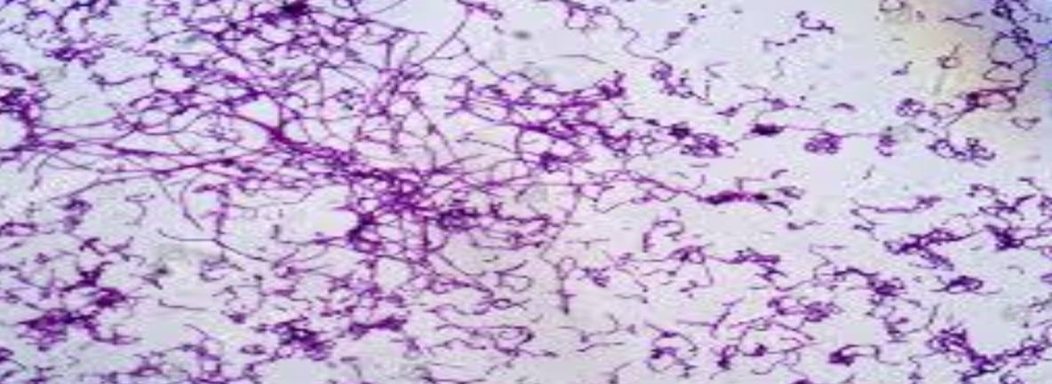
Specimen (microscopically slide):
• Name of organism:
• Stain:
• Sample from: culture or pus or in tissue
• Morphology
• Name of organism:
Actinomycetes
• Stain:
Gram stain
• Sample from:
culture
• Morphology:
Gram-positive branching, filamentous bacteria
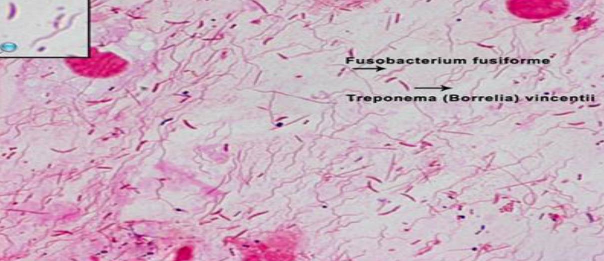
Specimen (microscopically slide):
• Name of organism:
• Stain:
• Sample from: culture or pus or in tissue
• Morphology
• Name of organism:
Fusobacterium fusiforme
and Gram-negative spirochetes
diagnostic of Vincent’s angina
• Stain:
Gram stain
• Sample from:
pus
• Morphology:
Gram-negative long fusiform bacilli together with gram-negative spirochetes and many pus cells
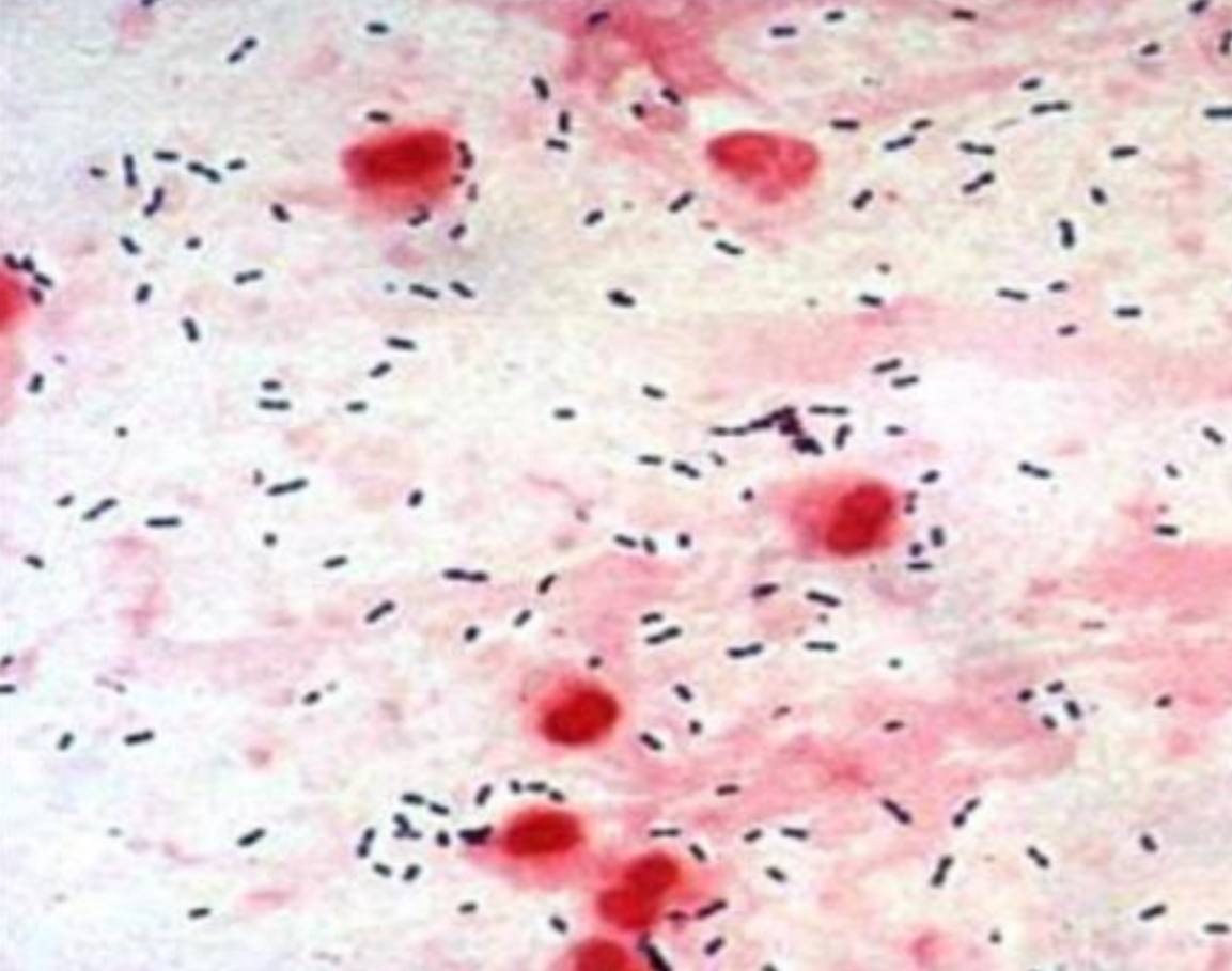
Specimen (microscopically slide):
• Name of organism:
• Stain:
• Sample from: culture or pus or in tissue
• Morphology
• Name of organism:
S. pneumoniae
(Streptococcus pneumoniae)
• Stain:
Gram stain
• Sample from:
in tissue
• Morphology:
Gram-positive cocci arranged in pairs
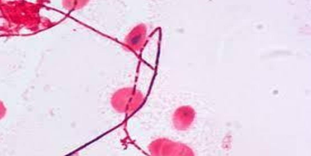
Specimen (microscopically slide):
• Name of organism:
• Stain:
• Sample from: culture or pus or in tissue
• Morphology
• Name of organism:
Leptotrichia
• Stain:
Gram stain
• Sample from:
Pus
• Morphology:
Gram-negative, slender, filamentous bacilli, usually with one pointed end
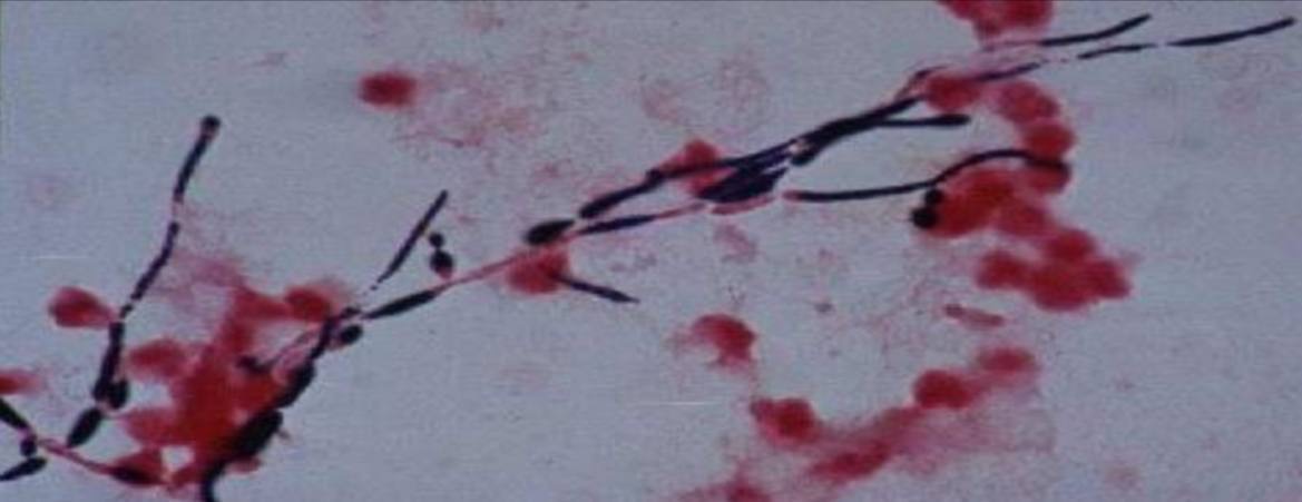
Specimen (microscopically slide):
• Name of organism:
• Stain:
• Sample from: culture or pus or in tissue
• Morphology
• Name of organism:
Candida albicans
• Stain:
Gram stain
• Sample from:
Pus
• Morphology:
Yeast cells with pseudohyphae
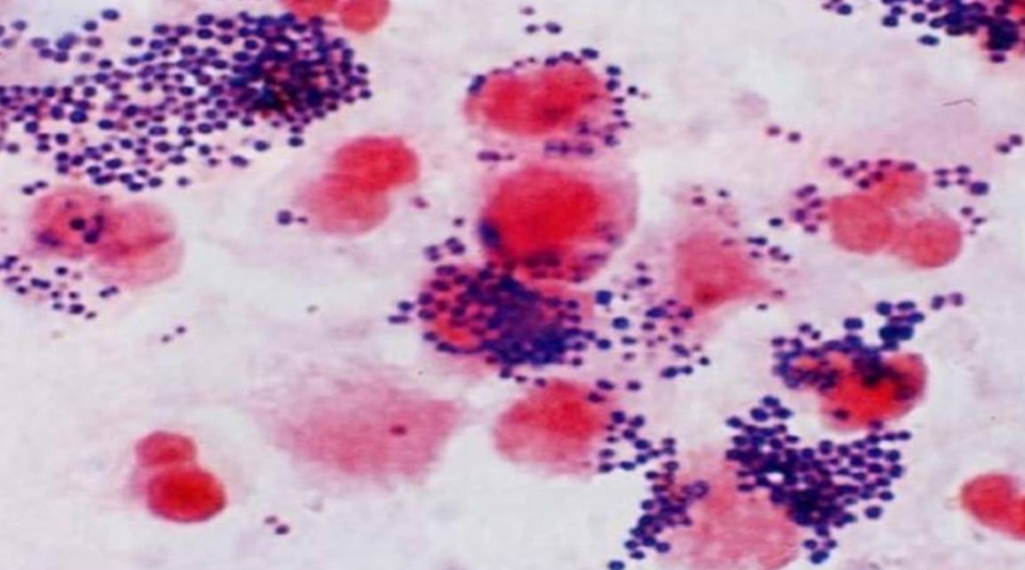
Specimen (microscopically slide):
• Name of organism:
• Stain:
• Sample from: culture or pus or in tissue
• Morphology
• Name of organism:
Staphylococci
• Stain:
Gram stain
• Sample from:
pus
• Morphology:
Gram-positive cocci arranged in grape-like clusters
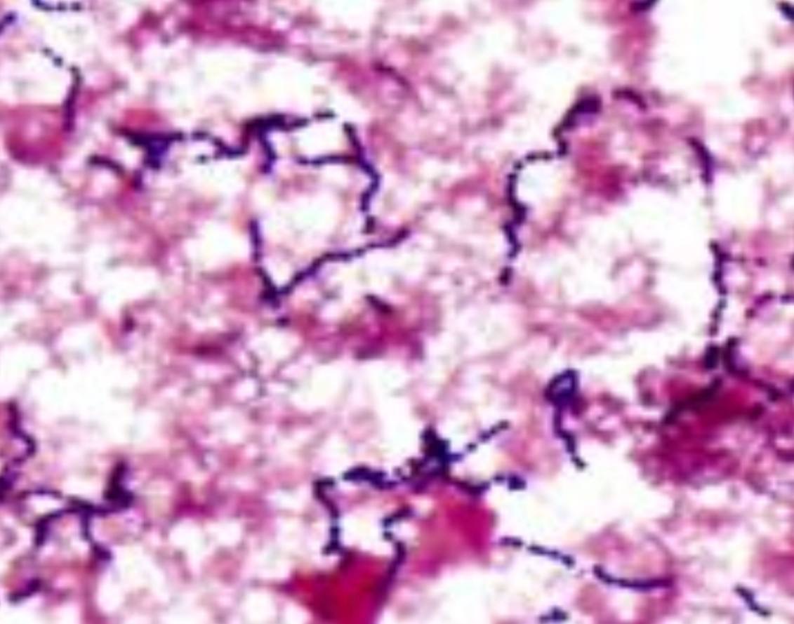
Specimen (microscopically slide):
• Name of organism:
• Stain:
• Sample from: culture or pus or in tissue
• Morphology
• Name of organism:
Streptococci
• Stain:
Gram stain
• Sample from:
pus
• Morphology:
Gram-positive cocci arranged in chains
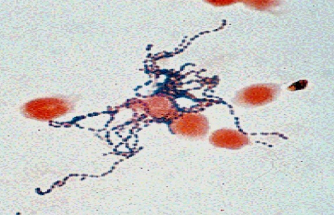
Specimen (microscopically slide):
• Name of organism:
• Stain:
• Sample from: culture or pus or in tissue
• Morphology
• Name of organism:
Streptococci
• Stain:
Gram stain
• Sample from:
pus
• Morphology:
Gram-positive cocci arranged in chains
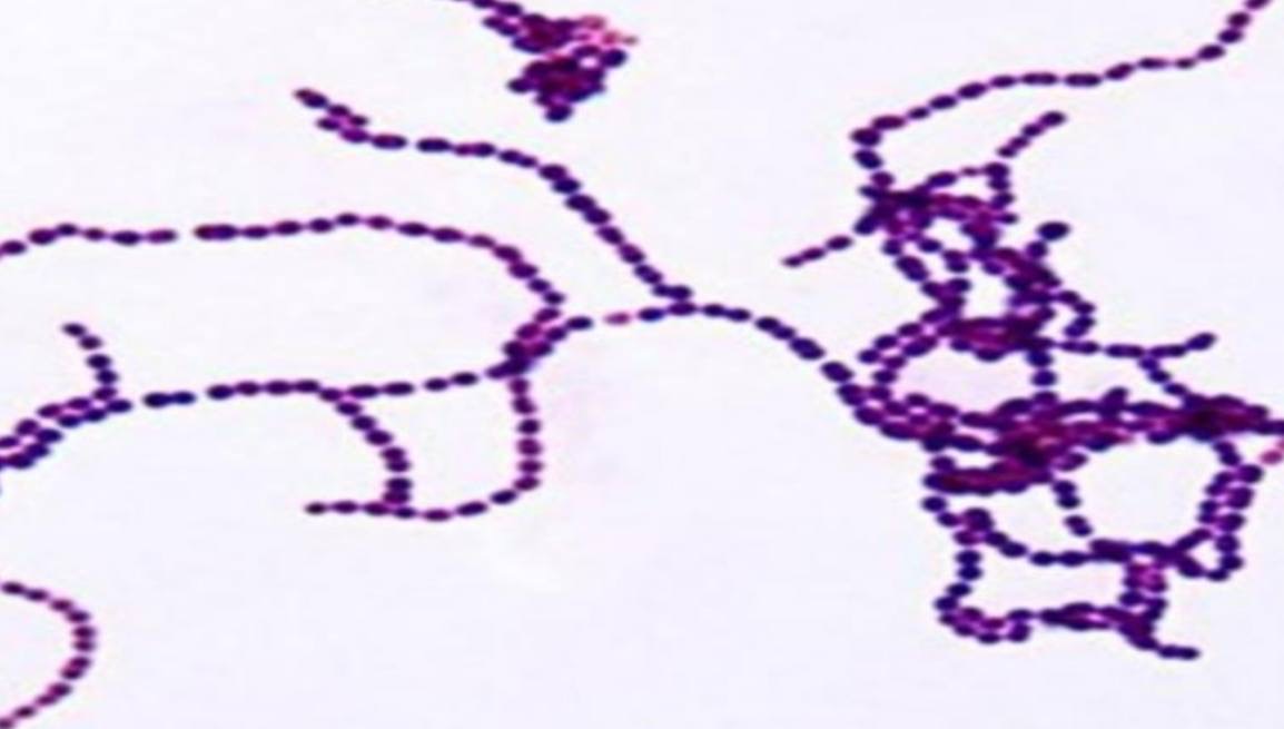
Specimen (microscopically slide):
• Name of organism:
• Stain:
• Sample from: culture or pus or in tissue
• Morphology
• Name of organism:
Streptococci
• Stain:
Gram stain
• Sample from:
culture
• Morphology:
Gram-positive cocci arranged in chains
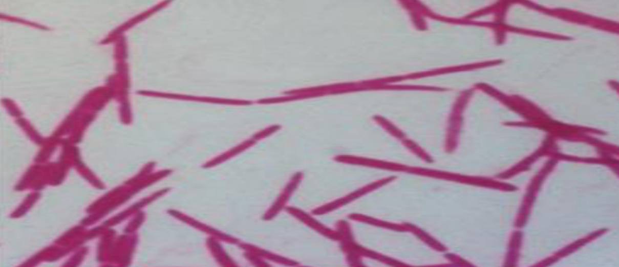
Specimen (microscopically slide):
• Name of organism:
• Stain:
• Sample from: culture or pus or in tissue
• Morphology
• Name of organism:
Fusobacteria
• Stain:
Gram stain
• Sample from:
culture
• Morphology:
Gram-negative, cigar-shaped cells with pointed ends
Specimen (microscopically slide):
• Name of organism:
• Stain:
• Sample from: culture or pus or in tissue
• Morphology
• Name of organism:
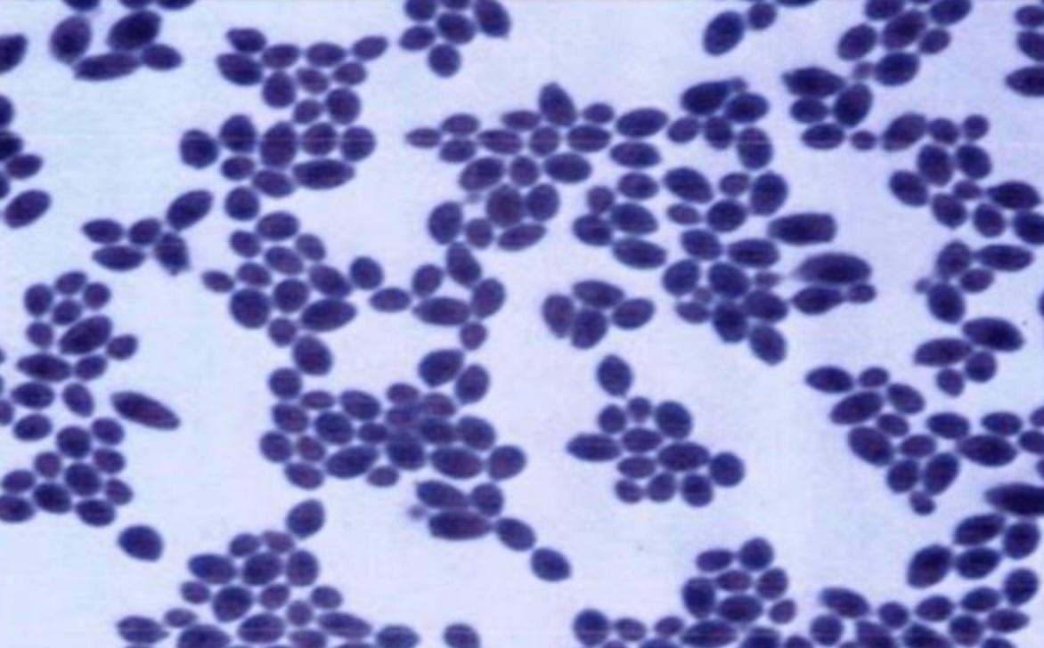
Candida
• Stain:
Gram stain
• Sample from:
culture
• Morphology:
Gram-positive budding yeast cells
Specimen (microscopically slide):
• Name of organism:
• Stain:
• Sample from: culture or pus or in tissue
• Morphology
• Name of organism:
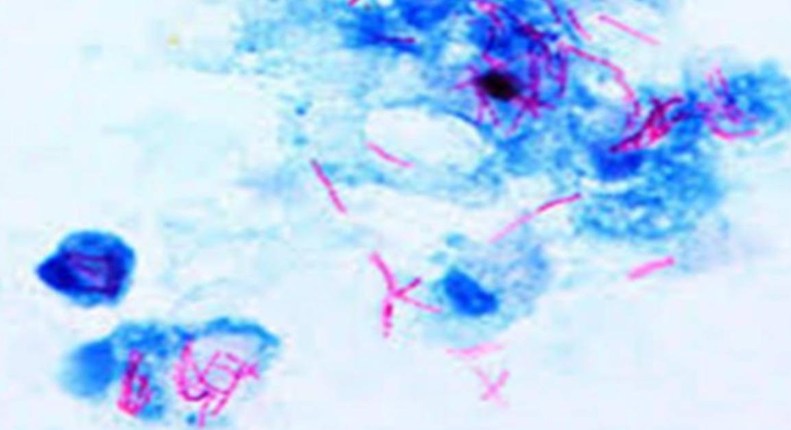
Acid-fast bacilli
(such as Mycobacterium tuberculosis)
• Stain:
Ziehl-Neelsen stain
• Sample from:
Sputum
• Morphology:
Acid-fast red bacilli against a blue background
Specimen (microscopically slide):
• Name of organism:
• Stain:
• Sample from: culture or pus or in tissue
• Morphology
• Name of organism:
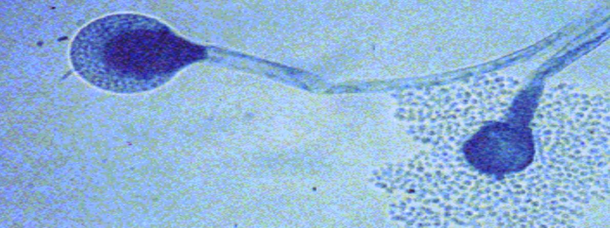
Mucor
• Stain:
Lactophenol blue stain
• Sample from:
culture
• Morphology:
Broad, aseptate (non-septate) hyphae with sporangiophores and sporangia.

Specimen (microscopically slide):
• Name of organism:
• Stain:
• Sample from: culture or pus or in tissue
• Morphology
• Name of organism:
S. pneumoniae
(Streptococcus pneumoniae)
• Stain:
Gram stain
• Sample from:
in tissue
• Morphology:
Gram-positive cocci arranged in pairs
Specimen (microscopically slide):
• Name of organism:
• Stain:
• Sample from: culture or pus or in tissue
• Morphology
• Name of organism:
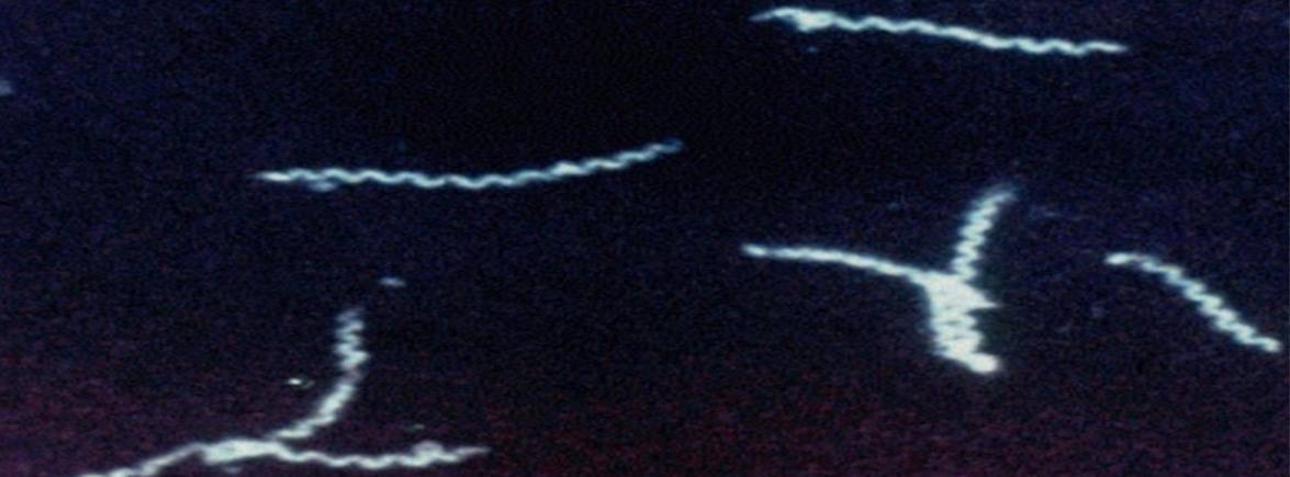
Treponema pallidum
• Stain:
No stain
• Sample from:
chancre fluid
• Morphology:
Refractile slender spirals with corkscrew movement

Specimen (microscopically slide):
• Name of organism:
• Stain:
• Sample from: culture or pus or in tissue
• Morphology
• Name of organism:
Treponema pallidum
• Stain:
Immunofluorescent stain
(or Fluorescent antibody stain)
• Sample from:
chancre fluid
• Morphology:
Apple-green fluorescent, slender spirochetes in dark background
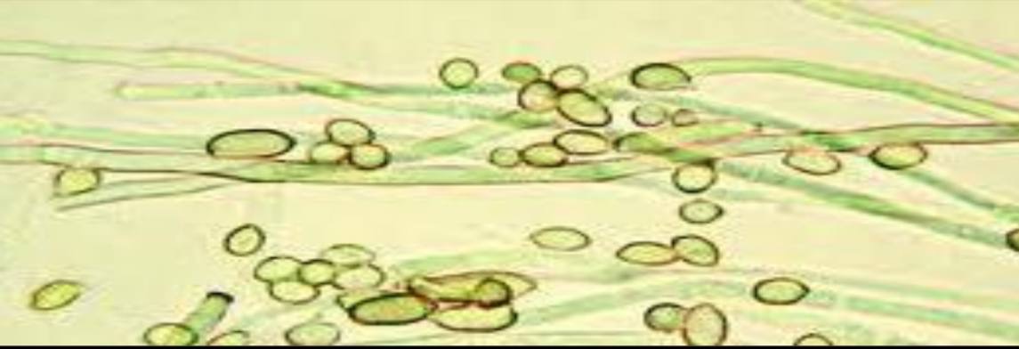
Specimen (microscopically slide):
• Name of organism:
• Stain:
• Sample from: culture or pus or in tissue
• Morphology
• Name of organism:
Aseptate hyaline fungus (hyphae)
• Stain:
unstained
• Sample from:
Sabouraud’s dextrose agar
• Morphology:
Aseptate hyaline hyphae
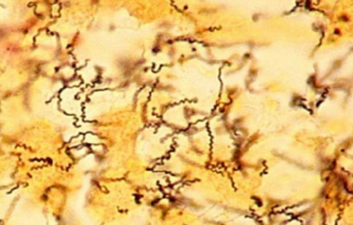
Specimen (microscopically slide):
• Name of organism:
• Stain:
• Sample from: culture or pus or in tissue
• Morphology
• Name of organism:
Treponema pallidum
• Stain:
Fontana stain
• Sample from:
in tissue
• Morphology:
Thin delicate spirochete made of 6–12 narrow regular coils, dark brown staining
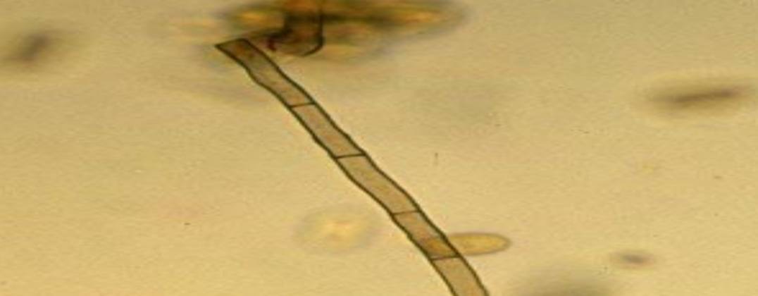
Specimen (microscopically slide):
• Name of organism:
• Stain:
• Sample from: culture or pus or in tissue
• Morphology
• Name of organism:
Dematiaceous fungus
• Stain:
Unstained
• Sample from:
Clinical specimen
• Morphology:
Dematiaceous septate hyphae
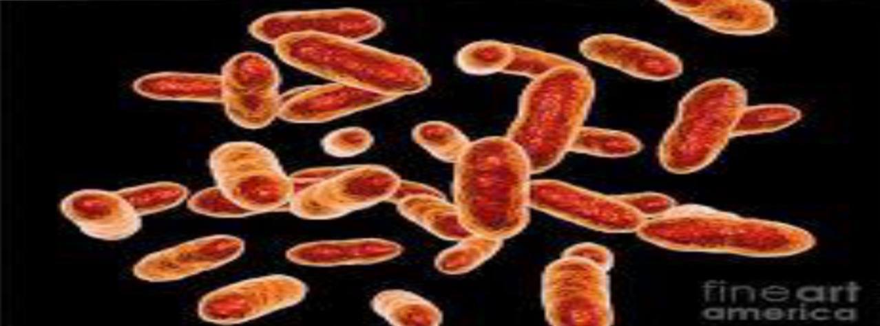
Specimen (microscopically slide):
• Name of organism:
• Stain:
• Sample from: culture or pus or in tissue
• Morphology
• Name of organism:
Tannerella forsythia
• Stain:
Gram stain
• Sample from:
culture
• Morphology:
Gram-negative, pleomorphic, spindle or fusiform-shaped rods