Parasitology Lab - Nematodes
 Call Kai
 Learn
Learn Practice Test
 Spaced Repetition
 Match
Match Flashcards
 Knowt Play
1/269

There's no tags or description
Looks like no tags are added yet.
Name | Mastery | Learn | Test | Matching | Spaced | Call with Kai | Chat |
|---|
No analytics yet
Send a link to your students to track their progress
270 Terms
Pseudocoel
false body cavity - no epithelial lining

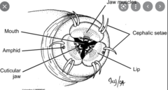
Pseudolabia
“false lips”bilateral lips around the mouth of the nematodes; they develop from the inner wall of the buccal cavity (found in those species that lost their “true lips”
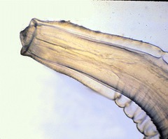
Papillae
small sensory organs located on the cuticle that act as mechanoreceptors and chemoreceptors detecting touch, water currents, and chemicals
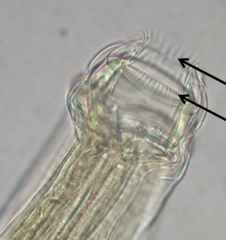
amphids
pair of bilaterally cephalic (head) chemosensory organs located on the side of the head, near the mouth
cordon/epaulets
circular structures found on the anterior end of nematodes, which are used for feeding and attachment, cephalic alae that unite anteriorly and ventrally
Bursa (nematodes)
a finger-like modification of the male posterior end and is used to grasp onto the body of the female during copulation

Phasmids
near the anus and also function in chemoreception
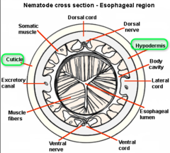
cuticle
flexible, multi-layered exoskeleton made of collagens and proteins that provide physical protection, maintain body shape, and enable movement

alae
ridged extensions of the cuticle that aids in movement, providing support, and act as sensory receptors

hypodermis
thin cellular layer beneath the outer cuticle that secretes and maintains it
bacillary band
band containing pores on ventral surface of esophageal region
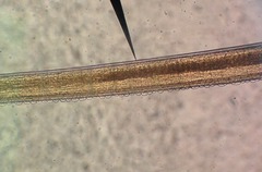
stoma/buccal cavity
cavity in which mouth opens to the start of the esophagus

isthmus
middle part of the pharynx between the corpus and the basal bulb
monodelphic
an adult female only has one functional ovary/uterus
lips
triangular structures surrounding mouths
amphidelphic
female has two functional ovaries/uteri
polydelphic
multiple ovaries/uteri
ovijector
muscular terminal portion of the uterus; used to force eggs out of the genital pore
ovoviviparous
The female retains fertilized eggs, which develop and hatch internally, with the larvae emerging from the mother's body, sometimes leading to the mother's death
giving birth to live larvae
what is the infectious stage of the nematodes
J3/M3
viviparous
female gives birth to larvae where the embroys develop and grow within the mother's uterus
oviparous
The female lays eggs which are fertilized inside the uterus and will hatch in the external environemtn
parthenogenesis
Asexual reproduction in which females produce offspring from unfertilized eggs.
gubernaculum (nematodes)
an organ in the male copulatory system that guides the spicules or bursa during copulation

ventriculus nematodes
region of the midgut or intestine that produces and secretes enzymes to initiate extracellular digestion
nerve ring
The main mass of the central nervous system, forming a ring around the pharynx

direct/monoxenous life cycle
A parasitic life cycle in which only a single host is used/the first and second stages may occur in the egg or in the soil
indirect/dixenous life cycle
Has two or more host organisms. Juveniles pass through an intermediate (vector). The 3rd stage is infective to the final host.
Sylvatic life cycles
involves wild animals and forest-dwelling vectors, with humans often becoming incidental hosts
urban life cycles
involves urban or domestic animals and humans as hosts, with urban-dwelling vectors
general structures of all nematodes
- pseudocoel
- lips
- papillae
- amphids
- pseudolabia
- bursa
- phasmids
- outermost cuticle
- middle hypodermis (dermis or epidermis)
- the interior somatic muscle layer
- alae
- cordons/epulates
- bacillary band
- alimentary canal
- stoma or buccal cavity
- esophagus
- isthmus
- bulb
- intestine
- cloaca
- nerve ring
Nematode head structures include:
lips
papillae
amphids
pseudolabia
Nematode tail structures include:
females: simple
males: varies
- bursa
- phasmids
Nematode body wall structure:
- cuticle
- hypodermis
- somatic muscles
- Alae
- cordons/ epaulets
- bacillary band
Nematode digestive structures include:
- alimentary canal
- buccal cavity
- esophagus
- isthmus
- bulb
- intestine
- cloaca
nematode nervous system structure includes:
- nerve ring
The body wall consists of three layers: the outermost _______________, the middle ___________, and the interior ________________.
cuticle
hypodermis
somatic muscles
cuticle
thin transparent elastic acellular outermost layer of a nematode
alimentary canal
a straight tube that runs the length of the organism
esophagus
muscular organ connecting the buccal cavity to the intestine
The male reproductive system consists of
testes
seminal vesicle
spicules
seminal vesicle is used for
sperm storage
Spicules
protrusible, cuticularized structures that function in mating
female reproductive system consists of
ovaries
oviduct
uterus
cuticular-lines vagina openeing to the external environment
ovijector
the _______________ makes up the central nervous system, forms a conspicuous ringsurround the esophagus and is usually the only visible portion of the nervous system.
nerve ring
A direct or monoxenous life cycle has ___________ host organism. The first and second stages may occur in the egg or in the soil. The _________ stage is infective to the final host.
single
third larvall (L3)
An indirect or dixenous life cycle has ____________ host organisms. Juveniles pass through an________________________, also called a vector. The __________ stage is infective to the finalhost.
two or more
intermediate host
adult stage
____________________ life cycles include wild carnivores such as bears, wild boars, and sealsas sources or human infection
sylvatic
_______________ life cycles involve swine, rodents, and humans, and these tend to be the primary sources of human infection.
urban
_______________ come in various sizes, shapes, and thicknesses and frequently are diagnostic
nematode eggs
nematode eggs all contain
an outer protein layer, thick middle eggshell, and a thin, vitelline membrane
microfilariae
The larval stage found in blood tissue involves an arthropod vector.
NEMATODES COVERED IN LAB
- Trichinella spiralis
- Trichuris trichiura
- Capillaria hepatica
- Strongyloides stercoralis
- Ancylotstoma duodenale
- Tococara canis
- Necator americanus
- Ascaris lumbricoides
- Enterboics vermicularis
- Dirofilaria immitis
- Wuchereria bancrofti
- Onchocerca volvulus
Trichinella spiralis =
pork worm
what is the most widely distributed nematode
Trichinella spiralis
trichinella spiralis is a small organism with a ________________________
posterior slightly thicker than their anterior
trichinella spiralis forms a cyst in
striated muscle tissue of host
The anterior portion of Trichinella spiralis consists of a short, musclulsr ________________________ followed by the
esophageal region terminating in a pseudobulb
stichisome
stichisome
long chain of cuboidal epithelial cells
males of Trichinella spiralis characteristics
no spicule
posterior end blunt with two large copulatory papilla
females of Trichinella spiralis characteristics
blunt posterior with terminal anus
vulva located in the stichosomal region
Trichinella spiralis is what?
ovoviviparous
life cycle of Trichinella spiralis through the domestic cycle
Pigs eat rats who are infected
Encysted larvae in the striated muscle of pigs
Human ingests undercooked meat
larvae released in small intestine
larvae mature in small intestine to adults
Adult females deposit larvae in the mucosa
Encysted larvae in striated muscle
Life cycle of Trichinella spiralis through the sylvatic cycle
Bears and boars eat rats who are infected
encysted larvae in the striated muscle of boars and bears
Humans ingest undercooked meat
larvae released in small intestine
larvae mature in small intestine to adults
Adult females deposit larvae in the mucosa
Encysted larvae in striated muscle
what is the infective stage of Trichinella spiralis
L1
First stage larvae
what is the diagnostic stage of Trichinella spiralis
larval stage
encysted larva in striated muscles
what kind of meat is ingested in Trichinella spiralis
undercooked meat (bear, boar, pig) with encysted larva
in Trichinella spiralis the eggs hatch __________________ and then the ____________________ are deposited into the ______________
within the female
juveniles
intestinal mucosa
newborn juveniles of Trichinella spiralis enter the ___________ and eventually encyst in ______________
bloodstream
striated skeletal muscles
Trichinella spiralis infection of new hosts occur when ___________________ or __________________ is consumed
raw or undercooked meat
after consuming the meat, juveniles of Trichinella spiralis reach sexual maturity within __________.
2-3 days
pathology of Trichinella spiralis within the first week
intestinal edema
nausea
vomitting
abdominal cramps
diarrhea
fever
pathology of Trichinella spiralis within one month
migration phase -
high fever
blurred vision
facial edema
cough
eosinophilia
muscular phase -
local inflammation
edema
pain
weakness
fatigue
diagnosis of Trichinella spiralis is made when
a group of people become symptomatic after a known exposure to undercooked pork products
Trichuris trichiura =
whipworm
Trichuris trichiura is more common where
warm regions with inadequate sanitation
what is the third most common nematode infection in humans
Trichuris trichiura
Trichuris trichiura characteristics
anterior two times thicker than posterior
no lips
tiny "spear" to penetrate the mucosa of the cecum, appendix, or rectum
what is the diagonstic stage of Trichuris trichiura
unembryonated eggs
what is the infective stage of Trichuris trichiura
embryonated eggs
Trichuris trichiura male characteristics
posterior coiled
spicule
Trichuris trichiura female characteristics
blunt posterior
vulva loctaed near posterior end of stichosome
life cycle of Trichuris trichiura is a __________ life cycle
monoxenous
Trichuris trichiura eggs can reach infective stage in _____________ or can remain viable for _____________
three weeks
several years
Ingested eggs of Trichuris trichiura hatch in the intestine of the host. Juveniles migrate to the _______________ and mature in one month.
rectum
life cycle of Trichuris trichiura
1. Embryonated eggs are ingested
2. larvae hatch in small inetstine
3. travels to lower part of the large intestine (rectum)
4. Unembryonated eggs passed in feces
5. 2-cell stage
6. advanced cleavage
7. Embryonated eggs are ingested
unembryonated eggs of Trichuris trichiura are always _________________ and are passed in ___________
plugged on both ends
feces
larvae of Trichuris trichiura hatch in the
small intestine
Juvenile Trichuris trichiura migrate to the ______________
large intestine (rectum)
Pathology of Trichuris trichiura: slight infections
asymptomatic
Pathology of Trichuris trichiura: heavy infections
ulcerative colitis
inflammatory bowel disease
abdominal pain
bloody mucoid diarrhea
rectal prolapse
weight loss
what is a heavy infection of Trichuris trichiura
500-5000 worms
how to diagnose Trichuris trichiura
Eggs in stool (plugged on both ends)
Capillaria hepatica is _____________ to animals however very few __________________
endemic
human infections have been recorded
Capillaria hepatica is most frequently found in
mammals
Capillaria hepatica does not require
an intermediate host
Capillaria hepatica life cycle
1. adults and eggs in liver
2. Unembryonated eggs may be released into the environment upon death and decomposition of host or predation or canibalism leads to another animal consuming it
3. unembryonated eggs released in feces
4. eggs embryonated in environment
5. eggs ingested by definitive host
6. humans infected by ingestion of host
Capillaria hepatica infective stage
embryonated eggs
Capillaria hepatica diagnostic stage
adults and eggs in liver
Capillaria hepatica adults inhabit the _________, where eggs accumulate without developing
liver