ANAT 321 Final
0.0(0)
Studied by 1 personCard Sorting
1/443
Last updated 5:25 PM on 11/18/22
Name | Mastery | Learn | Test | Matching | Spaced | Call with Kai |
|---|
No analytics yet
Send a link to your students to track their progress
444 Terms
1
New cards
Brain vs Mind
-the brain is a physical object that processes information
-the mind emerges from what occurs in the brain
-the mind emerges from what occurs in the brain
2
New cards
Marr's Book "Vision"
-big contribution to neuroscience and understanding of the visual system
-to understand how the brain or any other computational system works, need to approach it at 3 different levels: computational theory, representation & algorithm and hardware implementation
-to understand how the brain or any other computational system works, need to approach it at 3 different levels: computational theory, representation & algorithm and hardware implementation
3
New cards
Computational Theory
-to understand the brain, you must know what its trying to accomplish, why it's important and the logic behind it's strategies
-what is the goal of the brain's computation?
-why is the computation of the brain important?
-what is the logic behind the strategy of how computation of the brain is carried out?
-"trying to understand flight by studying feathers is like trying to understand perception by studying only neurons"
-what is the goal of the brain's computation?
-why is the computation of the brain important?
-what is the logic behind the strategy of how computation of the brain is carried out?
-"trying to understand flight by studying feathers is like trying to understand perception by studying only neurons"
4
New cards
Representation & Algorithm
-how can the computational theory be implemented?
-what is the representation for the input and output of computation?
-what is the algorithm for the transformation between input and output in the brain?
-the focus of the class
-focuses on action potentials (input), and connection made in the brain (transformation between input and output)
-what is the representation for the input and output of computation?
-what is the algorithm for the transformation between input and output in the brain?
-the focus of the class
-focuses on action potentials (input), and connection made in the brain (transformation between input and output)
5
New cards
Hardware Implementation
-everything has a physical substrate
-how can be representation and algorithm be understood physically?
-what is the brain made of?
-how can be representation and algorithm be understood physically?
-what is the brain made of?
6
New cards
Central Nervous System
-brain and spinal cord
7
New cards
Peripheral Nervous System
-sensory neurons carrying information from nose, ears, etc and output neurons carrying information to output organs
8
New cards
Efferent
-part of the peripheral nervous system
-output neurons
-leaving the brain
-2 types: to skeletal muscles (voluntary) and to internal organs (involuntary)= autonomic nervous system
-output neurons
-leaving the brain
-2 types: to skeletal muscles (voluntary) and to internal organs (involuntary)= autonomic nervous system
9
New cards
Function of Sensory Systems
-transform information about the external and internal environment into neuronal signals to the brain
-all do very different things
-all have an organizational theme
1. perception: conscious experience that guides behaviour
2. movement: we require constant sensory feedback in order to control movement
3. regulation of internal organs: sensory system measure body temperature, blood pressure, etc
4. maintenance of arousal: keep you awake and alert
10
New cards
Sensory Modalities
-vision, somatic sensation, hearing, taste, smell
-all share common features:
1. different forms of sensory input are transduced into a common neuronal code= the action potential
2. sub-modalities within them get encoded by specific labelled lines (different neurons) that travel in parallel to one another
3. perception often involves combinatorial processing of sensory information= combining all labelled lines!
4. sensory information destined for the cortex must first pass through the thalamus
5. sensory information is processed at multiple hierarchal levels so that the most complex information is processed at the highest levels
6. initial cortical processing occurs in primary sensory areas, and then in higher order unimodal and multimodal association regions
7. sensory perceptions are not replications of the real world- only the brain's abstraction!
11
New cards
Sensory Sub-Modalities
-specific things within 1 modality
-information is carried by different sets of neurons
-example: pressure vs warmth of skin are different sub-modalities within the somatic sensation modality
-information is carried by different sets of neurons
-example: pressure vs warmth of skin are different sub-modalities within the somatic sensation modality
12
New cards
Somatic Sensory System
-a modality of sensation
-includes many sub-modalities
-involves touch, vibration, pain and all other information from the skin's surface in addition to the feedback from joints and muscles (proprioception)
-includes many sub-modalities
-involves touch, vibration, pain and all other information from the skin's surface in addition to the feedback from joints and muscles (proprioception)
13
New cards
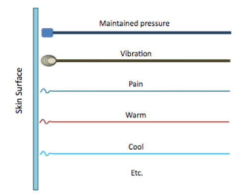
Types of Axon Endings in the Somatic Sensory System
-we have very different endings at the skin's surface to detect different types of sensory input= sub-modalities!
1. Merkel Cells
2. Ruffini Endings
3. Meissner Corpuscles
4. Pacinian Corpuscles
5. Free Nerve Endings
-different ion channels are involved for each different axon type
1. Merkel Cells
2. Ruffini Endings
3. Meissner Corpuscles
4. Pacinian Corpuscles
5. Free Nerve Endings
-different ion channels are involved for each different axon type
14
New cards
Merkel Cells
-a type of axon ending in the somatic sensory system that brings sensory information to the CNS
-also called SA1 or slowly adapting 1
-fire action potentials in response to constant pressure on the skin's surface
-also called SA1 or slowly adapting 1
-fire action potentials in response to constant pressure on the skin's surface
15
New cards
Ruffini Endings
-a type of axon ending in the somatic sensory system that brings sensory information to the CNS
-also called SA2 or slowly adapting 2
-fire action potentials in response to constant pressure on the skin's surface
-also called SA2 or slowly adapting 2
-fire action potentials in response to constant pressure on the skin's surface
16
New cards
Meissner Corpuscles
-a type of axon ending in the touch and proprioception somatic sensory system that brings sensory information to the CNS
-also called RA1 or rapidly adapting 1
-fire action potentials in response to changing pressure on the skin's surface (pressure being turned on or off)
-also called RA1 or rapidly adapting 1
-fire action potentials in response to changing pressure on the skin's surface (pressure being turned on or off)
17
New cards
Pacinian Corpuscles
-a type of axon ending in the somatic sensory system that brings sensory information to the CNS
-also called RA2 or rapidly adapting 2
-fire action potentials in response to changing pressure on the skin's surface (pressure being turned on or off)
-also called RA2 or rapidly adapting 2
-fire action potentials in response to changing pressure on the skin's surface (pressure being turned on or off)
18
New cards
Free Nerve Endings
-a type of axon ending in the somatic sensory system that brings sensory information to the CNS
-endings have no specialized organization- they are just bare
-fire action potentials in response to pain or temperature on the skin's surface- why they're the ending found on a-delta and c fibers
-endings have no specialized organization- they are just bare
-fire action potentials in response to pain or temperature on the skin's surface- why they're the ending found on a-delta and c fibers
19
New cards
Combinatorial Processing
-sensory systems rarely activate only 1 labelled line/sub-modality
-we usually combine many labelled lines at once
-how sensations such as wetness, colour and the taste and smell of food are generated
-we usually combine many labelled lines at once
-how sensations such as wetness, colour and the taste and smell of food are generated
20
New cards
Types of Primary Somatic Sensory Afferent Neurons
-4 different types of primary somatic sensory afferent neurons
-all propagate action potentials at different rates and are anatomically distinct
-also relay different kinds of somatic sensory information to the CNS (touch and proprioception vs pain and temperature)
-cell bodies are in dorsal root ganglia
1. a-alpha fibers
2. a-beta fibers
3. a-delta fibers
4. c fibers
21
New cards
a-alpha Fibers
-the thickest sensory afferent because covered in a thick layer of myelin
-very fast propagation of action potentials
-the sensory fibers that innervate muscles and joints
-often have specialized sensory endings such as Pacinian Corpuscles
-responsible for fine touch and proprioception
-very fast propagation of action potentials
-the sensory fibers that innervate muscles and joints
-often have specialized sensory endings such as Pacinian Corpuscles
-responsible for fine touch and proprioception
22
New cards
a-beta Fibers
-thinner than a-alpha fibers but also very myelinated
-fast propagation of action potentials but slower than a-alpha fibers
-low-threshold mechanoreceptors
-often have specialized sensory endings such as Pacinian Corpuscles
-responsible for fine touch and proprioception
-fast propagation of action potentials but slower than a-alpha fibers
-low-threshold mechanoreceptors
-often have specialized sensory endings such as Pacinian Corpuscles
-responsible for fine touch and proprioception
23
New cards
a-delta Fibers
-much thinner than a-alpha and a-beta fibers
-hardly myelinated or not myelinated at all
-slower action potential propagation
-have free nerve endings
-responsible for pain and temperature sensation
-detect the initial pain we feel when we touch something hot that causes us to pull away
-hardly myelinated or not myelinated at all
-slower action potential propagation
-have free nerve endings
-responsible for pain and temperature sensation
-detect the initial pain we feel when we touch something hot that causes us to pull away
24
New cards
c Fibers
-very thin and non-myelinated
-slowest action potential propagation
-have free nerve endings
-responsible for pain and temperature sensation
-responsible for the delayed throbbing pain we feel after burning ourselves
-slowest action potential propagation
-have free nerve endings
-responsible for pain and temperature sensation
-responsible for the delayed throbbing pain we feel after burning ourselves
25
New cards
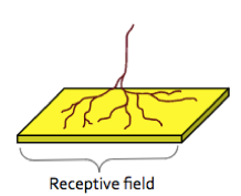
Receptive Field
-the specific region on the body's surface that primary somatic sensory afferents innervate
-a stimulus only within the region will excite the cell and cause it to fire an action potential
-stimulus at the centre will result in the most action potentials
-stimulus at the edge will result in fewer action potentials
-the neurons it synapses with higher up in the peripheral nervous system are also specific for this receptive field
-can have different sizes: very small in the fingertip and large on the back
-the smaller the receptive field= the higher acuity and precision
-higher acuity/precision DOES NOT MEAN HIGHER SENSITIVITY
-the smaller the receptive field= the more axons that innervate the receptive field
-neurons in primary sensory cortexes will have small receptive fields
-neurons in association cortexes will have larger receptive fields by convergence
26
New cards
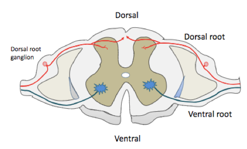
Dorsal
-back
-top of the brain
-top of the brain
27
New cards
Ventral
-stomach
-bottom of the brain
-bottom of the brain
28
New cards
Caudal/Posterior
-back
29
New cards
Rostral/Anterior
-front
30
New cards
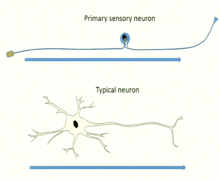
Primary Somatic Sensory Afferents
-the first sensory neurons in the pathway
-includes a-alpha fibers, a-beta fibers, a-delta fibers and c fibers
-have a different morphology than typical neurons- have a long axon and a small cell body that sits off to the side
-flow of information: skin > dendrites (endings) (action potential is generated) > axon (action potential is propagated) > pre-synaptic terminal where it synapses with the next neuron in the pathway
-different flow of information than a typical neuron- action potential is usually generated in the cell body
-enter the spinal cord at the dorsal horn
-have different endings that innervate the skin's surface to respond to different kinds of sensory information
31
New cards
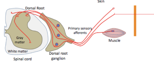
Dorsal Root Ganglia
-a bundle of primary somatic sensory afferent cell bodies (a-alpha, a-beta, a-delta and c-fibers) in the peripheral nervous system
-axons from dorsal root ganglia become the dorsal roots and enter the spinal cord at 31 different locations on either side and carry in sensory information
-axons from dorsal root ganglia become the dorsal roots and enter the spinal cord at 31 different locations on either side and carry in sensory information
32
New cards
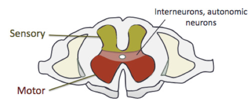
Gray Matter of the Spinal Cord
-at the center of the spinal cord
-in the shape of an H
-consists of cell bodies, dendrites and synapses
-in the shape of an H
-consists of cell bodies, dendrites and synapses
33
New cards
White Matter of the Spinal Cord
-at the periphery of the spinal cord
-consists of myelinated axons
-acts as the "highway" of the spinal cord- where information is carried up and down the spinal cord
-consists of myelinated axons
-acts as the "highway" of the spinal cord- where information is carried up and down the spinal cord
34
New cards
Dorsal Roots
-axons of dorsal root ganglia neurons
-enter the spinal cord at the dorsal horn
-either enter the dorsal part of the gray matter or the white matter
-enter the spinal cord at the dorsal horn
-either enter the dorsal part of the gray matter or the white matter
35
New cards
Ganglia
-an organized group of neuron cell bodies, dendrites and synapses in the peripheral nervous system
36
New cards
Nuclei
-an organized group of neuron cell bodies, dendrites and synapses in the central nervous system
37
New cards
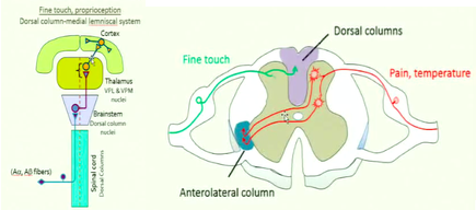
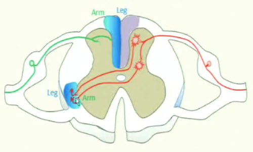
Dorsal Column-Medial Lemniscal System
-the pathway for fine touch and proprioception information to get to the brain
1. a-alpha and a-beta fibers coming from the skin's surface enter the spinal cord at the dorsal horn
2. axons move into the white matter at the center of the dorsal horn= the dorsal column
3. then their axons turn and begin travelling up towards the brain stem= axons must be very long
4. axons reach the brain stem and make synapses with second order neurons here (dorsal column nuclei)
5. excitatory neurotransmitter glutamate gets released
6. the axons of excited dorsal column nuclei cross the midline in the brain stem
7. the axons of second order neurons travel up to the thalamus in the medial lemniscus
8 axons of dorsal column nuclei make more excitatory synpases with thalamic neurons in the VPL and VPM nuclei
9. excited thalamic neurons send their axons to the primary somatic sensory cortex for initial somatic sensory information processing
-NOTE: other branches of a-alpha and a-beta fibers enter the gray matter of the dorsal horns= involved in reflexes
-will lose touch and proprioception sensation on the same side of the body as the lesion in the spinal cord
38
New cards
Ipsilateral
-axons that travel towards the brain on the same side of the body as where they endings innervate the skin's surface
-primary somatic sensory afferents are ipsilateral
-primary somatic sensory afferents are ipsilateral
39
New cards
Contralateral
-axons that cross the midline of the body and travel towards the brain on the opposite side of the body as where their endings are
-second order neurons are contralateral
-second order neurons are contralateral
40
New cards
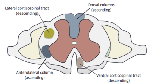
Dorsal Column
-the white matter in the dorsal horn of the spinal cord
-how a-alpha and a-beta fibers carrying fine touch and proprioception information travel up to brain stem
-an ascending tract
-somatotopically organized: have 2 different pathways for information coming from legs vs arms to travel along
-how a-alpha and a-beta fibers carrying fine touch and proprioception information travel up to brain stem
-an ascending tract
-somatotopically organized: have 2 different pathways for information coming from legs vs arms to travel along
41
New cards
Dorsal Horns
-the dorsal part of the "H" of gray matter
-where somatic sensory information enters the spinal cord
-contains mainly sensory neurons
-where somatic sensory information enters the spinal cord
-contains mainly sensory neurons

42
New cards
Ventral Horns
-the ventral part of the "H" of gray matter
-where motor information leaves the spinal cord
-mainly contains motor neurons
-where motor information leaves the spinal cord
-mainly contains motor neurons

43
New cards
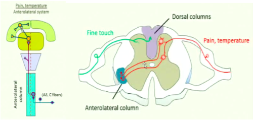
Anterolateral System
-the pathway for pain and temperature information to get to the brain
-the pathway allows for pain to be precisely located
1. a-delta and c fibers coming from the skin's surface enter the spinal cord at the dorsal horn
2. axons move into the gray matter of the dorsal region of the spinal cord and make excitatory synapses with second order neurons here
3. axons of second order neurons cross the midline of the spinal cord and enter the ventral region of the spinal cord gray matter on the contralateral side
4. axons of second order neurons enter the anterolateral column to travel up to the thalamus (VPL)
5 second order neurons synpase with thalamic neurons
6 the axons of thalamic neurons travel to the primary somatic sensory cortex in the internal capsule
-NOTE: other branches of thalamic neurons are what control the emotion and feelings associated with pain in other areas of the cortex
-will lose pain and temperature sensation on the side of the body opposite to the lesion in the spinal cord
44
New cards
Anterolateral Column
-a region in the white matter of the ventral region of the spinal cord
-travels up to brain stem and thalamus (an ascending tract)
-carries pain and temperature information from a-delta and c fibers
-somatotpically organized: 2 different pathways from information coming from the legs vs arms to travel along
-travels up to brain stem and thalamus (an ascending tract)
-carries pain and temperature information from a-delta and c fibers
-somatotpically organized: 2 different pathways from information coming from the legs vs arms to travel along

45
New cards
Somatotopical Organization
-touch and proprioception and pain and temperature information from the arms and legs is organized in the spinal cord, dorsal columns, anterolateral column and all the way up to the primary somatic sensory cortex
-axons carrying touch and proprioception information from the legs get layered more medially and axons carrying touch and proprioception information from the arms get layered more laterally
-axons carrying pain and temperature information from the legs get layered more laterally and axons carrying pain and temperature information from the arms get layered more medially
46
New cards
Information Processing of the Nervous System
-inputs and outputs are different due to processing that occurs in between
-what comes out is much more complex and integrated than what comes in
-happens as a result of convergence, divergence and lateral inhibition
-what comes out is much more complex and integrated than what comes in
-happens as a result of convergence, divergence and lateral inhibition
47
New cards
Convergence
-second order neurons receive information from many primary afferent neurons
-means there are usually a lot less second order neurons
-also why second order neurons have larger receptive fields than primary neurons
-means there are usually a lot less second order neurons
-also why second order neurons have larger receptive fields than primary neurons
48
New cards
Divergence
-primary afferent neurons branch and make synapses with many second order neurons
49
New cards
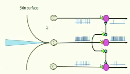
Lateral Inhibition
-neurons with long axons that relay information between different regions of the brain are excitatory (projection neurons)
-neurons with short axons that relay information within a localized region of the brain are inhibitory (interneurons)
-allows for strong excitatory input signals to pass through and reach the brain but the filtering out of weaker input signals
-strong excitatory input signals will synapse with inhibitory neurons to excite them so that they will filter out weaker signals
-this generates more precise and localized signals
-inhibits the excitatory neurons laterally- to the periphery of a receptive field
50
New cards
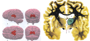
The Thalamus
-like the "gatekeeper" of the cortex
-all sensory information destined for the cortex must first pass through the thalamus with FEW EXCEPTIONS
-located deep inside the brain
-a collection of nuclei- broken down into 3 types: specific relay, association and midline/intralaminar
-one of each side of the brain and just barely touch each other at the midline
-forms the floor of the lateral ventricle
-the cerebral cortex also sends projections back to thalamic neurons and this tells the thalamus what inputs its receiving from sensation of the body, etc to filter out and what info to allow to be relayed to the cortex
51
New cards
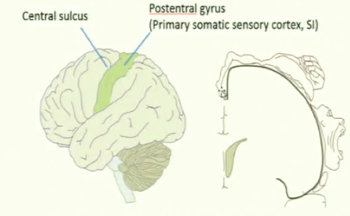
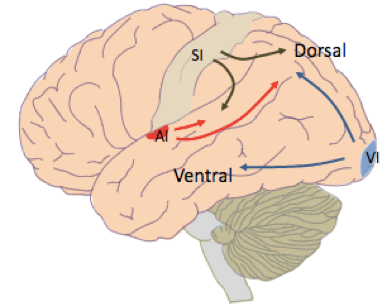
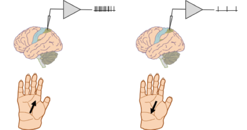
Primary Somatic Sensory Cortex (SI)
-the region of the cerebral cortex that receives all initial somatic sensory information from the thalamus
-the body's surface is "mapped" onto it: the area of it controlling sensation of the legs is small and medial, arms and face are much larger and more lateral= topographic organization
-means that a larger region and more neurons of the primary somatic sensory cortex are dedicated to processing information from the face and hands= higher precision and acuity in these areas
-only a small region and number of axons are involved in processing somatic sensory information from the legs= low precision and acuity in the legs
-after processing here, somatic sensory information then gets sent to higher level cortical regions (unimodal and multimodal association cortexes)
-somatic sensory information flows out of the primary somatic sensory cortex dorsally and ventrally- applies to all primary cortexes!
-also called the post-central gyrus
-divided into areas 1, 2 and 3 according to Brodmann's organization of the cerebral cortex
52
New cards
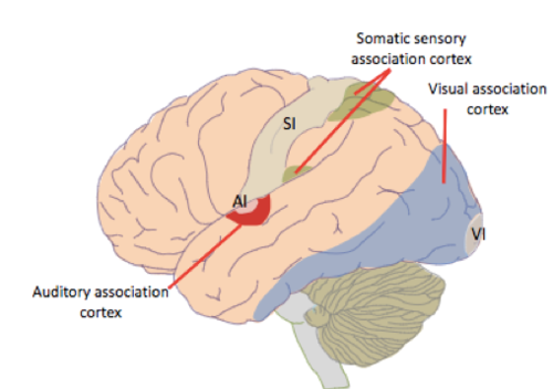
Primary Sensory Cortexes of the Brain
-includes primary somatic sensory cortex (SI), primary visual cortex (VI) and primary auditory cortex
-involved in initial processing of sensory information
-small receptive fields
-respond to simple stimuli of sensation (pressure, vibration)
-information then gets passed along to association cortexes
-sensory information flows out of the primary sensory cortexes dorsally and ventrally to reach association cortexes
-damage to these areas is very specific to the modality (ex: losing vision when VI is damaged)
53
New cards
Association Cortexes of the Brain
-involved in processing higher order information
-unimodal or multimodal
-have larger receptive fields
-experience divergence
-respond to more complex properties of sensation
-can even respond to inputs coming from either hand
-damage to these regions results in deficits that are much more complex and unexpected than damage to primary cortexes
-unimodal or multimodal
-have larger receptive fields
-experience divergence
-respond to more complex properties of sensation
-can even respond to inputs coming from either hand
-damage to these regions results in deficits that are much more complex and unexpected than damage to primary cortexes
54
New cards
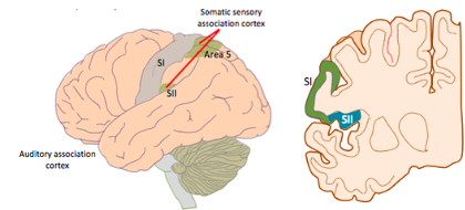
Unimodal Association Cortexes
-involved in processing information from one modality (ex: somatic sensory)
-will respond to more complex stimuli than primary cortexes, like something moving across the hand in a specific direction
-includes the premotor cortex & supplemental motor area, somatic sensory association cortexes (area 5 and SII), visual association cortex (VII) and the auditory association cortex
-will have larger receptive fields than primary sensory cortexes
55
New cards
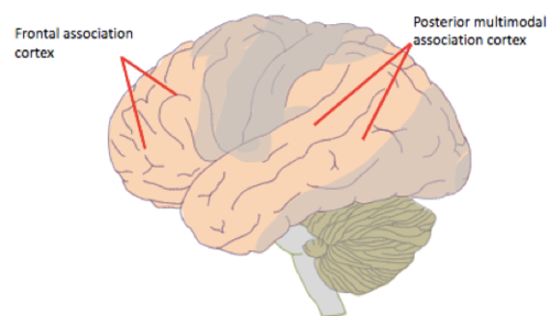
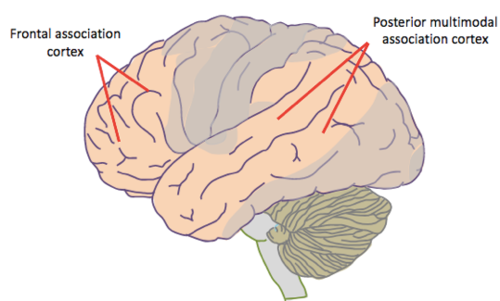
Multimodal Association Cortexes
-are not dedicated to one specific sensory modality
-involved in the most complex cognitive processes like integrating information and achieving goals
-modalities get combined together here to create 1 unified perception of the world
-very large in the human brain but small in other species
-includes the Frontal Association Cortex and the Posterior Multimodal Association Complex
56
New cards
Unimodal Somatic Sensory Association Cortexes
1. Area 5
2. SII (Somatic Sensory II)
2. SII (Somatic Sensory II)
57
New cards
Primary Visual Cortex (VI)
-dedicated to visual processing
-information from here then gets sent to visual association cortexes
-layer 4 is large and layer 5 is small because must receive a lot of input but doesn't need to send much output to sub-cortical areas
-mainly inside the brain- can be seen in the medial surface
-area 17 according to Brodmann's organization of the cerebral cortex
58
New cards
Frontal Association Cortex
-a multimodal association cortex
-responsible for planning multi-step processes and decision making
-responsible for planning multi-step processes and decision making

59
New cards
Posterior Multimodal Association Cortex
-a multimodal association cortex
-where the different sensory modalities converge
-involved in the ability the use sensory information to guide action
-involved in higher order cognitive processing such as language
-where the different sensory modalities converge
-involved in the ability the use sensory information to guide action
-involved in higher order cognitive processing such as language
60
New cards
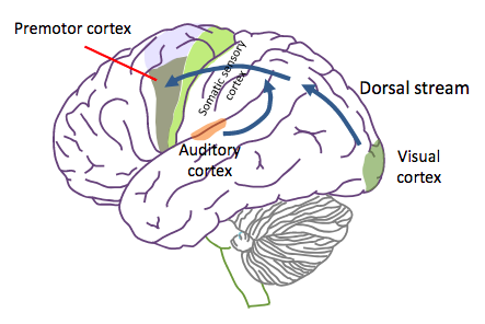
Dorsal Stream of Sensory Information Flow from Primary to Association Sensory Cortexes
-uses sensory information (somatic sensation, vision and audition) to guide action and movement
-information ultimately ends up in the premotor cortex
-information ultimately ends up in the premotor cortex
61
New cards
Ventral Stream of Sensory Information Flow from Primary to Association Sensory Cortexes
-involved in conscious perception and memory
62
New cards
Neglect Syndrome
-caused by damage to the posterior multimodal association cortex, generally on the right side
-therefore, results in people "neglecting" everything on the left side of their visual field, although they can still see it
-patients with severe cases experience asomatognosia
-therefore, results in people "neglecting" everything on the left side of their visual field, although they can still see it
-patients with severe cases experience asomatognosia
63
New cards
Asomatognosia
-a severe case of neglect syndrome
-when the patient also "neglects" the left side of their body and don't even acknowledge it as part of their body
-when the patient also "neglects" the left side of their body and don't even acknowledge it as part of their body
64
New cards
The Spinal Cord
-runs inside the backbone, up to the 1st lumbar vertebra (doesn't run the entire length)
-less than 1 cm in diameter
-axons going in and out of the spinal cord travel together in spinal nerves
-spinal nerves emerge from 31 different segments in between vertebrae on either side of the spinal cord
-has cervical and lumbar enlargements
-enters the skull at the foramen magnum
-continuous with the medulla of the brain stem- becomes it once it enters the brain
65
New cards
Spinal Cord Segments
-where spinal nerves enter and exit the spinal cord in between vertebrae
-31 different paired segments
-can be cervical, thoracic, lumbar, sacral or coccygeal depending their location along the backbone
-31 different paired segments
-can be cervical, thoracic, lumbar, sacral or coccygeal depending their location along the backbone
66
New cards
Cervical & Lumbar Enlargements of the Spinal Cord
-bulges on the spinal cord in the cervical and lumbar regions
-where spinal nerves carrying sensory information from the legs and arms enter the spinal cord
-where spinal nerves carrying motor information for the legs and arms leave the spinal cord
-gray matter is much larger here than thinner regions of the spinal cord because lots of neurons and synapses located here
-spinal cord much thinner where spinal nerves going to or from the chest or back enter or leave
67
New cards
Lumbar Cistern
-spinal fluid that fills the empty space in the backbone where the spinal cord does not reach
-contains sacral spinal nerves that are projecting downwards to exit the backbone at the sacral location
-best retrieved by inserting a needle near the bottom of the vertebrae, where the spinal cord does not reach
-contains sacral spinal nerves that are projecting downwards to exit the backbone at the sacral location
-best retrieved by inserting a needle near the bottom of the vertebrae, where the spinal cord does not reach
68
New cards
Cauda Equina
-sacral spinal nerves in the lumbar cistern that project downwards to exit the backbone
-look like a horse's tail
-look like a horse's tail
69
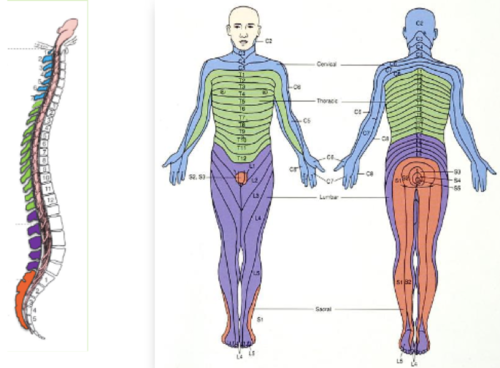
New cards
Dermatomes
-regions of the body's surface that get innervated by a specific pair of spinal nerves
-don't need to memorize certain ones but should know the general areas of the body
-don't need to memorize certain ones but should know the general areas of the body
70
New cards
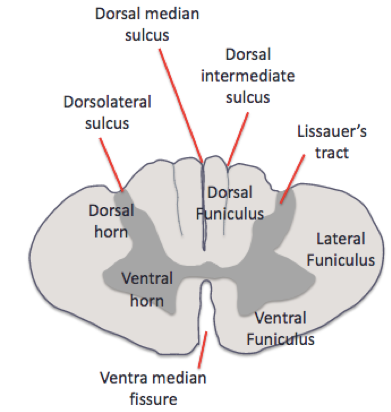
Funiculi of the Spinal Cord
-how white matter of the spinal cord is organized
-there are lateral, ventral and dorsal funiculi
-there are lateral, ventral and dorsal funiculi
71
New cards
Sulci/Fissures of the Spinal Cord
-deep grooves in the spinal cord, fissures are just deeper than sulci
-form landmarks
-includes the dorsolateral sulcus, dorsal median sulcus, dorsal intermediate sulcus and ventral median fissure
-form landmarks
-includes the dorsolateral sulcus, dorsal median sulcus, dorsal intermediate sulcus and ventral median fissure

72
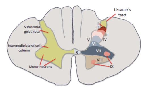
New cards
The Numbering System of the Gray Matter of the Spinal Cord
-roman numerals
-dorsal parts numbered the lowest and work their way up in ventral regions
-from I-X (1-10)
-don't need to memorize
-dorsal parts numbered the lowest and work their way up in ventral regions
-from I-X (1-10)
-don't need to memorize
73
New cards
Intermediate Neurons of the Gray Matter of the Spinal Cord
-located in between dorsal and ventral horns
-connect sensory and motor neurons and other neurons that are located between dorsal and ventral horns
-where the pre-ganglionic neurons of the sympathetic nervous system are found
-connect sensory and motor neurons and other neurons that are located between dorsal and ventral horns
-where the pre-ganglionic neurons of the sympathetic nervous system are found
74
New cards
Lissauer's Tract
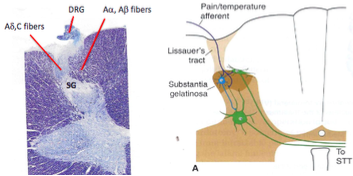
-at the lateral portion at the start of the dorsal horns of the gray matter of the spinal cord
-where primary somatic sensory afferents carrying pain and temperature information (a-delta and c fibers) enter the spinal cord gray matter
-primary somatic sensory afferents carrying touch and proprioception information (a-alpha and a-beta fibers) enter the dorsal horn more medially than Lissauer's tract because they're too large in diameter due to myelination

75
New cards
Substantia Gelatinosa
-just after Lissauer's tract in the dorsal horns of the spinal cord
-contains a lot cell bodies of neurons that are receiving information from pain and temperature primary afferents (a-delta and c fibers)
-contains a lot cell bodies of neurons that are receiving information from pain and temperature primary afferents (a-delta and c fibers)
76
New cards
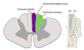
Fasciculus Gracilis
-one of 2 pathways in the dorsal column for touch and proprioception information to travel along up to the thalamus
-the pathway for a-alpha and a-beta fibers carrying somatic sensory information from the legs
-more medial
-as the spinal cord becomes the medulla, myelinated axons in the pathway get replaced by the nucleus gracilis of the brainstem= switch from white matter to gray matter
-the pathway for a-alpha and a-beta fibers carrying somatic sensory information from the legs
-more medial
-as the spinal cord becomes the medulla, myelinated axons in the pathway get replaced by the nucleus gracilis of the brainstem= switch from white matter to gray matter

77
New cards
Fasciculus Cuneatus
-one of 2 pathways in the dorsal column for touch and proprioception information to travel along up to the thalamus
-the pathway for a-alpha and a-beta fibers carrying somatic sensory information from the arms
-more lateral
-as the spinal cord becomes the medulla, myelinated axons in the pathway get replaced the nucleus cuneatus=switch from white matter to gray matter
-the pathway for a-alpha and a-beta fibers carrying somatic sensory information from the arms
-more lateral
-as the spinal cord becomes the medulla, myelinated axons in the pathway get replaced the nucleus cuneatus=switch from white matter to gray matter
78
New cards
Dorsal Intermediate Sulcus
-the sulcus between the fasciculus gracilis and fasciculus cuneatus in the dorsal column of the white matter of the spinal cord

79
New cards
Spinal Reflexes
-the link between sensory input to motor output
-involuntary but coordinated patterns of muscle contraction and relaxation, elicited by peripheral stimuli
-stimuli that cause them come from receptors in the muscles, joints and skin
-the movements they produce are often protective
-one specific stimulus can cause contraction of many muscles= example of divergence
-the extent and force of muscle contraction depends on the intensity of the stimulus causing it
-regulated and modified by descending inputs from higher brain areas that gate out inappropriate reflexes for a given context
-not just automatic- must be appropriate for the context!
-complex, voluntary movements like walking use existing reflex circuits
-evolved to keep track of the body in space- maintain body posture and body position
-2 types: withdrawal and stretch
80
New cards
Withdrawal Reflex
-a type of spinal reflex
-includes pulling your hand away from something hot
-includes pulling your hand away from something hot
81
New cards
Stretch Reflex
-a type of spinal reflex
-includes the knee reflex at the doctor
-includes the knee reflex at the doctor
82
New cards
Central Pattern Generators
-more complex neural circuits contained in the spinal cord gray matter and brain stem
-generate the motor commands for rhythmic movements like walking, swimming and chewing
-generate the motor commands for rhythmic movements like walking, swimming and chewing
83
New cards
Pre-Ganglionic Neurons of the Autonomic Nervous System
-cell bodies located in the brain stem or the intermediate zone of the spinal cord
-send their axons to and make synapses with post-ganglionic neurons outside of the brainstem and spinal cord
-send their axons to and make synapses with post-ganglionic neurons outside of the brainstem and spinal cord
84
New cards
Sympathetic Pre-Ganglionic Neurons
-cell bodies are located in the intermediate zone of the gray matter of the spinal cord
-axons are short so synapse with post-ganglionic neurons just outside of the spinal cord in the sympathetic chain
-axons are short so synapse with post-ganglionic neurons just outside of the spinal cord in the sympathetic chain
85
New cards
Sympathetic Post-Ganglionic Neurons
-cell bodies located just outside the spinal cord, in the sympathetic chain
-axons are long and travel all the way from the sympathetic chain to the target organ
-axons are long and travel all the way from the sympathetic chain to the target organ
86
New cards
Sympathetic Chain
-2 chains of ganglia that run down either side of the spinal cord
-where sympathetic post-ganglionic neuron cell bodies are located and where sympathetic pre and post-ganglionic synapses occur
-where sympathetic post-ganglionic neuron cell bodies are located and where sympathetic pre and post-ganglionic synapses occur
87
New cards
Parasympathetic Pre-Ganglionic Neurons
-cell bodies mainly located in the brain stem, but a few in the sacral region of the spinal cord
-long axons so travel almost all the way to the target organ to make synapses with post-ganglionic neurons
-long axons so travel almost all the way to the target organ to make synapses with post-ganglionic neurons
88
New cards
Parasympathetic Post-Ganglionic Neurons
-cell bodies located just outside of the target organ
-axons are short so travel only a short distance to the target organ
-axons are short so travel only a short distance to the target organ
89
New cards
Vagus Nerve
-one of the most important cranial nerves
-the main parasympathetic output to control the internal organs
-sensory and motor functions
-innervates the heart, lungs, bronchial tubes, digestive tract, pharynx and larynx and other internal organs by releasing acetylcholine onto them and controlling their motor function
-also carries sensory information from these internal organs to monitor and control their function
90
New cards
Nerve
-a bundle of axons in the peripheral nervous sytem
91
New cards
Fiber Tract/Projection
-a bundle of axons in the central nervous system
92
New cards
Projection Neurons
-neurons with long axons
-contribute to fiber tracts
-connect parts of the nervous system together
-usually excitatory
-contribute to fiber tracts
-connect parts of the nervous system together
-usually excitatory
93
New cards
Interneurons
-neurons with short axons
-connects neurons in localized regions
-usually inhibitory
-includes the neurons in the intermediate region of the gray matter of the spinal cord
-connects neurons in localized regions
-usually inhibitory
-includes the neurons in the intermediate region of the gray matter of the spinal cord
94
New cards
Coronal Section of the Brain
-cut the brain in half, looking at it face on
95
New cards
Horizontal Section of the Brain
-cut the brain in half, looking at it from above
96
New cards
Sagittal Section of the Brain
-cut the brain in half, looking at it from the side
97
New cards
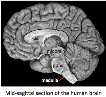
The Brainstem
-part of the CNS
-a continuation of the spinal cord
-divided into 3 parts: the medulla, pons and midbrain (from bottom up)
-can't function with any missing or damaged pieces (unlike the cerebral cortex)
-has conduit, cranial nerve and integrative functions
-contain 6 different types of nuclei: somatic sensory, special sensory, visceral sensory, visceral motor, somatic motor and brachial that are well organized
98
New cards
The Medulla
-continuous with the spinal cord
-the lowest part of the brain stem
-the most caudal (towards the back of the brain) of the brainstem
-can be seen on the ventral surface of the brain
-very similar in shape to the spinal cord (round) because before the fourth ventricle
-the lowest part of the brain stem
-the most caudal (towards the back of the brain) of the brainstem
-can be seen on the ventral surface of the brain
-very similar in shape to the spinal cord (round) because before the fourth ventricle
99
New cards
The Pons
-the middle section of the brainstem
-looks like an enlargement of the brain stem
-can be seen on the ventral surface of the brain
-after the fourth ventricle, therefore no longer round in shape like the medulla, has a depression due to the fourth ventricle
-looks like an enlargement of the brain stem
-can be seen on the ventral surface of the brain
-after the fourth ventricle, therefore no longer round in shape like the medulla, has a depression due to the fourth ventricle

100
New cards
The Midbrain
-the top of the brain stem
-the deepest within the center of the brain
-cannot be seen on the ventral surface of the brain
-the most rostral of the brainstem (towards the front of the brain)
-contains the super and inferior colliculi and the cerebral aqueduct
-the deepest within the center of the brain
-cannot be seen on the ventral surface of the brain
-the most rostral of the brainstem (towards the front of the brain)
-contains the super and inferior colliculi and the cerebral aqueduct
