Posterior thoracic wall
1/38
There's no tags or description
Looks like no tags are added yet.
Name | Mastery | Learn | Test | Matching | Spaced | Call with Kai |
|---|
No analytics yet
Send a link to your students to track their progress
39 Terms
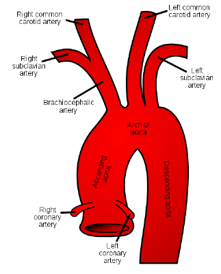
Where does Ascending aorta Originate?
The ascending aorta originates from the left ventricle of the heart, where it carries oxygenated blood to the rest of the body.

What are the branches of Ascending aorta?
Coronary artery left and right

What is the path of Ascending aorta?
behind the sternum (short segment Ascending)
What are the branches of of Aortic Arch?
Brachiocephalic trunk on right side (this then branches off into the right common carotid and subclavian artery)
Common Carotid artery on left side
Subclavian artery on left side
The "Aortic Knob" is a key landmark on a routine Chest X-ray.
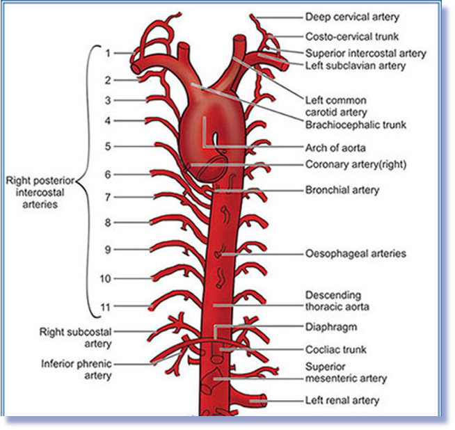
Where does thoracic Aorta Begins?
Begins at the T4/T5 level after the aortic arch and descends on the left side of the vertebral Bodies.
It enters the aortic hiatus of diaphragm at T12

What are the branches of Thoracic Aorta?
○Posterior intercostal arteries
○Subcostal arteries
○Bronchial arteries
○Esophageal arteries - supply the thoracic esophagus
○Mediastinal arteries - supplies mediastinal structures
○Pericardial arteries - supplies the pericardium
○Superior phrenic arteries - run to superior surface of the diaphragm
A continuous H- shaped network of veins in the posterior thorax?
Azygos venous system, which drains the thoracic wall and is formed by the azygos, hemiazygos, and accessory hemiazygos veins.
What is found in the right side of the azygos system?
Azygos vein arches over the root of the right lung to drain into the superior vena cava

What is found in the left side of azygos system?
Accessory hemiazygos vein (Space 4-8)
Hemiazygos vein (Space 9-12)
Cross to the right side of the thorax to join the azygos vein
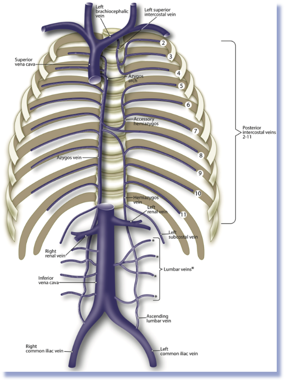
Formed by the union of right and left brachiocephalic veins
Descends vertically on the right side of the ascending aorta
Azygos vein joins the SVC prior to it interesting the pericardium
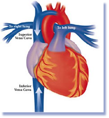
Superior Vena Cava- is a major vein that collects deoxygenated blood from the upper body and returns it to the heart.
Formed by the union of common iliac veins
Ascends to the right of the abdominal aorta through the caval opening of the diaphragm into the lower portion of the right atrium
Inferior Vena Cava - a large vein responsible for carrying deoxygenated blood from the lower body to the heart.

Network composed fluid, vessel, and organs distributed throughout the entire body?
Lymphatic system- a crucial part of the circulatory system that helps maintain fluid balance, transport nutrients, and defend against pathogens.
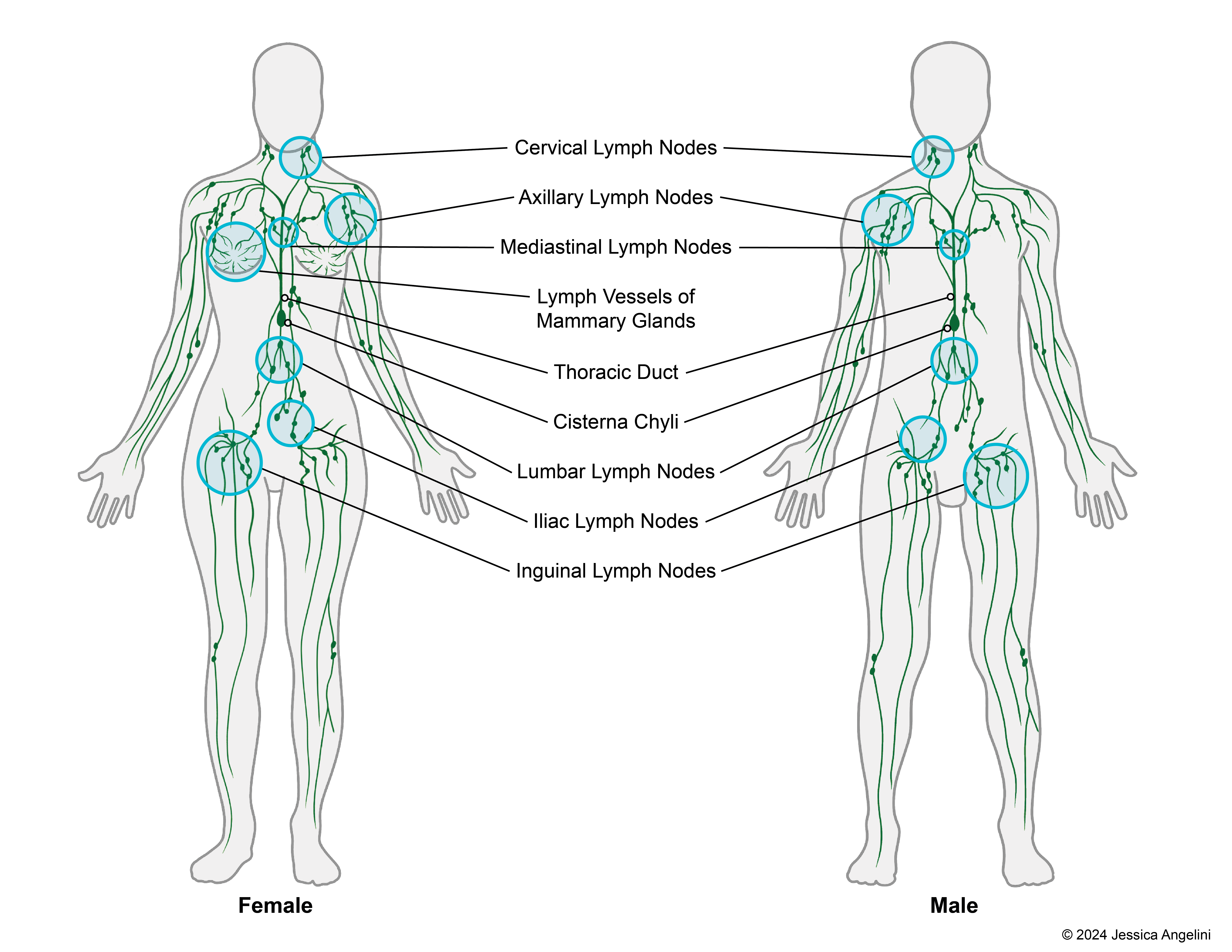
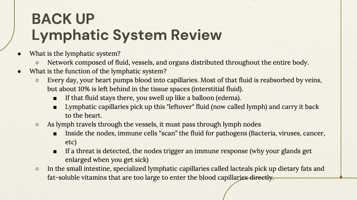
What is the function of Lymphatic system?
The lymphatic system is responsible for maintaining fluid balance, transporting nutrients, and playing a key role in immune defense by aiding in the removal of pathogens from the body.
A clear to milky fluid form the interstitial fluid
Lymph
Carries lymph to the heart (capillaries to the thoracic duct)
Lymphatic vessels
What are the Lymphoid organs?
Spleen (filters blood) Thymus (matures T-cells) / Bone Marrow (matures B-cells)
Small concentrations of lymphoid tissue found in various submucosal membrane sites of the body, such as the gastrointestinal tract, nasopharynx, thyroid, breast, lung, salivary glands, eye, and skin.
Mucosa-associated lymphoid tissue (MALT)
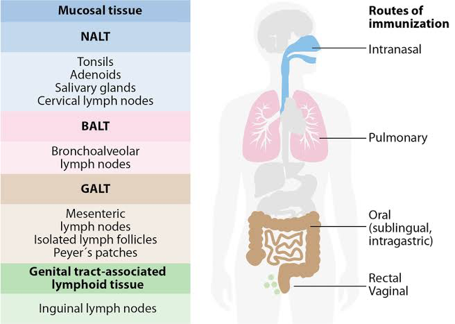
Clumps of lymphoid tissue in the Gut
(GALT) Gut-associated lymphoid tissue found in the lining of the gastrointestinal tract.
Clumps of lymphoid tissue in the lung?
(BALT) Bronchus-associated lymphoid tissue found in the lining of the respiratory tract.

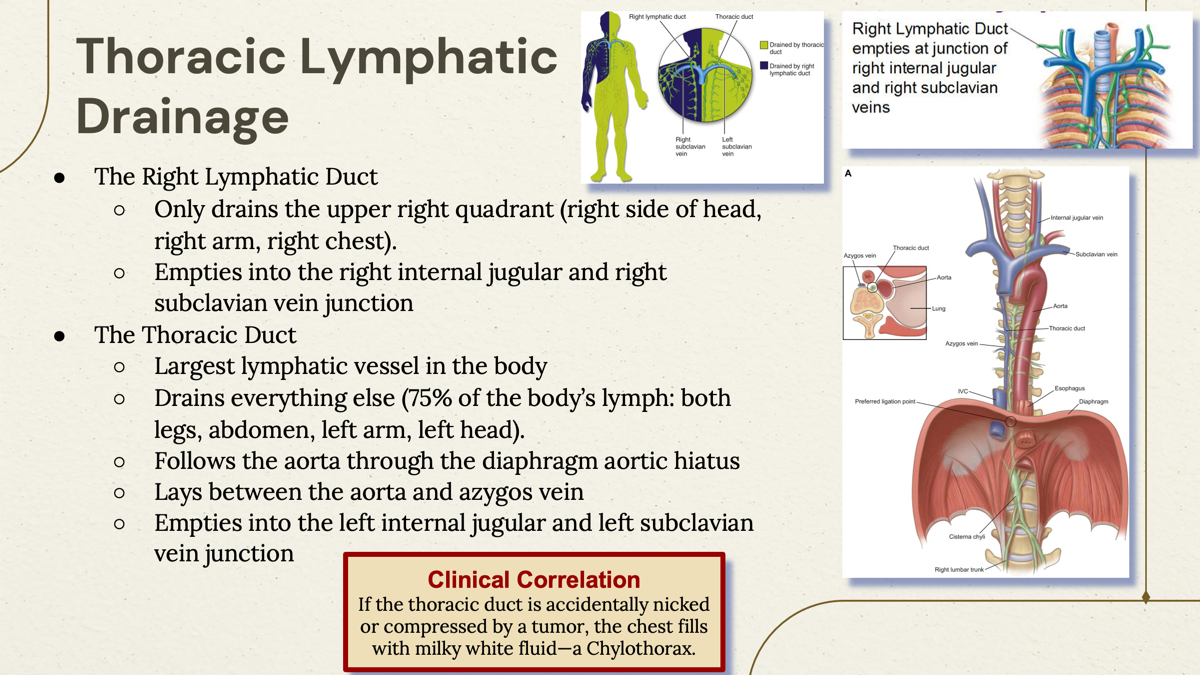
Right Lymphatic Duct
Only drains the upper right quadrant (right side of the head, right arm, right chest)
Empties into the right internal jugular and right subclavian vein junction
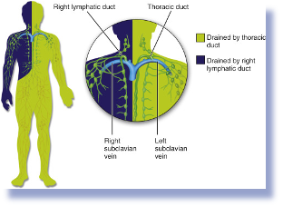
Thoracic Duct
Main lymphatic vessel draining the left side of the body and right lower quadrant. It empties into the left internal jugular and left subclavian vein junction.
○Largest lymphatic vessel in the body
○Drains everything else (75% of the body’s lymph: both legs, abdomen, left arm, left head).
○Follows the aorta through the diaphragm aortic hiatus
○Lays between the aorta and azygos vein
○Empties into the left internal jugular and left subclavian vein junction
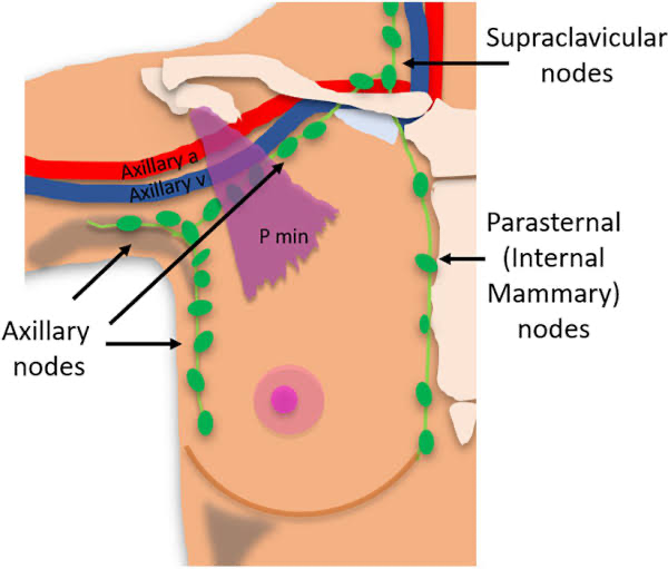
Parasternal nodes
Along the internal thoracic arteries
Key route for breast cancer metastasis.
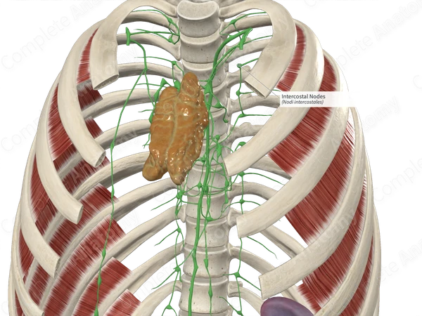
Intercostal nodes
In the posterior Intercostal spaces
Phrenic nodes
Around the diaphragm
Pulmonary nodes
Around the lungs

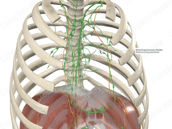
Bronchopulmonary nodes
located in the helium
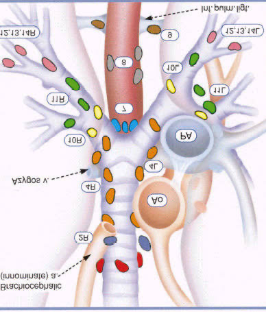
Tracheobronchial nodes
Located near the bifurcation of the trachea
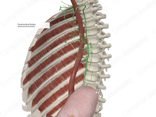
Paratracheal nodes
around the trachea
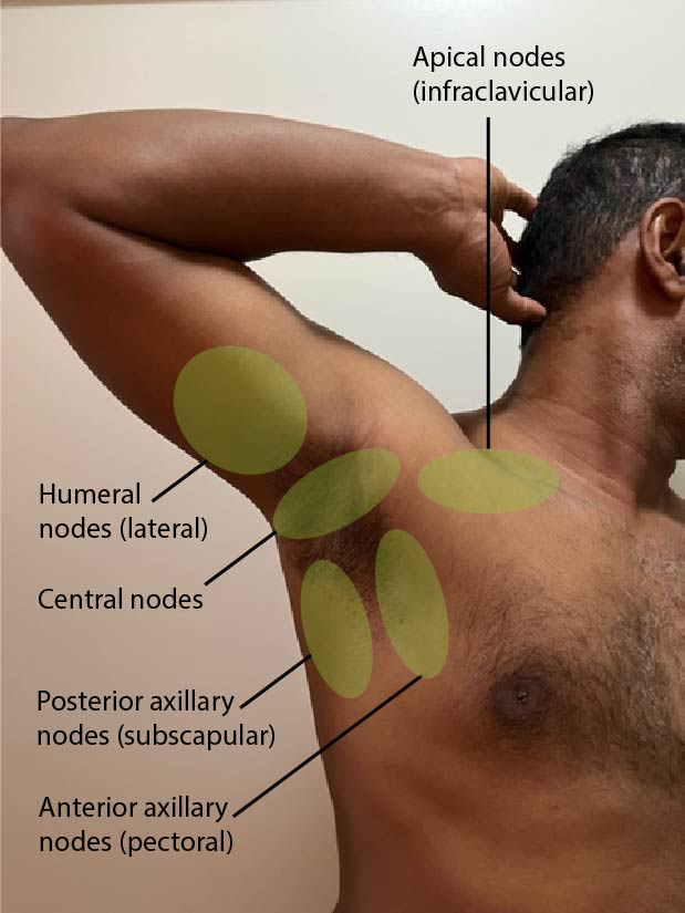
axillary nodes:
5 different groups of axillary nodes
Drains the skin and superficial fascia of everything above the umbilicus (like the breast tissue)
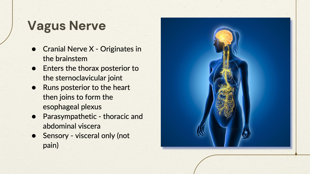
Vagus nerve
The vagus nerve is a cranial nerve that innervates various structures in the neck, thorax, and abdomen, playing a crucial role in autonomic control, including heart rate and digestive processes.
It is also involved in parasympathetic regulation and provides sensory information from internal organs (visceral only not pain)
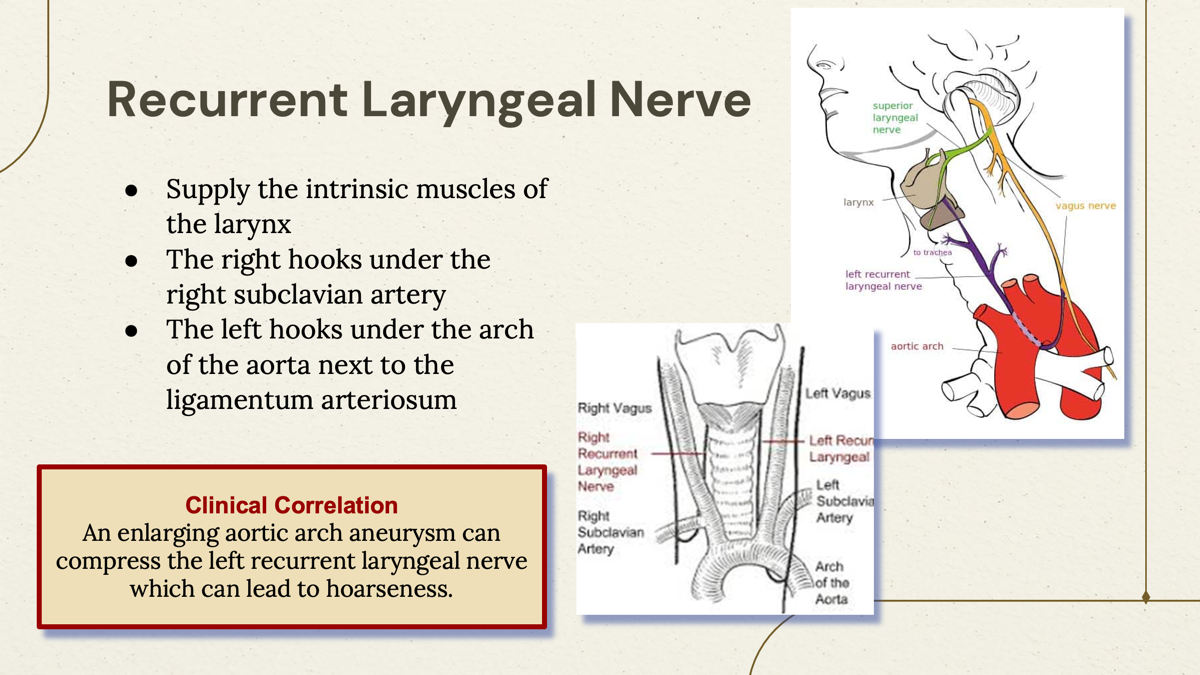
Controls the intrinsic muscles of larynx and is responsible for voice production. It loops under the aortic arch on the left and subclavian artery on the right before ascending to the larynx.
Recurrent Laryngeal Nerve
What is the location of the Long thoracic nerve?
Outside of the rib cage
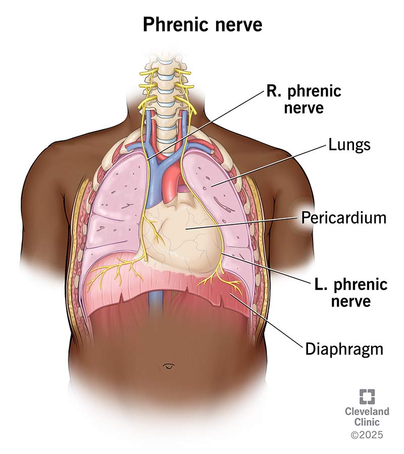
Location of the phrenic nerve?
Around the diaphragm

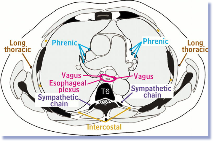
Location of the Vagus nerve
Hides behind the heart with the esophagus

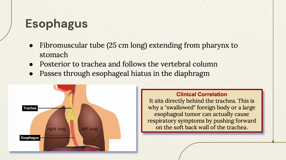
What organ is posterior to the trachea, passes through esophageal hiatus, and fibromascular tube (25 cm long) from pharynx to stomach?
Esophagus
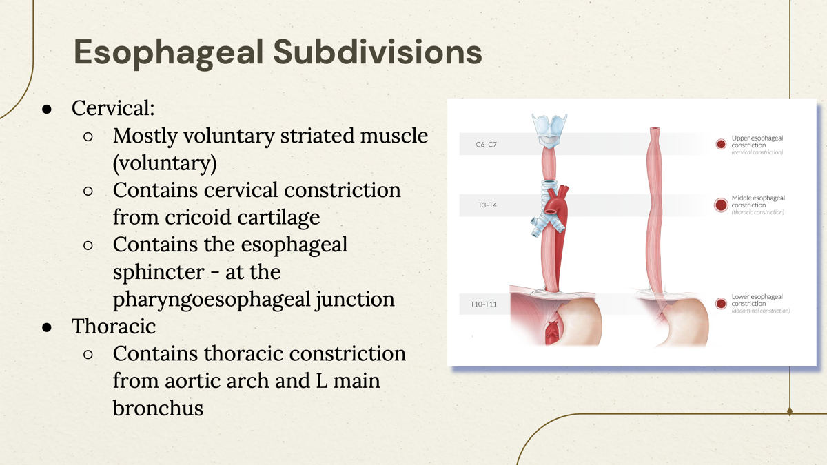
What are the three types of Esophageal subdivisions and their features?
Cervical-
Mostly voluntary and striated
Contains cervical constriction from cricoid cartilage
Contains the esophageal sphincter - at the pharyngoesophageal junction
Thoracic-
Contains thoracic constriction from aortic arch and L main bronchus
Abdominal-
Involuntary smooth muscle (involuntary)
Contains abdominal constriction due to going through the diaphragm at the esophageal hiatus.
Lower (inferior) esophageal sphincter (LES) - Gastroesophageal junction
Network of autonomic nerves that surrounds esophagus in the thorax
Esophageal Plexus
Parasympathetic part of Esophageal Plexus
Vagus nerve- Posterior and anterior vagal trunks
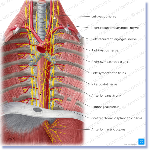
Sympathetic part of Esophageal Plexus
○Branches of splanchnic nerves of
○Cervical / Thoracic sympathetic trunk
