Measurement Colour Vision
1/56
Earn XP
Description and Tags
The essential elements and techniques of assessing colour vision.
Name | Mastery | Learn | Test | Matching | Spaced | Call with Kai |
|---|
No study sessions yet.
57 Terms
Colour Vision Function
Colour is set of perceptions elicited from the spectral distribution of light (wavelengths)
Found in many species
Evolved in primates
Chromatic contrast allows us to see objects that would be otherwise indistinguishable from the background – helps distinguish borders
Colour is set of perceptions elicited from the spectral distribution of light (wavelengths). True or False?
True
___________ is visible to humans due to human physiology!
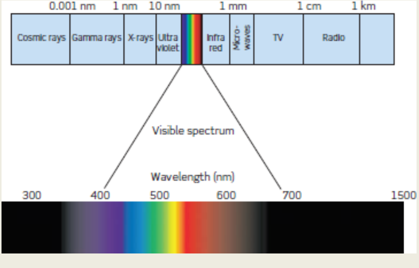
Visible Spectrum
Visible light encompasses wavelengths roughly from ______ to _____ nanometers.
380nm; 780nm
Snakes have __________ and birds and insects have ________.
Infrared Sensitivity; Ultraviolet Sensitivity
The Colour Stimulus:
Isaac Newton (1704) - stimuli of different wavelengths within the visible range produce different colour sensations
Prism split into “rainbow of colours” - index of refraction inversely proportional to wavelength (diffraction)
ROY G BIV
Issac Newton figured of the colour stimulus in _______.
1704
Colour Perception:
Colours are generated by the visual system
Color is not a physical property but a psychophysical property
There is no red in a 700 nm light! … its what we perceive
The colour of an object is due to:
The wavelength reflected to the eye
The reaction of the visual pigments to those wavelengths
Neural processing of those signals
Colour is _______ and is only due to how wavelengths reflect to the eye, reaction of the visual pigments to those wavelengths and neural processing.
Psychophysical
Trichromatic Theory
Proposed by Thomas Young (1802) and Hermann von Helmholtz (1866)
Theory of color vision that says eye has three kinds of receptors that are “tuned” to be sensitive to red, green and blue light
All the colors we see are a combination of these three colors
Relative activities of these different receptors encode colour
James Maxwell (1831 – 1879) developed a colour-matching technique that is still used today (Nagel Anomaloscope)
The eye has _____ kinds of receptors that are tuned to red, green and blue lgiht.
3
Trichromacy
Mixing Short, Medium and Long Wavelength Light can match (nearly) any colour
A person with trichromatic vision can match lights with 3 lights on one side of matching field
The two matched fields of light are known as metamers
Two matched fields of light are known as ________. Colors that appear identical but are physically different (they emit or reflect physically different light wavelengths) are ________.
Metamers; Metamers
Evidence of Trichromacy:
Neurophysiological experiments confirmed rods and three different types of cones
Micro spectrophotometry
Suction electrode technique
The three classes of cones are termed Short (S), Medium (M) and long (L) wavelength and also referred to as Blue, Green and Red
There are three different types of ________.
Cones
Distribution of Cones:
few “S” cones at fovea and only about 10% of cones “S” – centre of fovea
Trichromatic colour vision – extends 20-30 deg from fovea
S-pigment – response to short wavelengths (BLUE)
M-pigment – response to middle wavelengths (GREEN)
L-pigment – response to long wavelengths (RED)
Only _____% of cones at the centre of the fovea
10
Cones:
Conical in shape
Look like rods at the fovea
Colour Vision
Visual pigment
Three types Sensitive to three different wavelengths of light
Photopic vision
High levels of illumination
High levels of visual acuity
Tightly packed cones at the fovea
Only green and red at fovea
______ are tightly packed at the fovea and have only _____ and _____ vision at the fovea.
Cones; Red; Green
A single ____ shows different responses to lights of different wavelengths but the same intensity.
Photoreceptor
Just one ________ cannot discriminate colour.
Photoreceptor
Principle of Univariance:
When a photopigment molecule absorbs a quantum of light, it can only signal information about how much light is absorbed and not what the wavelength of the light is
A single cone cannot give any indication of the colour of an object because its response can only vary in one dimension
A ________ molecule can only signal information about how much light is absobred not the wavelength of light
Photopigment
An individual with only one photopigment is know as a ________. Monochromats are unable to make wavelength based discrimination – two patches of light can always be made to appear identical by adjusting their intensities so number of quanta absorbed is equal – do not see colours!
Monochromat
The two wavelengths that produce the same response from one type of _____produce different patterns of responses across the three types of cones (S, M, and L).
Cone (M)
Trichromatic Theory Phenomena
Many visual phenomena that are not easily explained by the trichromatic theory - no greenish red or yellowish blue perceived
Complementary colours – produce a grayscale colour
Red - green
Blue - yellow
Complementary Afterimages
A red stimulus elicits a green after-image and a blue stimulus elicits a yellow afterimage (and vice versa)
Red and _____ are complimentary and ______ and yellow are complimentary.
Green; Blue
Colour Opponent Theory:
Herring (1892) - trichromatic signals from the cones fed into subsequent neurons and processed by three “opposing” channels:
red vs green
blue vs yellow
black vs white
Channels can signal only one of the two colours at any given time
There are ______ opposing channels.
Three
Electrophysiological Evidence:
Single unit recording in retina and LGN found colour opponent cells*
Short wavelength stimulus causes inhibition, while long wavelength stimulus produces excitation
Colour opponent cells occur first at level of the bipolar retinal cells in primates
Short wavelengths can cause ________, while long wavelengths produce _______.
Inhibition; Excitation
Opponency Neurons - Channels
Red-Green Channel - Compares output from L and M cones
Blue-Yellow Channel - Coompares output from S cones with combined output of L and M cones.
Black-White Channel - Luminance Channel adds the cone outputs
_________ refers to a receptive field structure in the visual system where the central area of a cell is excited by light, while the surrounding area is inhibited
Centre - Surround Organisation
Stages of Colour Vision:
Trichromatic photoreception (1st Stage)
Opponent - process neural coding (Retina and lateral geniculate nucleus (LGN)) (2nd Stage)
Higher level cortical processing (3rd Stage)
There are ______ stages to colour vision.
Three
Parallel Processing of Colour Signals - Retina to LGN:
R-G "P" ganglion cells → LGN parvocells, which show R-G opponency
Y-B ganglion cells → LGN Konio cells, which show Y-B opponency
Spectrally opponent LGN cells about 60% of LGB neurons
Koniocellular (K) cells and parvocellular (P) cells are two distinct types of neurons found in the lateral geniculate nucleus (LGN) of the thalamus, which plays a crucial role in relaying visual information to the visual cortex.
P ganglion cells got to __________ in R-G opponency, meanwhile Y-B ganglion cells go to _______ in Y-B opponency.
LGN parvocells; LGN koniocells
Cortical Processing:
Visual cortex (V1) – cortical area where most LGN axons synapse
Concentrically organised
double colour-opponent neurons
Parvo input – RG
V1 to prestriate cortex V4
V1 is where______ LGN axons synapes are.
Most
Colour Constancy
A blue shirt looks blue under vastly different lighting condition:
Indoors
Natural sunlight (e.g. day time, sunset)
Fluorescent light
The apparent colour of a reflective surface remains constant even when changes in the illuminance alter the wavelengths reflected from it
Adelson’s illusion - an optical illusion where two squares of the same color and brightness appear different due to the perceived presence of shadows.
Visual system able to discount the illumination by “looking at lots of objects at once”
If all reflecting long wavelengths, probably means objects are being illuminated by red light and system compensates for this…blue shirt appears blue as it is bluer than things around it!
Lighting affects ________.
Perceived Object Colour
Describing Color Perception:
Hue - Lay person’s term for colour – the colour we see
Saturation
Purity of colour - a desaturated colour appears as though it has been mixed with white
Brightness
Perceived intensity of the coloured surface or source
Primary colours – Red, Green and Blue lights – mixed together see white
Complementary colours- two colours mixed produce white
Color space: A three dimensional space that describes all colors.
There are several possible color spaces
RGB color space: defined by the outputs of long, medium, and short wavelength lights (i.e., red, green, and blue).
Hue, Saturation, Brightness color space
_______ is a three dimensional space that describes all colours.
Color Space
CIE Colour System:
This system was developed by the International Commission of Illumination (CIE – Commission Internationale de L’Eclairage)
Standard graphical representation of the hue and saturation attributes of color, based on color matching data obtained from large groups of observers
Important colour metric used internationally in industry for colour specification - defines colour experience in quantitative way
CIE stands for ____________.
International Commission of Illumination (CIE – Commission Internationale de L’Eclairage)
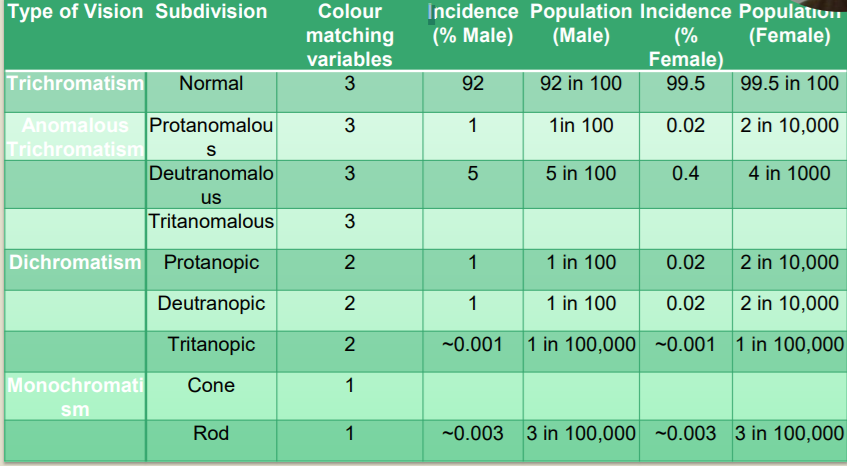
Anomalies of Colour Vision:
Occur in around 8% of males and 0.4% of females
Colour matching divides colour anomalies into:
Anomalous trichromat require three primaries to achieve metameric matches
Dichromats require only two primaries to achieve metameric matches
Monochromats require only one primary
Classified according to the number of primary colours required to match all the spectral hues
Means the number of primary colours required to match to the number of photopigments present
Opponent theory of excitation and inhibition allows us to make those assumptions according to the hue presented
_______ only require two primaries to achieve metameric matches.
Dichromats
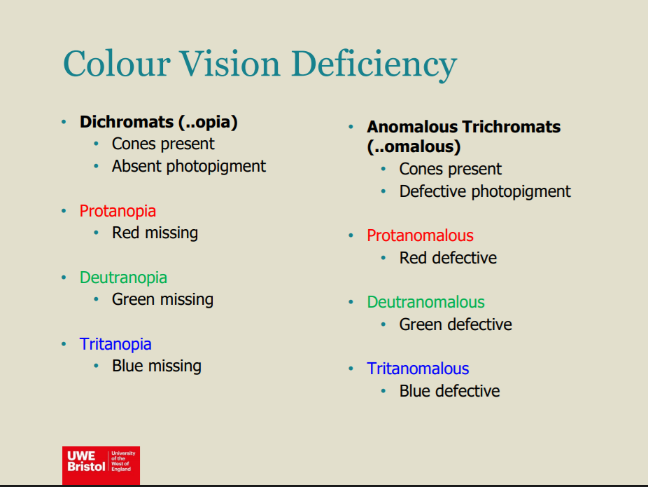
Anomalous Trichromacy:
Trichromatic vision as has three types of photopigment in their cones
One of the pigments is shifted from the normal position
The greater the displacement the more severe is the colour vision anomaly
Require a different mixture of primaries to a normal in order to obtain a colour match
In ______ one of the pigments is shifted from the normal position.
Anomalous Trichromacy
Protanomalous Trichromacy:
Mutation of L-cone photopigment gene
L cone pigment (erythrolabe) spectrum is displaced toward shorter wavelengths –
Hereditary – X-linked recessive trait; Approx. 1 % males
In ______ the L cone pigment spectrum is displaced towards _____ wavelengths.
Protanomalous Trichromacy; Shorter
Deuteranomalous Trichromacy
M-cone photopigment gene replaced by hybrid L/M gene
M cone pigment (Chlorolabe) - spectrum is displaced towards longer wavelengths.
Most common form of congenital colour deficiency – about 5% population
Hereditary – X-linked recessivecyn
In _________ the M cone pigment (chlorolabe) is displaced towards longer wavelengths.
Deuteranomalous Trichromacy
In ___________ the S cone pigment (cyanolabe) spectrum is displaced towards longer wavelengths (Rare).
Tritanomalous Trichromacy
The defect in dichromatic colour vision tends to be more severe than in ______ with Dichromats lacking ______ type of pigment.
Anomalous Trichromacy; One
________ is dichromacy with a missing L cone and reduced sensitivity to long-wavelength.
Protanopia
_______ is dichromacy with a missing M cone.
Deuteranopia
_______ is dichromacy with a missing S cone.
Tritanopia
Colour Labelling:
Individuals with red-green dichromacy are essentially monochromatic for wavelengths beyond approximately 545nm, yet do surprisingly well at labelling colours, especially when other cues present such as
Brightness
Context
____________ dichromacy is essentially monochromatic for wavelengths beyond _____.
Red-Green; 545nm
______ classifications end with _______
‘-anomalous’; '-opia'
Achromatopsia
Extremely rare condition presenting as monochromatic vision either as:
Cone monochromatic
Characterised by presence of only one primary colour and thus person is truly colour blind
Such patients usually have normal visual acuity
Defect in post receptor processing
Rod monochromatic
Autosomal recessive trait, very rare
Characterised by total colour blindness, poor visual acuity, nystagmus, photophobia
There are two types of monochromaticism _______ and ______.
Cone; Rod
Acquired Colour Vision Anomalies
• Secondary to disease or toxicity and may be R-G or BY.
B-Y anomalies rarely inherited, assume acquired until proven otherwise
Acquired colour anomalies often unstable and may not produce clean test results
May be unilateral or asymmetric
Important to perform colour vision tests monocularly
B-Y anomalies are _______ and is assumed acquired until proven otherwise.
Rarely Inherited
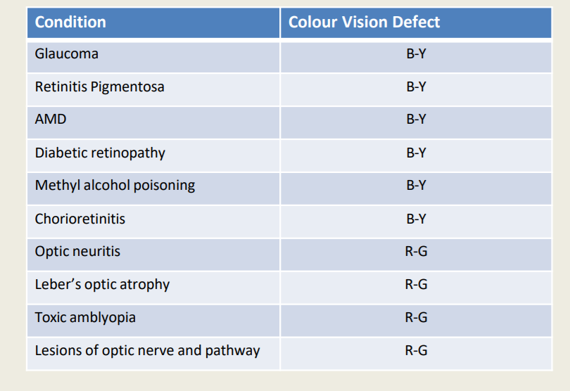
Optic neuritis, Leber’s Optic Atrophy, Toxic Amblyopia or Leisions of Optic Nerve and Pathway are ______ colour vision defect.
R-G
Colour psychophysical phenomenon … how we perceive a stimulus … normal colour vision = trichromatism
Colour perception has 3 aspects: wavelength reflected to eye, visual pigments react, neural processing of information
Colour contrast helps to distinguish objects (finding fruit …)
At least two receptors needed to detect colour due to principle of univariance
Trichromacy (Young & Helmholtz):
three primary colours can be mixed (different proportion) to achieve any colour to be perceived; two colours can be perceived identical despite different wavelength composition (i.e., metamer)
There are 3 different types of receptors sensitive to Short (S), Medium (M) and Long (L) wavelengths that encode the wavelength signal perceived
Does not explain complementary colours or complementary afterimages
Colour Opponent theory (Herring): trichromatic signals from the cone receptors feed into subsequent neurons and are processed by opposing channels (R/G, B/Y, B/W)
Colour vision anomalies/deficiencies can be congenital (x-linked recessive, rare: 8% males and 0.4% female) or acquired (monocularly or asymmetric)
anomalous trichromats, dichromats, monochromats
Normal colour vision = Trichomatism. True or False?
True
Perception of Colour
Perception of colours depends on wavelength, amount of light absorbed, threshold stimulus + frequency of cone impulses
Orange = 99% Red: 42% green: 0%
Yellow = 50% red: 50% green: 0% blue
Blue = 0% red: 0% green: 97% blue
Yellow = 50% red: 50% green: 0% blue. True or False?
True
Colour Opponets
Opponents are processed within the same channel
Not possible to see both in a channel at the same time
When cones are excited their opposing colour is inhibited
No reddish green
No yellowy blue
Activation of one channel inhibits the other
Inhibition
Works within the channels and can result in fatigue of a cone’s excitatory ability
Fatigue of the channel’s processing ability
The channels also compete for processing space
When the red and green cones are stimulated at the same time the blue-yellow channel is inhibited
We see yellow even though there is no yellow cone…
When cones are excited their opposing colour is _______.
Inhibited
Colour Vision
Trichromatism
Normal colour vision
All three cone types work as they should
Congenital Anomalies
Lack of functional cones
Cones all present, absorption characteristics abnormal
One or two photopigments rather than three
Acquired Anomalies
Secondary to pathology
Something selectively causes the cones to not work
Something selectively causes the channel to not work
Trichromatism is ____________.
Normal Colour Vision
Colour Vision Deficiency
Monochromats
Rod monochromatic
No functioning cones
Poor visual function and acuity
Nystagmus
Photophobia
Cone monochromatic
Single cone response
Otherwise normal visual function
Dichromats
Absent photopigment
Red
Green
Blue
Anomalous Trichromats
Defective photopigment
Red
Green
Blue
Rod monochromats have no functioning _______.
Cones
Pro- means missing ______. Deutr- means missing _____. Tritan- means missing _____.
Red; Green; Blue
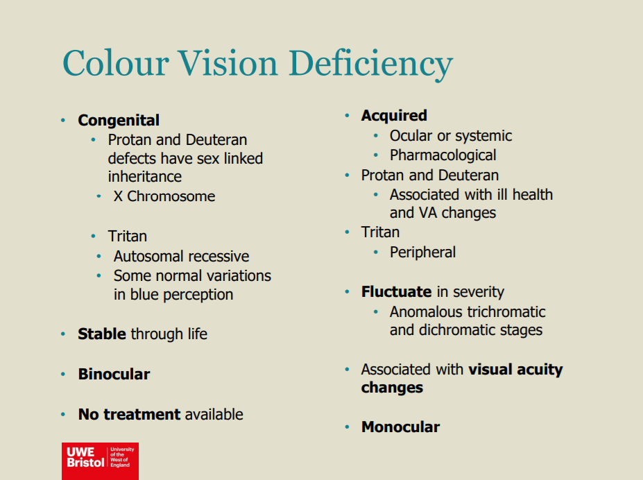
Protan and Deuteran defects have ___________ inheritance.
Sex-linked
Colour Vision Deficiency Screening Tests
Ishihara
Standard Pseudoisochromatic Plates Part 2
Red Desaturation1
The City University Colour Vision Test Part 1
Colour Vision Deficiency Grading Tests
The City University Colour Vision Test Part 2
Fansworth D-15 Test
The ________ can be used for screening and grading.
The City University Colour Vision Test
Take _______ measurement for testing congenital colour vision defect. Take ______ measurement if testing for acquired colour vision tests.
Binocular; Monocular
Ishihara Test
Screening for red green colour deficiency at 75cm test distance
25 plates (abbreviated version) containing numerals and lines
Pseudoisochromatic
First plate is an introductory plate
Demonstration of the task
Detecting malingerers
Next twenty plates
Screening for protan/deuteran
Four for classification
Protan/deuteran
Screening for red green colour deficiency
Transformation Plates (Confusion plates)
One number is seen by colour normals (8 and 5)
A different number is seen by those with colour deficiency (3 and 2)
Vanishing number plates
A number is seen by colour normal
No number is seen by those with colour deficiency
Classification of red green colour deficiency
Two numbers or lines presented
Protan observers
See the right hand number (6)
bottom line
Deuteran observers
See the left hand number (2)
top line
When no number seen severe red-green defect
When both numbers seen with significant errors previously ask if one of the numbers look clearer
Ishihara should be done at _______.
75cm
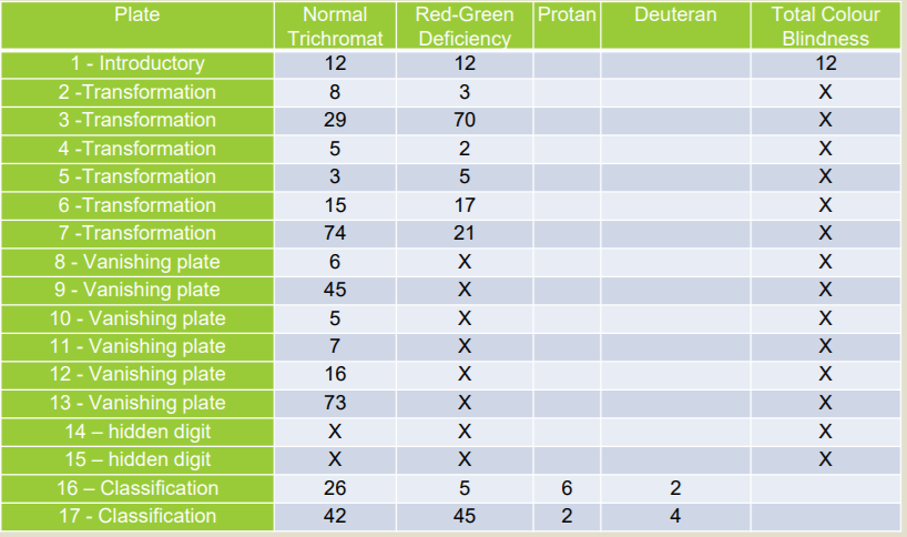
Ishihara has transformation, _______, hidden digit and classification plates.
Vanishing
The City University Test
Adjunct to a screening test
Monitoring of acquired colour vision deficiency
Tests for blue-yellow deficiency as well as red-green deficiency
Passing the City University Test after failing the Ishihara Test is likely to indicate a mild colour deficiency which is unlikely to impact on occupational choices
Part 2
Derived from the Farnsworth D-15
grading
10 charts
Central colour with four surrounding colours
Patient is asked to select which of the surrounding colours best matches the central colour
Options displayed
Normal
Protan
Deuteran
Tritan
Interpretation
Protan/Deuteran
Patients can select colours in both categories
Differentiating between protan and deuteran can be difficult
‘Borderline’
One or two mistakes
‘Fail’ (not clear if n/p/d/t)
More than two mistakes
Score out of 10
Number of mistakes in the normal column indicates the severity of the deficiency
Part two of the city university test is derived from ________ the test.
Farnsworth D-15
Farnsworth D15
Developed to assist in vocational guidance
Farnsworth D-15
Farnsworth-Munsell 100 hue
Farnsworth D-15
Indication significant defects
The patient arranges the 15 colours in sequence as they see it
This sequence is then plotted on a circular chart
The shape of the pattern plotted indicates the type of defect
Protan
Deuteran
Tritan
Number of errors indicates the severity of the defect
Scoring
This sequence is then plotted on a circular chart
The shape of the pattern plotted indicates the type of defect
Protan
Deuteran
Tritan
Number of errors indicates the severity of the defect
Normal Colour vision
Trichromatism
Protan
Severe (r)
Moderate (l)
Deficient with red
Deuteran
Severe
Moderate
Deficient with green
Tritan
Severe
Moderate
Deficient with blue
The two types of Farnsworth are the _________ and ______ tests.
Farnsworth D-15; Farnsworth-Munsell 100 Hue
Farnworth-Munsell 100 Hue
Similar to D-15
More colour discrimination options
85 Hues
Approximately equal steps
Four boxes with a reference hue at each end of each box
Tests the hue discrimination in normal trichromatic
Tests the hue discrimination in those with colour deficiencies
Scoring on a circular graph
Error score created by comparing the numerical differences of adjacent colours
Congenital defects are shown by errors in defined poles
Axis of confusion
Degree of defect estimated from the total error score
Good for acquired defects
Monitoring change over time
Fatigue effects
Learning effects
Farnsworth-Munsell 100 Hue colour vison test is good for __________ defects.
Acquired
Other Colour Vision Tests:
Nagel Anomaloscope
Red-green defects
Normal or deficient (screening)
Dichromatism or anomalous trichromatism
Colour matching by altering luminance of a luminance control
Colour Vision Lanterns
Red-green defects
Screening or grading
Patients identify the colour of the light emitted by the lantern
The ________ test is used for screening and the _______ test is used for screening or grading.
Nagel Anomaloscope; Colour Vision Lanterns
Colour Vision Tests for Occupation:
Aviation - Ishihara
Navi - Colour blindness does not restrict but limits progression, Ishihara or Holmes-Wright Lantern
Fire Service - Farnsworth
Train Drivers - Ishihara
Police - Farnworth or 2nd Edition City
Fire and Police are ______, TAN is ________, and Police is _____ and Navi is ______.
Farnworth; Ishihara; City; Holmes-Wright Lantern
Red Desaturation
Testing the sensitivity to the colour red
Acquired colour defect
Reduced VA
Pupil function
Adjunct test for rapidly assessing the likelihood of neurological changes
optic nerve and visual pathway pathologies
Comparison of how a red object is perceived with each eye separately
Red colour appears washed out with the affected eye
The Red Desaturation test is used for ________ defects and should be tested ________.
Acquired; Monocularly