The Fetal Heart and Chest
 Call Kai
 Learn
Learn Practice Test
 Spaced Repetition
 Match
Match Flashcards
 Knowt Play
1/108

There's no tags or description
Looks like no tags are added yet.
Name | Mastery | Learn | Test | Matching | Spaced | Call with Kai | Chat |
|---|
No analytics yet
Send a link to your students to track their progress
109 Terms
The embryonic heart begins as two tubes. These two tubes ultimately fuse and fold to form into four chambers,
two atria and two ventricles.
The heart begins to contract at 36 to 37 days of gestation and is fully formed by 10 weeks. A heart rate using M-mode should be sonographically obtainable with endovaginal (EV) imaging when the crown rump length (CRL) measures
4-5mm
During a biophysical profile assessment in the third trimester, an average fetal heart rate is
150 beats per minute (bpm), with a range of 110 to 180 bpm considered normal after 1st trimester
what is biophysical profile
a method of fetal monitoring with sonography to produce a numeric scoring system that predicts fetal well-being
A normal fetal heart will fill about 1/3 of the fetal chest. If it is suspected that the heart is enlarged,
then a cardio/thoracic diameter ratio can be obtained.
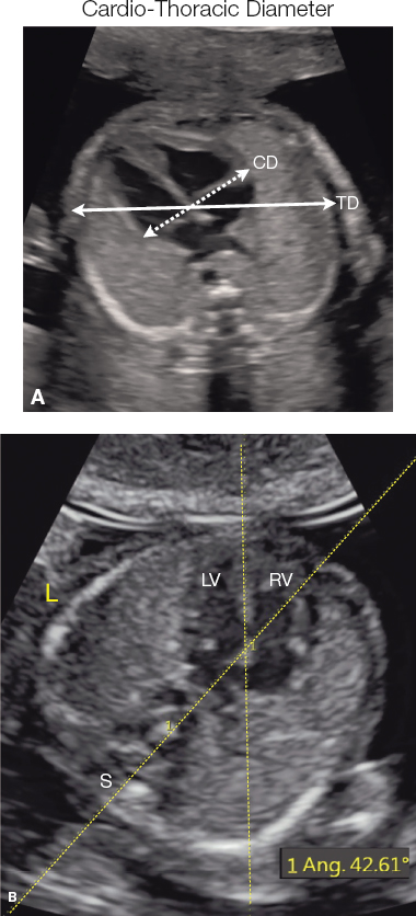
4 chamber heart view will show
The apex of the heart will be angled to the left of the midline (same side as stomach as well), with the base closest to the spine.
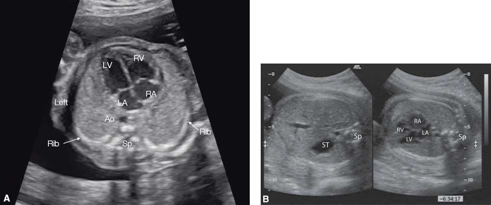
the apex of the heart forms a __ angle with the spine
45 degree
The two atria are separated by the ____ septum, and the two ventricles are separated by the _______ septum.
atrium, ventricular
The ventricular septum should be uninterrupted and of equal thickness to the
left ventricular wall,
the atrial septum is open only at the
foramen ovale
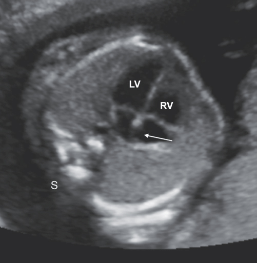
Within the right ventricle can be seen the __________ ________ a normal structure that appears as an echogenic focus
moderator band
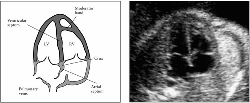
Between the right ventricle and the right atrium, one should visualize the
tricuspid valve,
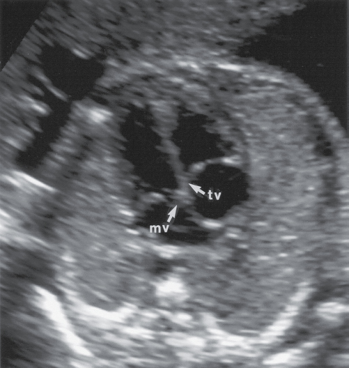
between the left ventricle and the left atrium,
mitral valve should be noted
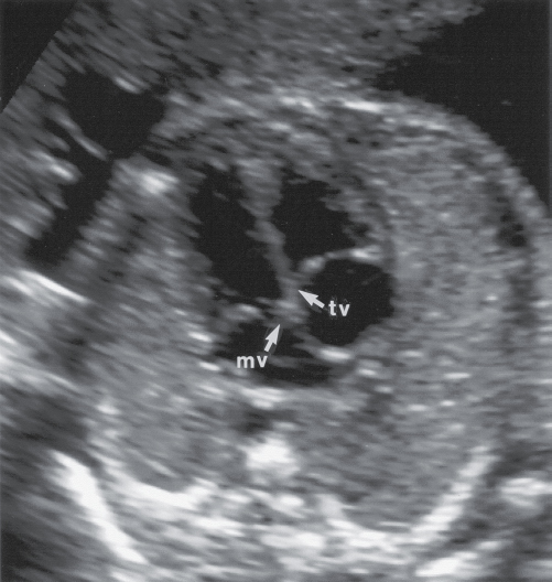
Normally, the tricuspid valve is positioned closer to the
cardiac apex than the mitral valve
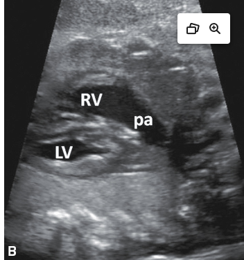
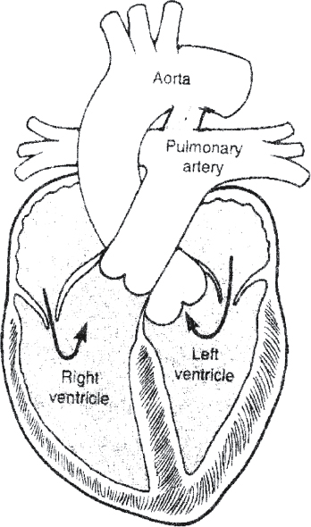
The right ventricular outflow tract leads to the
pulmonary artery and branches
the left ventricular outflow tract leads to the
aorta.
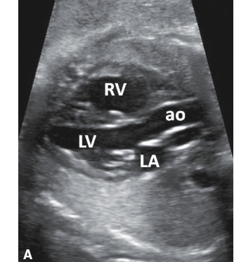
One important anatomic finding is that the normal pulmonary artery should be positioned
anterior to the aorta and should be visualized crossing over it
The three-vessel view (3-VV) shows the
main pulmonary artery, ascending aorta, and superior vena cava
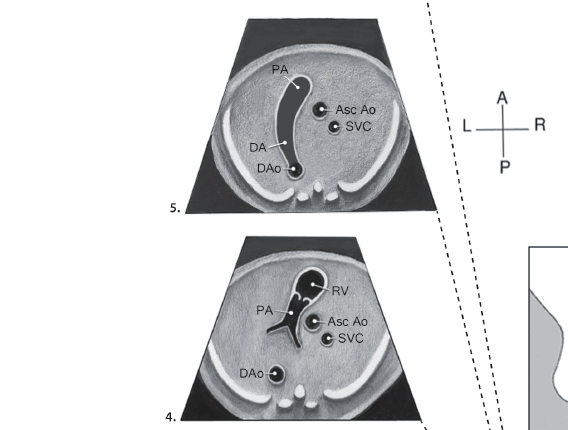
The 3VV is used to detect
abnormal vessel number, abnormal vessel caliber, abnormal course or alignment of the vessels, and abnormal flow
The three-vessel trachea view (3VT) is used to evaluate the major vessels in the fetal mediastinum as well. In the 3VT, a
“V” is formed by the merging of the transverse aortic arch and isthmus and the pulmonary trunk and ductus arteriosus
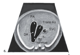
The 3VT is also used to assess the
number, size, and alignment of the main pulmonary artery, aorta, and superior vena cava.
Representative scan planes for fetal echocardiography
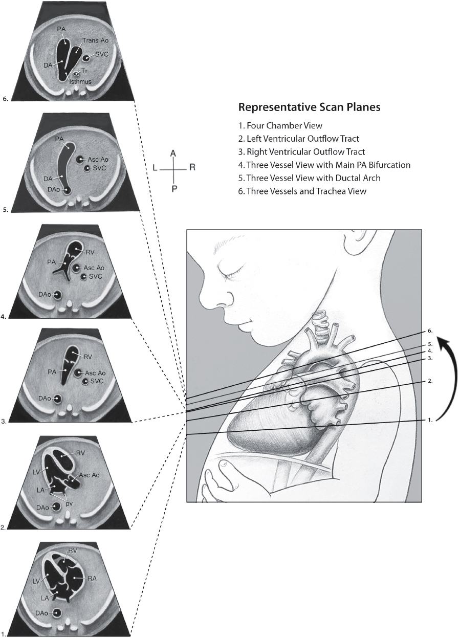
Basic assessment of the fetal outflow tracts

fetal circulation
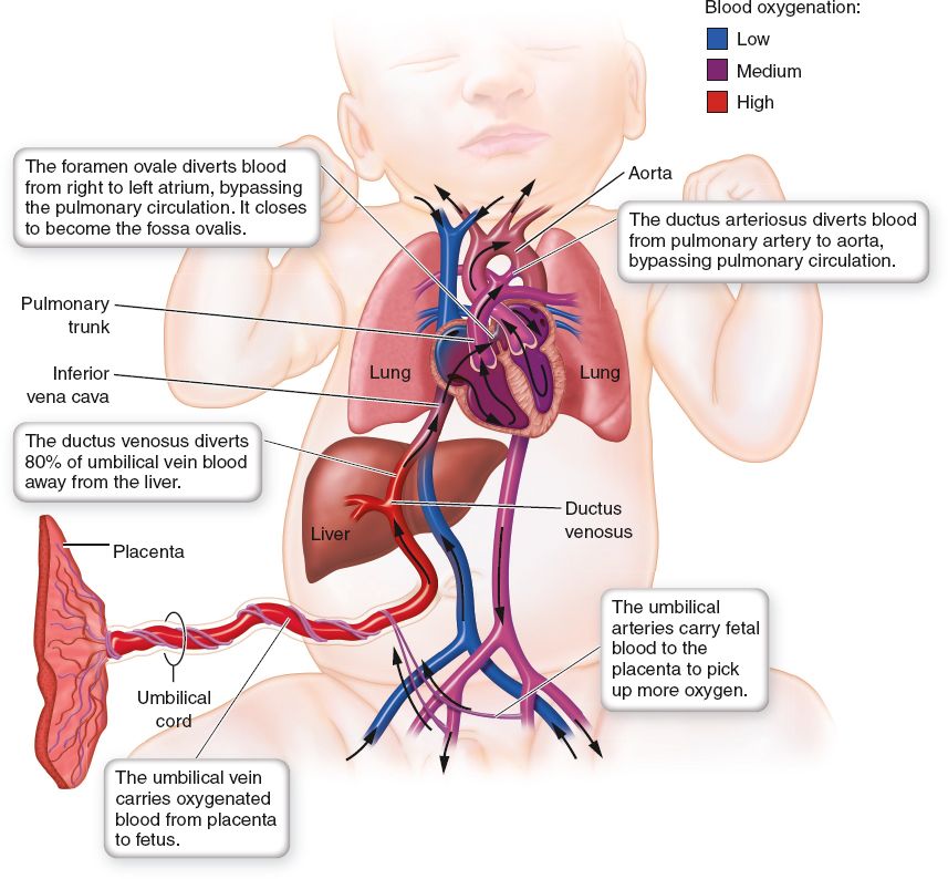
what is ductus arteriosus
a fetal shunt that connects the pulmonary artery to the aortic arch
what is ductus venosus
a fetal shunt that connects the umbilical vein to the inferior vena cava
The normal umbilical cord contains two arteries and one vein. The umbilical vein brings oxygen-rich blood from the placenta to the
fetus
It travels superiorly and connects to the left portal vein. Half of the blood goes to the liver through the left portal vein, whereas the other half is shunted directly into the
Inferior vena cava (IVC) via a small branch of the umbilical vein called the ductus venosus.
The blood that was taken to the liver is used to oxygenate the liver and is then returned back to the
IVC via hepatic veins
The existing oxygen-rich blood in the IVC travels up to the heart and enters the right atrium. Blood can then travel across the
foramen ovale into the left atrium, or it can enter the right ventricle through the tricuspid valve.
The blood then leaves the right ventricle through the main pulmonary artery. The main pulmonary artery bifurcates into
right and left, thus allowing a small amount of blood to travel to the respective lung.
Blood from the right ventricle can also flow through the
ductus arteriosus and into the descending aorta
The blood returning from the lungs through the pulmonary veins enters into the
left atrium
Blood then travels from the left atrium into the left ventricle via the
mitral valve
From the left ventricle, it travels to the ascending aorta and into the aortic arch, where it exits into the
brachiocephalic artery, left common carotid artery, and left subclavian artery on its way to the thorax, upper extremities, and head.
The blood will return from the head and upper torso via the
superior vena cava to the right atrium
The blood that flows through the ductus arteriosus and into the descending aorta travels inferiorly to either exit the abdomen via the
umbilical arteries or travel to the abdomen and lower extremities to replenish those regions
Therefore, the umbilical arteries return the deoxygenated blood from the fetus back to the
placenta
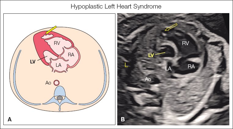
What is Hypoplastic left heart syndrome
group of anomalies characterized sonographically as a small or absent left ventricle
Hypoplastic left heart syndrome is the leading cause of
cardiac death in the neonatal period, with 95% dying within the first month of life if surgery is not performed
To distinguish this anomaly from complete absence of the left side of the heart,
a small or normal left atrium must be visualized
When found in girls,
Turner syndrome should be suspected. There is also a connection with trisomy 18.
Sonographic Findings of Hypoplastic Left Heart Syndrome
Absent or small left ventricle
No communication between the left atrium and the left ventricle
Aortic atresia (possibly)
Aortic stenosis (possibly)
Coarctation of the aorta (possibly)
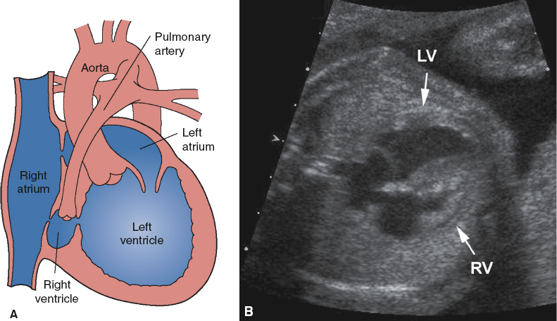
what is Hypoplastic right heart syndrome
sonographically identified as a small or an absent right ventricle
Hypoplastic heart syndrome most often results from
pulmonary stenosis or pulmonary atresia, but it may result from stenosis or atresia of the tricuspid valve.
Sonographic Findings of Hypoplastic Right Heart Syndrome
Absent or small right ventricle
Enlarged left ventricle
Fetal hydrops (secondary to cardiac failure)
Narrowing of the pulmonary valve
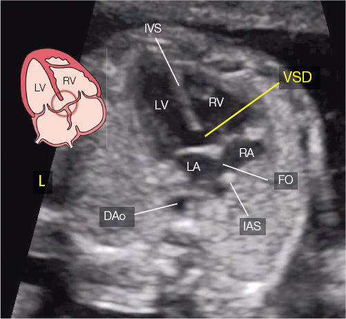
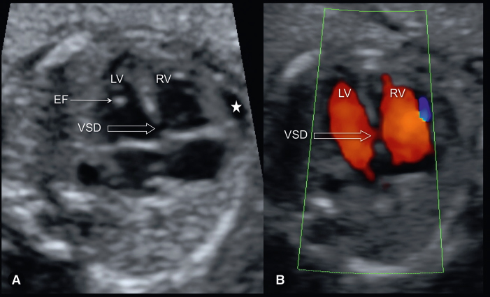
A ventricular septal defect (VSD) is an
abnormal opening in the septum between the two ventricles of the heart
The VSD is the
most common form of cardiac defect
Sonographic Findings of Ventricular Septal Defects
Absence of part of the ventricular septum
Color Doppler is helpful at detecting small defects
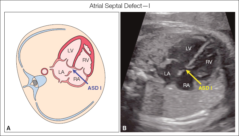
An atrial septal defect (ASD) is an
abnormal opening in the septum between the two atria of the heart. (types 1-5)
Sonographic Findings of Atrial Septal Defects
Absence of part of the atrial septum
Color Doppler is helpful at detecting small defects
The combination of both ASD and VSD is termed
atrioventricular septal defect (AVSD) or atrioventricular canal.
An AVSD results from the
abnormal development of the central portion of the heart.
The central portion of the heart is referred to as the
“endocardial cushion”; this is the reason why the AVSD may be referred to as an endocardial cushion defect.
AVSDs are commonly associated with
aneuploidy, trisomy 21, and trisomy 18.
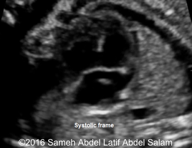
Sonographic Findings of Atrioventricular Septal Defects
Absence of the atrial and ventricular septum
Color Doppler findings are helpful at showing mixture of flow patterns
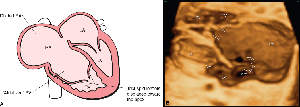
what is Ebstein anomaly
Malformation or malpositioning of the tricuspid valve
The prognosis is poor, with
80% of infants dying in the perinatal period.
Sonographic Findings of Ebstein Anomaly
Malpositioned tricuspid valve
Right and left atrial shunting
Tricuspid regurgitation
Enlarged right atrium
Deviation of the atrial septum to the left
Fetal hydrops (secondary to cardiac failure)
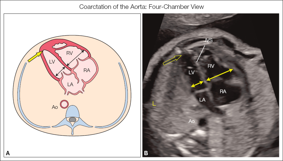
Coarctation of the aorta is the
narrowing of the aortic arch
The most common location of coarctation of aorta is between the
left subclavian artery and the ductus arteriosus
Associated findings with coarctation of aorta consist of
right ventricle enlargement, pulmonary artery enlargement, and disproportion in the size of the ventricles in the four-chamber view
Sonographic Findings of Coarctation of the Aorta
Narrowing of the aortic arch
Right ventricular enlargement
Pulmonary artery enlargement
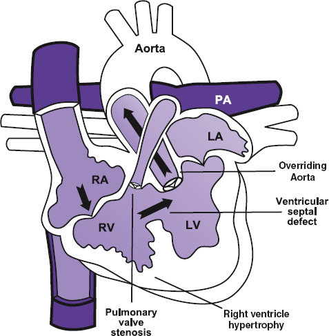
what is Tetralogy of Fallot
an overriding aortic root, subaortic VSD, pulmonary stenosis, and right ventricular hypertrophy (not always noted in utero but manifests after birth)
Sonographic Findings of Tetralogy of Fallot
Overriding aortic root
VSD
Pulmonary stenosis
Right ventricular hypertrophy
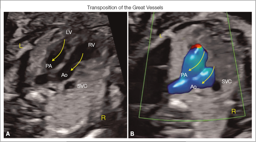
With transposition of the great vessels, the outflow tracts are
reversed
With transposition of the great vessels, the 4 chamber view will appear
normal
Sonographic Findings of Transposition of the Great Vessels
The pulmonary artery abnormally arises from the left ventricle, and the aorta abnormally arises from the right ventricle
The outflow tracts will be positioned parallel to each other rather than crisscrossing
VSD may be present
An echogenic intracardiac focus (EIF) is most often seen within the left ventricle of the heart. This is thought to represent the
calcification of the papillary muscle or chordae tendineae
An EIF may be seen in the normal fetus. However, there have been studies that have linked the incidence of an EIF with
trisomy 21, particularly if there is more than one EIF detected.
Sonographic Findings of an Echogenic Intracardiac Focus
Echogenic structure most commonly located within the left ventricle
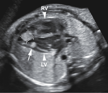
what is a Rhabdomyoma
tumors, located within the myocardium of the heart
Rhabdomyomas are associated with
tuberous sclerosis, eventual cardiac failure, and subsequent development of fetal hydrops
The most common fetal cardiac tumor is the
rhabdomyoma
Sonographic Findings of a Rhabdomyoma
Echogenic tumor(s) within the myocardium of the heart
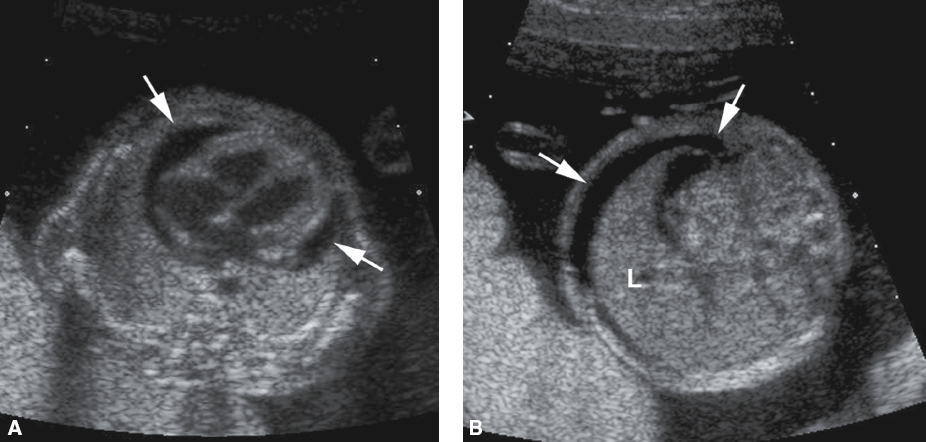
what is Pericardial effusion
fluid located around the heart
Pericardial effusion can be isolated or associated with
fetal hydrops
It is important to note that the normal hypoechoic appearance of the myocardium can mimic the sonographic appearance of small
pericardial effusions.
Sonographic Findings of Pericardial Effusion
Anechoic fluid surrounding the heart
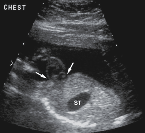
what is ectopic cordis
heart is located either partially or completely outside the chest
what is Pentalogy of Cantrell
group of anomalies that combine ectopic cordis and an existing omphalocele, prognosis is poor
Sonographic Findings of Ectopic Cordis
Heart located either partially or completely outside the chest
what is Pulmonary hypoplasia
underdevelopment of the lungs, is caused by a decreased number of lung cells, airways, and alveoli.
Pulmonary hypoplasia is often associated with
major structural and chromosomal abnormalities
The most common lesion that occupies the chest, resulting in pulmonary hypoplasia, is the
diaphragmatic hernia
Amniotic fluid plays an important role in the development of the fetal lungs; therefore, the fetus, surrounded by little or no amniotic fluid, is at
increased risk of pulmonary hypoplasia
Consequently, pulmonary hypoplasia is a common finding with
oligohydramnios
pulmonary hypoplasia is also associated with
bilateral renal agenesis and the abnormal facial features in the condition known as Potter syndrome.
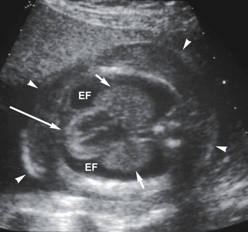
what is pleural effusion
aka hydrothorax; Fluid surrounding the lungs
Pleural effusions that occur in utero may spontaneously resolve or may be found in the presence of
fetal hydrops, other chest abnormalities, and Turner syndrome
Fetal pleural effusions can be treated with an ultrasound-guided
thoracentesis
Sonographic Findings of a Pleural Effusion
Anechoic fluid surrounding the fetal lung(s)—“bat-wing” sign
Other signs of hydrops may be present
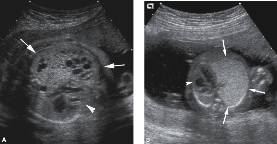
what is Cystic adenomatoid malformation (CAM),
also referred to as congenital cystic adenomatoid malformation (CCAM), is actually a mass consisting of abnormal bronchial and lung tissue.
Sonographic Appearance of Cystic Adenomatoid Malformations
Lung mass with varying degrees of cystic and solid components
Completely echogenic mass within the lungs
Pleural effusion may be present
what is Pulmonary sequestration
or bronchopulmonary sequestration, is a separate mass of nonfunctioning lung tissue with its own blood supply. The fetal form of this disease is specifically referred to as extrapulmonary sequestration
Sonographic Findings of Pulmonary Sequestration
Echogenic, triangular-shaped mass within the fetal chest
Pleural effusion may be present
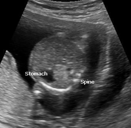
The most common reason for fetal cardiac malposition is the existence of a
diaphragmatic hernia
what is a diaphragmatic hernia
abnormal opening in the fetal diaphragm that allows the herniation of abdominal contents into the chest cavity.
The most common location of a diaphragmatic hernia is on the
left side; may also be referred to as a Bochdalek hernia
The foramen of Bochdalek is located in the
left posterolateral portion of the diaphragm