Lecture 3: Equine Diagnostic Imaging
 Call Kai
 Learn
Learn Practice Test
 Spaced Repetition
 Match
Match Flashcards
 Knowt Play
1/35

There's no tags or description
Looks like no tags are added yet.
Name | Mastery | Learn | Test | Matching | Spaced | Call with Kai | Chat |
|---|
No analytics yet
Send a link to your students to track their progress
36 Terms
indication for radiographs
lameness localized to a specific region
and you suspect a bony abnormality
often first line imaging in equine practice
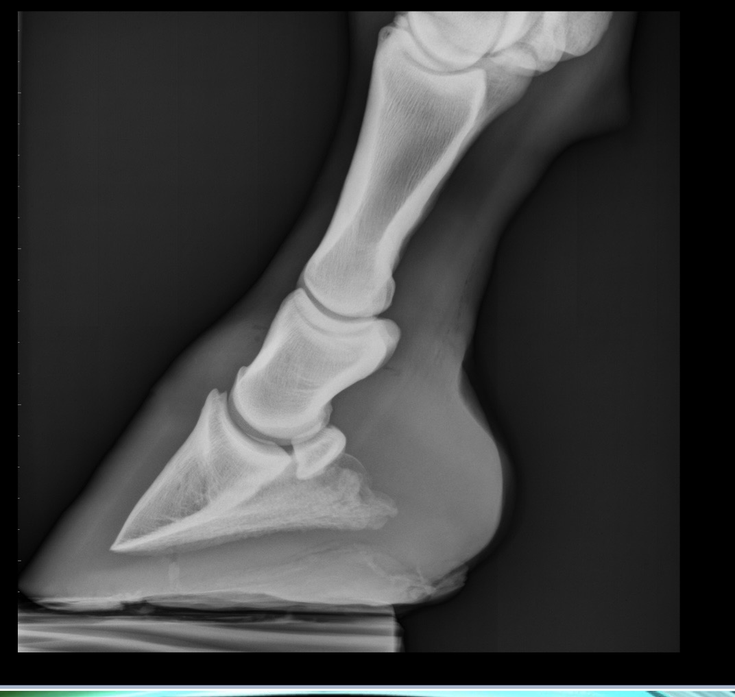
what is the name of this view of a front limb?
lateromedial
radiograph positioning
horse must stand squarely on the limb being radiographed
convention → plate is always medial and almost always palmar/plantar
R vs L markers for radiographs
lateral or dorsal → always lateral when that is an option, if not, then dorsal
label hind vs fore if hock or carpus is not in view
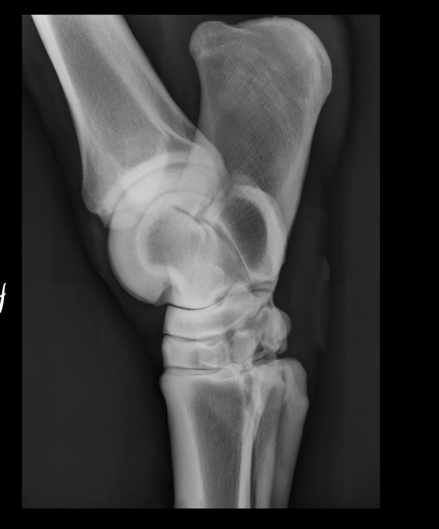
what view is this?
LM → talus/trochlear ridges are perfectly superimposed
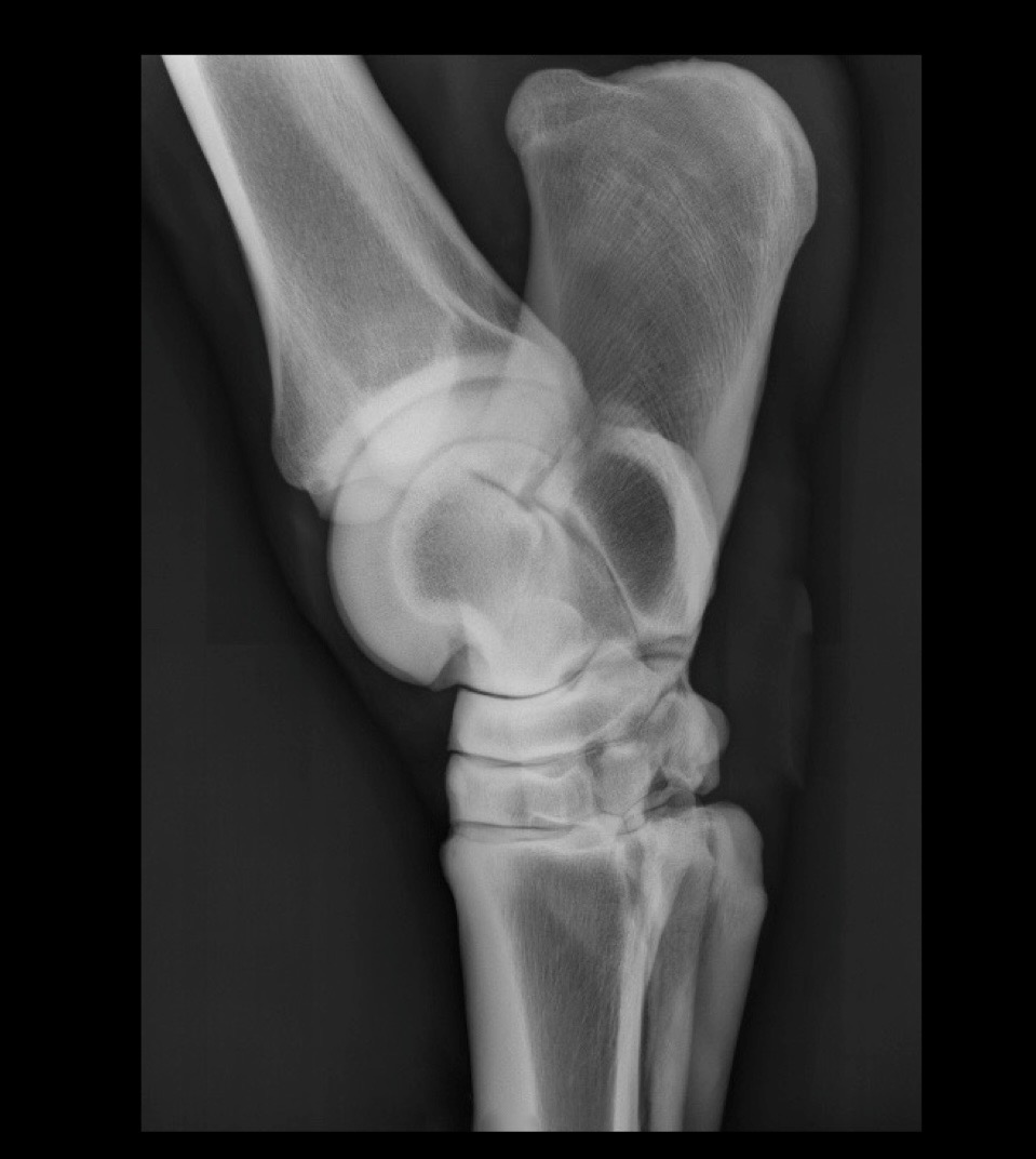
is this image diagnostic quality?
yes
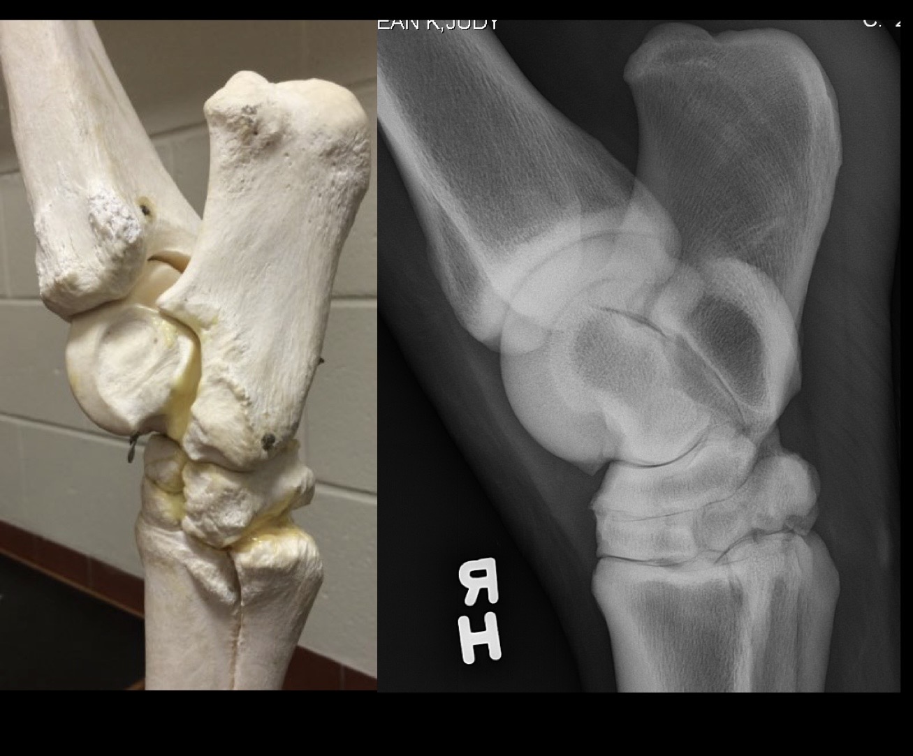
what view is this?
lateromedial (LM)
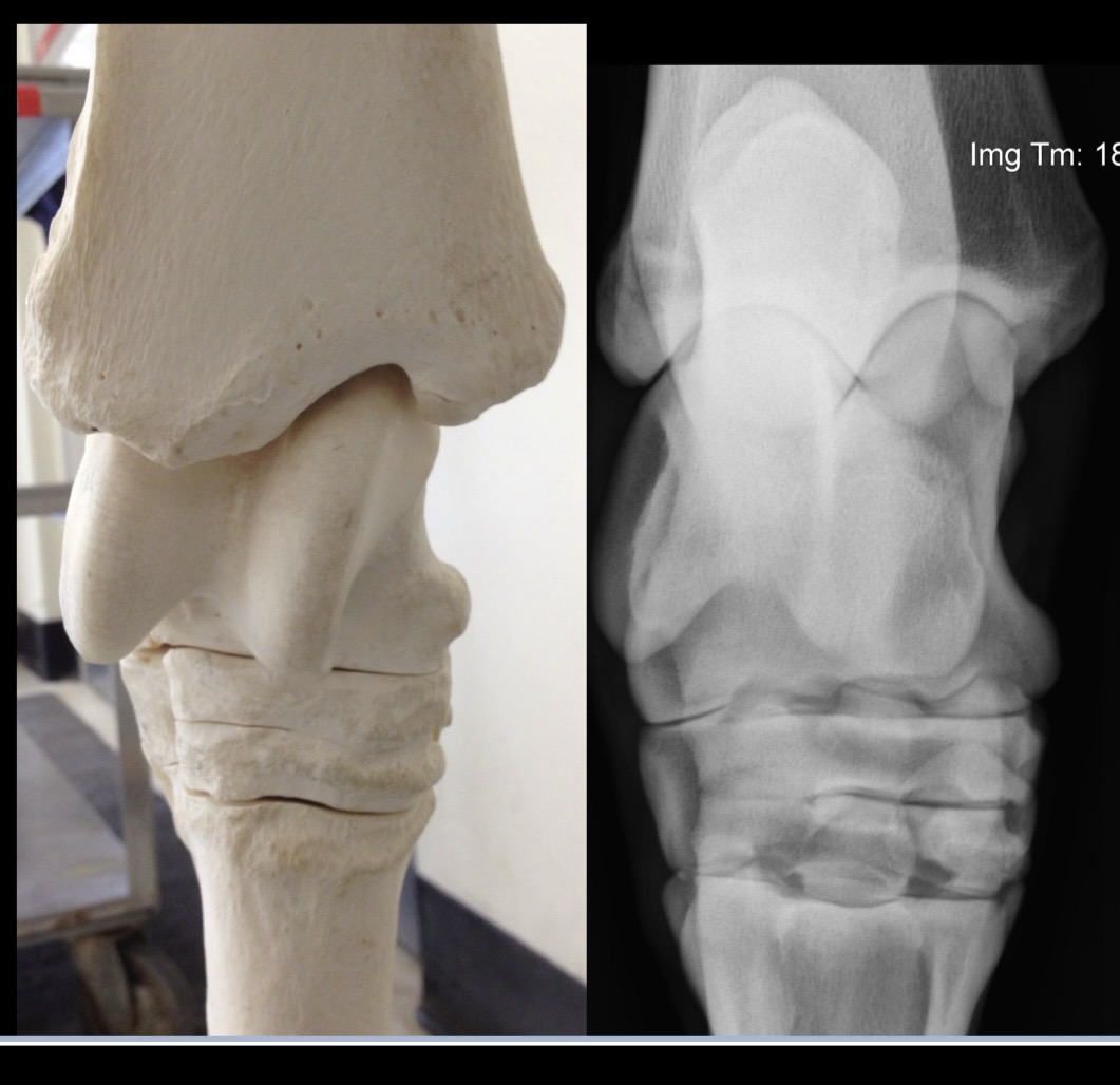
what view is this?
dorsoplantar (DP)
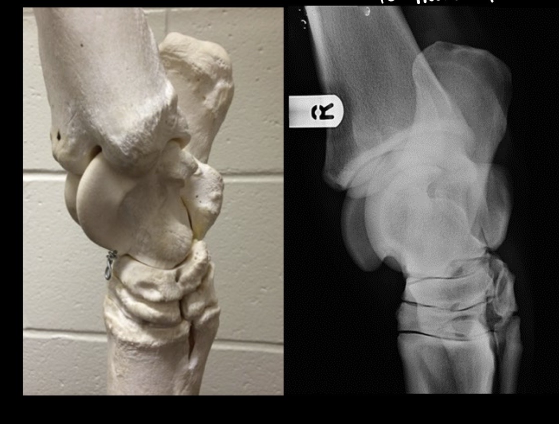
what view is this?
dorsomedial-plantarolateral oblique (DMPLO)
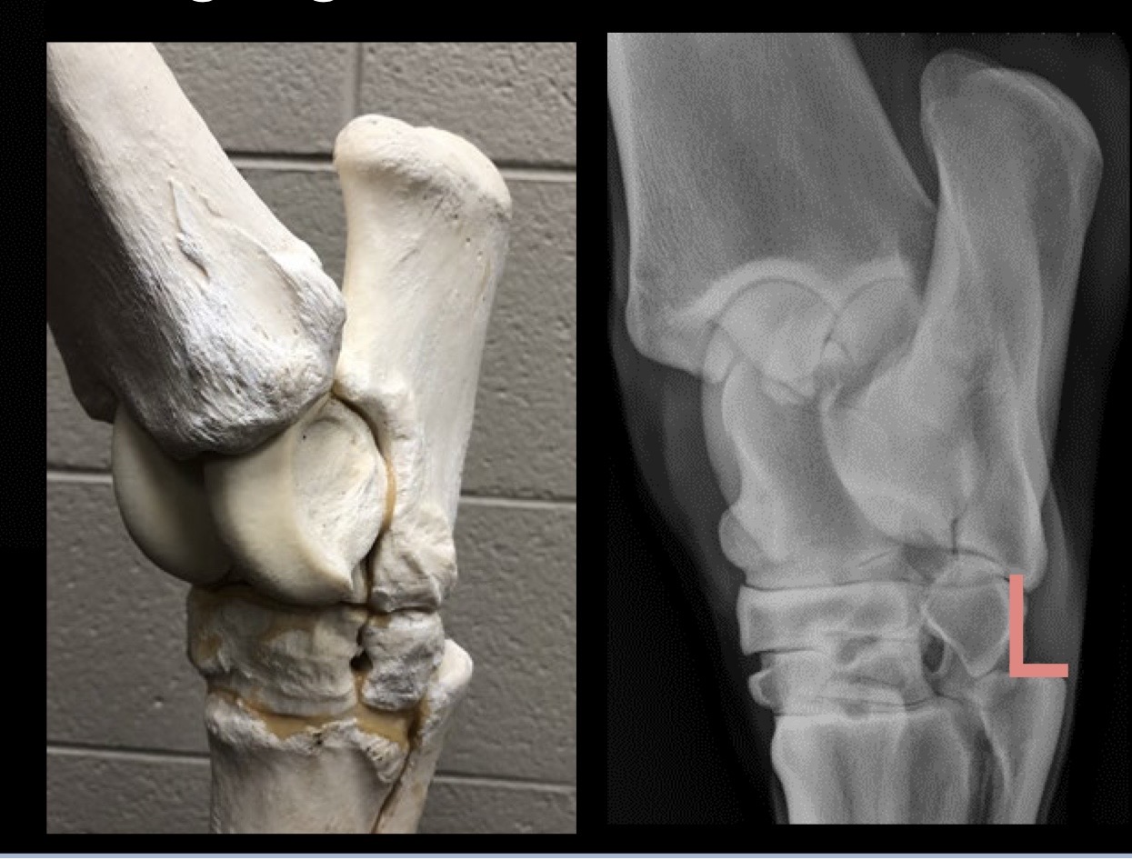
what view is this?
dorsolateral-plantaromedial oblique (DLPMO)
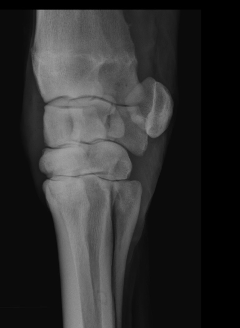
what view is this?
DLPMO
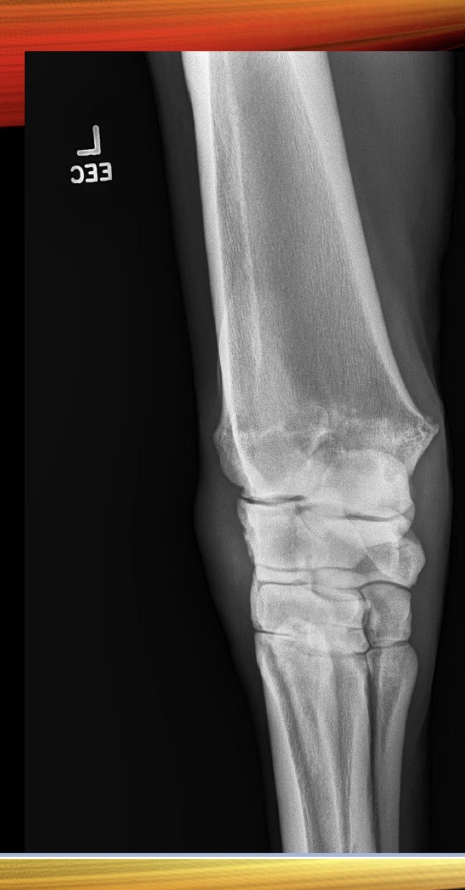
what view is this?
DMPLO → can’t see accessory carpal
what if no radiologic abnormalities, but suspect bone injury?
wait 10 days → callus formation, resorption at fracture line
additional imaging
what imaging is not done under GA if suspect fracture?
MRI, CT
ultrasonography principles
high-frequency sound waves penetrate tissue and bounce back to transducer
crystals in transducer convert sound waves to electric current
computer in ultrasound machine converts electric current to image
ultrasonography indications
image soft tissue lesions → tendons, ligaments, joints
image surface of bone
ultrasonography of the equine limb
must clip hair
remove grease with alcohol
lots of gel
8-15 mHz transducer
standoff provides better detail in areas with less soft tissue
with increasing mHz of transducer, how does the image change?
decrease depth and increase resolution
well-defined areas for equine distal limb are measured in
zones or distance from accessory carpal bone or point of hock
obvious increase in hypoechogenicity indicates
core lesion
fluid = edema = inflammation
increased size of structure in ultrasound
diffuse lesion
correlated with increased fluid
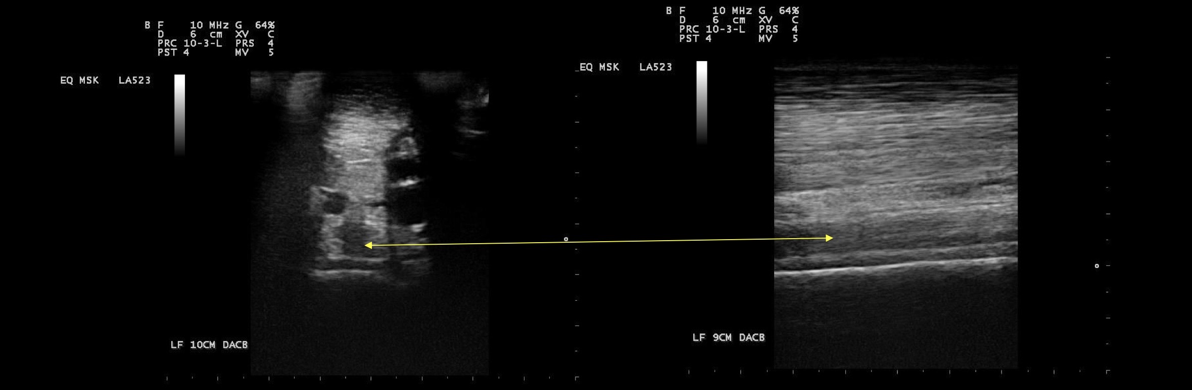
what is this?
fiber alignment and echogenicity on longitudinal view
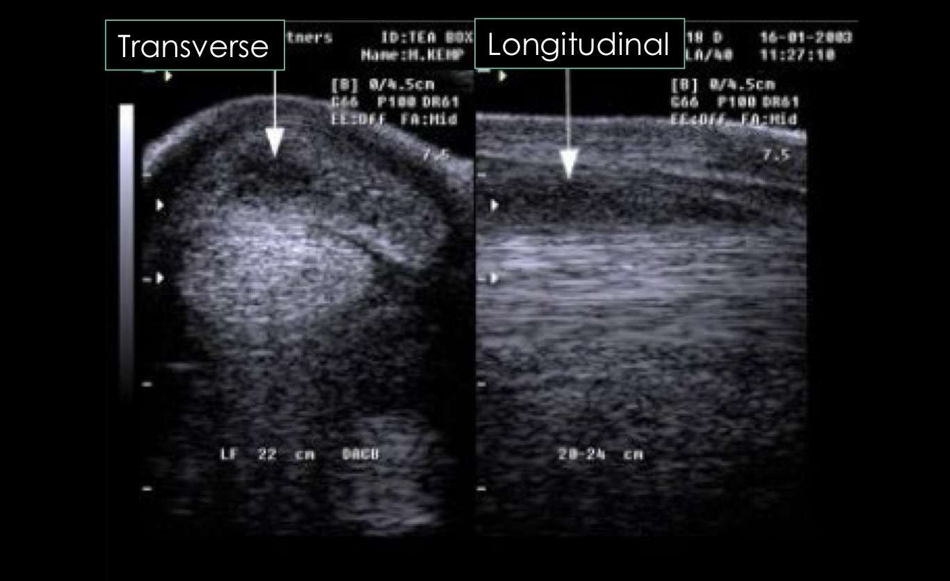
what is this showing?
lesions in 2 different views
indications for nuclear scintigraphy
fail to localize lameness with blocks
localize lameness but no lesions on radiographs or ultrasound
multiple limb lameness
upper limb or axial musculoskeletal issue
suspect fracture not imaged on radiographs
principles of nuclear scintigraphy
inject a drug that is bound to rapidly decaying radioactive atom
drug has propensity for certain tissue → methylene diphosphonate - hydroxyapatite in bone
gamma camera detects decay of radioactive atom
increased bone activity → increased uptake → increased atoms decaying → increased signal
what is the most common radioactive atom used in nuclear scintigraphy?
technetium-99m
hydroxyapatite
formed by osteoblasts in areas of active bone formation
proportional to bone resorption
indications for computed tomography
detailed evaluation of bone → limited soft tissue detail so requires injection of contrast agent
fracture repair planning
image navicular apparatus
image the head
principles of CT
x-ray tube in circle
x-rays detected
intensity of x-ray allows for differentiation of structures
computer reconstructs the data acquired from detectors to make a slice image
software programs can reconstruct slices into a 3D image
application of CT
horse is anesthetized
imaging limited to → carpus/tarsus and distal limb, head
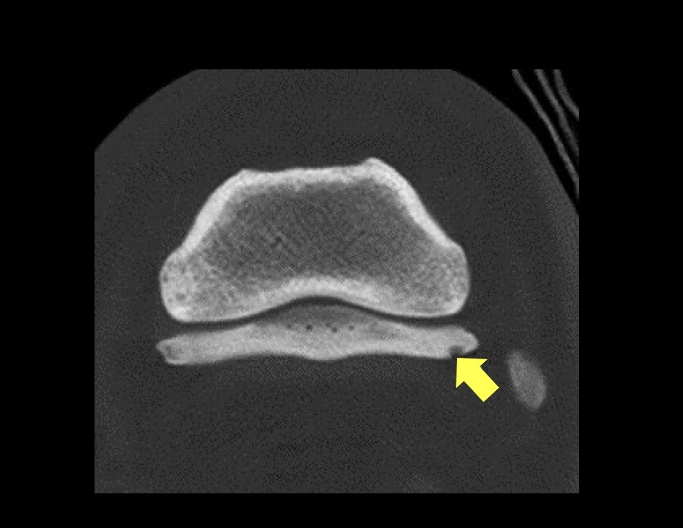
what is this?
lateral collateral ligament insertional lesion in the navicular bone
MRI indications
imaging soft tissue and bone lesions → areas where ultrasound is not possible = the foot
similar limitations in structures that may be imaged as CT
MRI principles
all tissues have lots of hydrogen protons because they are made of H2O
magnet is applied and protons are excited by a radiofrequency pulse
pulse removed → protons relax → emits signal
protons in different tissues relax differently → superior contrast in tissues
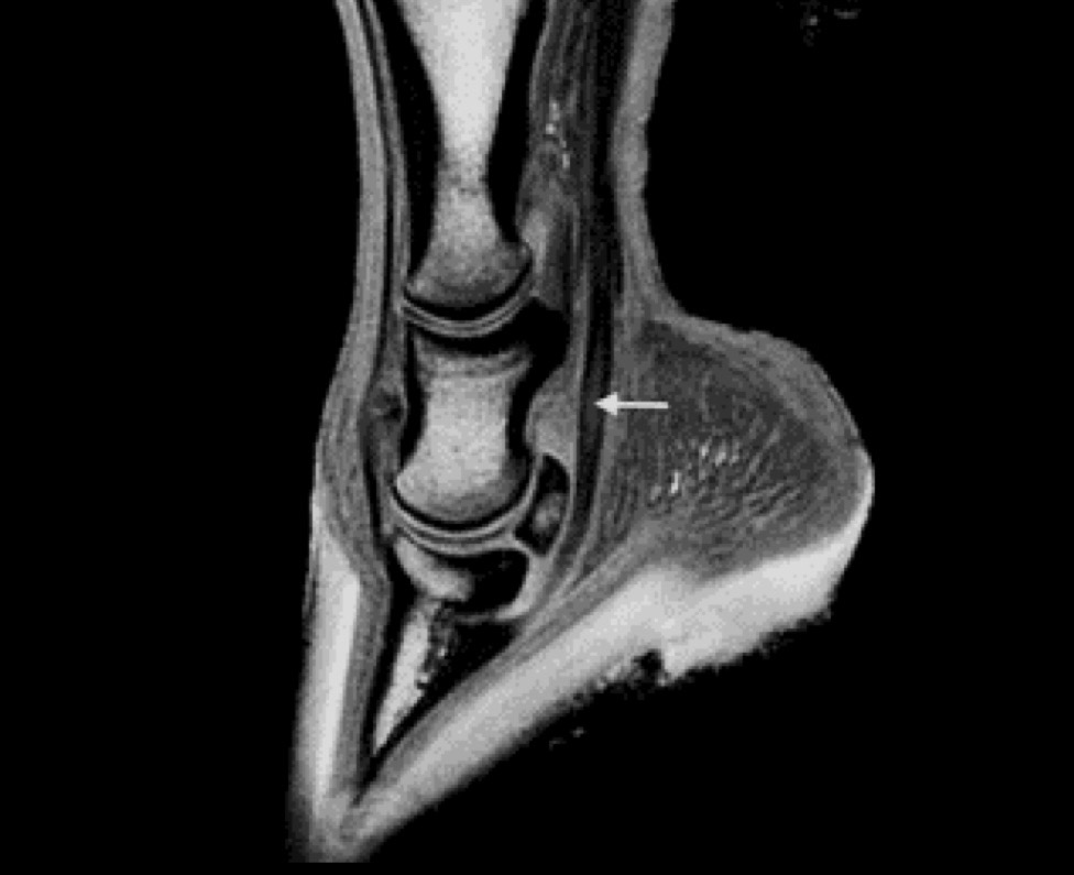
what is this showing?
core lesion in the DDF tendon → desmitis
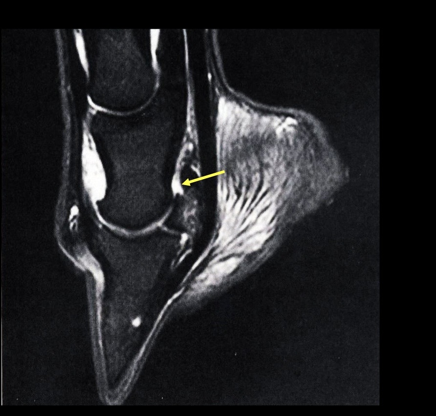
what is the arrow pointing to?
navicular suspensory ligament
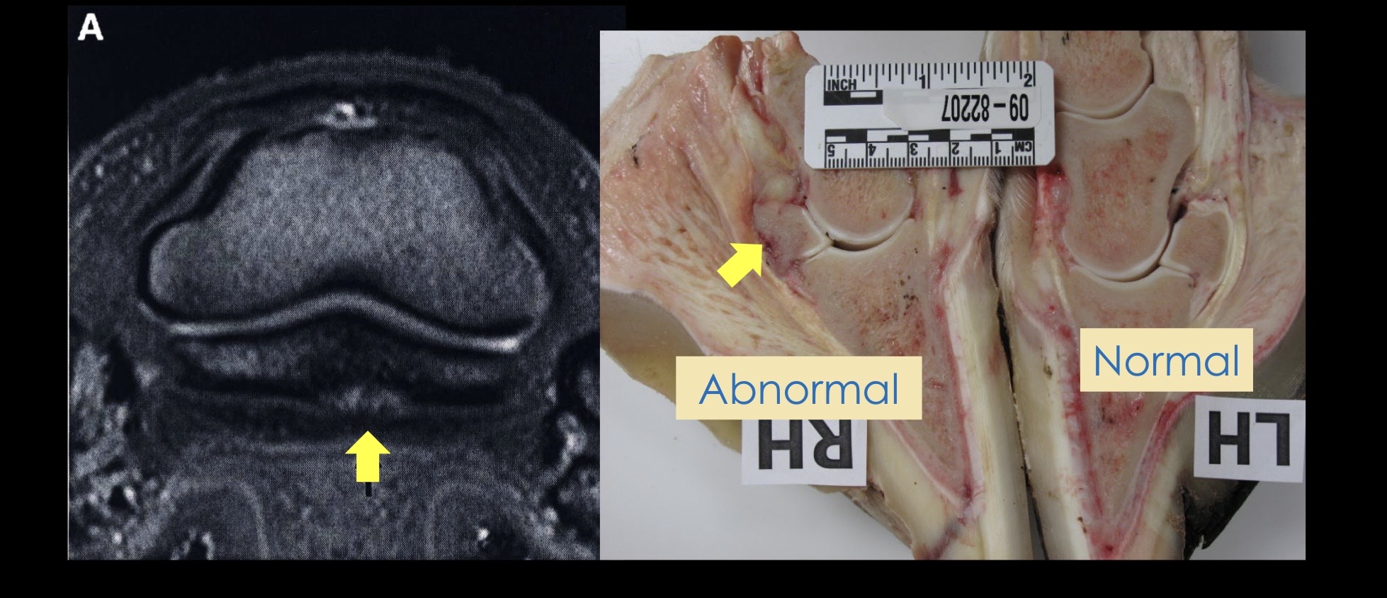
what is this?
adhesion between the navicular bone and the DDFT