Functional Anatomy 1 - Lecture 15: Ankle and Foot
 Call Kai
 Learn
Learn Practice Test
 Spaced Repetition
 Match
Match Flashcards
 Knowt Play
1/93

There's no tags or description
Looks like no tags are added yet.
Name | Mastery | Learn | Test | Matching | Spaced | Call with Kai | Chat |
|---|
No analytics yet
Send a link to your students to track their progress
94 Terms
tarsal bones (tarsus)
talus, calcaneus, navicular, cuneiforms, cuboid
talus
articulates with the tibia and fibula superiorly
articulates with the calcaneus inferiorly
articulates with navicular bone anteriorly
talocrural joint
articulates with the tibia and fibula superiorly
most superior
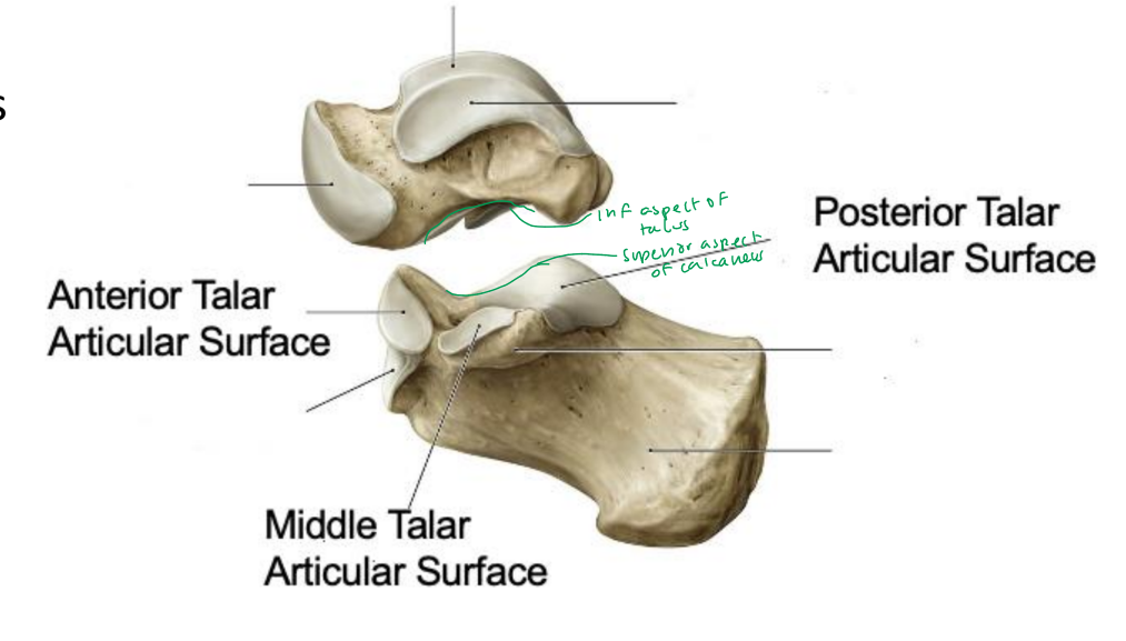
subtalar joint
articulates with the calcaneus inferiorly
talonavicular joint
articulates with navicular bone anteriorly
calcaneus
inferior and posterior to talus
articulates with talus superiorly
articulates with cuboid anteriorly
insertion for Achilles tendon
subtalar joint
articulates with talus superiorly
calcaneocuboid joint
articulates with cuboid anteriorly
navicular bone
medial, anterior to talus
tuberosity of navicular
cuneiforms
medial, intermediate, lateral
anterior to navicular
cuboid
lateral
anterior to calcaneus
metatarsals
1-5
base (proximal)
shaft
head (distal)
phalanges
digit 1: proximal and distal
digits 2-5: proximal, middle and distal
rearfoot
talus and calcaneus
midfoot
navicular, cuboid, cuneiforms (3)
forefoot
MTs 1-5, phalanges 1-5
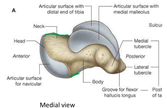
talus
head: articulates with navicular
neck: projects medially and anteriorly
body
wider anterior (medial/lateral talar domes)
narrower posteriorly
groove for flexor hallucis longus (FHL) tendon
no muscle attachments
essential in transmitting motion through rearfoot/ankle to LE
stability via shape of body in talocrural joint
calcaneal tuberosity
attachment for Achilles tendon
weight-bearing surface
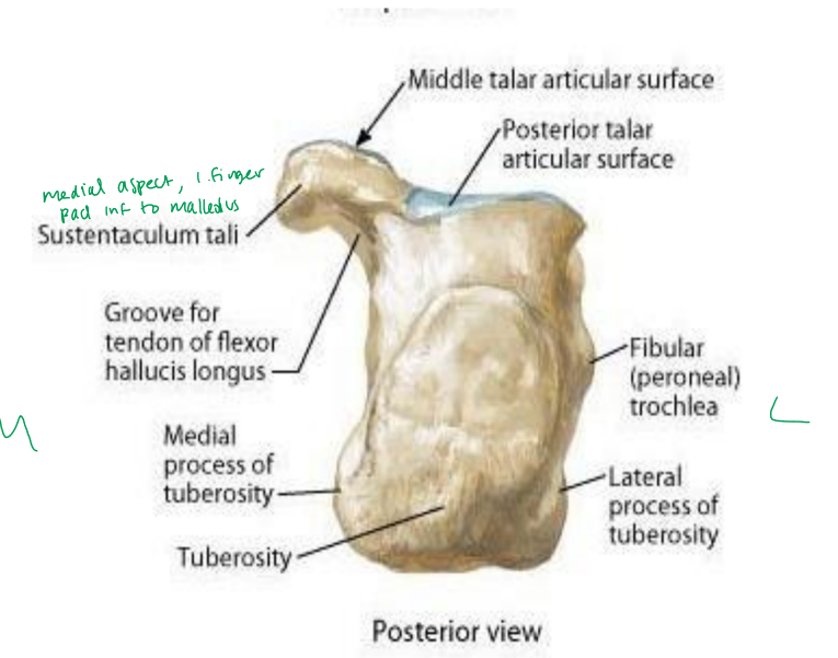
sustentaculum tali
groove for FHL tendon inferior
fibular (peroneal) trochlea
peroneus brevis superior
peroneus longus inferior
lateral process of calcaneus
origin for abductor digiti minimi
medial process of calcaneus
origin for plantar fascia and abductor hullucis
navicular
posterior articulation: talus
anterior articulation: cuneiforms (all 3)
lateral articulation: cuboid
navicular tuberosity
attachment for tibialis posterior
keystone of medial longitudinal arch
cuboid
posterior articulation: calcaneus
medial articulations
navicular (posteriorly)
lateral cuneiform (anteriorly)
anterior articulation: base of MT 4 and 5
medial cuneiform
anteriorly articulates with base MT 1
intermediate cuneiform
anteriorly articulates with base with MT 2
lateral cuneiform
anteriorly articulates with base with MT 3
laterally articulates with cuboid
sesamoid bones
protect FHL tendon in weight-bearing positions
medial and lateral
metatarsals
5 metatarsals
base (proximal), shaft, head (distal)
head proximal side of metatarsophalangeal joint
tuberosity of 5th MT
attachments for peroneus brevis
at lateral base of 5th mt
jones fracture
fracture at the base of 5th metatarsal
near the tuberosity
not very well vascularized, will take longer to heal
1st digit
proximal phalanx
interphalangeal joint
distal phalanx
digits 2-5
proximal phalanx
proximal interphalangeal joint
middle phalanx
distal interphalangeal joint
distal phalanx
ankle motions
eversion/inversion
dorsiflexion/plantarflexion
adduction/abduction
talocrural joint
medial and lateral malleolus grip talus firmly (ankle mortise)
requires distal tibiofibular joint stability
talocrural joint motion
primarily ankle dorsiflexion and plantarflexion
axis is slightly anterior/superior on medial side and posterior/inferior on lateral side
location of medial malleolus and lateral malleolus
subtalar joint
posterior articulation of the talus and calcaneus (primarily)
subtalar joint motion
inversion and eversion (of the subtalar joint)
subtalar joint motion in closed kinetic chain
stj inversion transmits to tibial ER
stj eversion transmits to tibial IR
transverse tarsal joint
aka midtarsal joint
includes talonavicular and calcaneocuboid joints
transverse tarsal joint functions
allows for flexibility of the forefoot based on the position of the rearfoot
two joint axes working together
transverse tarsal joint motion
tri-planar, allows movement in all 3 planes
transverse tarsal joint motion, open chain
pronation: dorsiflexion, eversion, abduction
supination: plantarflexion, inversion, adduction
transverse tarsal joint motion, closed chain
pronation: more flexible foot
supination: more rigid foot
tarsometatarsal joints
medial cuneiform with 1st metatarsal
intermediate cuneiform with 2nd metatarsal
lateral cuneiform with 3rd metatarsal
cuboid with 4th and 5th metatarsal
all plane joints, limited motion in all!
metatarsophalangeal joints
MT 1-5 with respective proximal phalanx 1-5
synovial, condyloid joints
metatarsophalangeal joint motion
flexion/extension, abduction/adduction, 1st MTP extension plays significant role during gait
interphalangeal joints
1st interphalangeal joint
2-5: DIP and PIP
synovial, hinge joints
interphalangeal joint motion
flexion/extension
medial longitudinal arch
calcaneus, talus, navicular, 3 cuneiforms, medial 3 metatarsal bones
arches of the foot
medial longitudinal, lateral longitudinal, transverse
lateral longitudinal arch
calcaneus, cuboid, lateral 2 metatarsal bones
transverse arch
tarsometatarsal joints
pes cavus
increased medial longitudinal arch height, higher arch
pes planus
decreased medial longitudinal arch height, lack of arch
lateral collateral ligaments
anterior talofibular ligament, calcaneofibular ligament, posterior talofibular ligament
anterior talofibular ligament (ATFL)
flat, weaker band
lateral malleolus to neck of talus
calcaneofibular ligament (CFL)
round, cord-like
lateral malleolus to lateral calcaneus
posterior talogibular ligament (PTFL)
thick, strong band
malleolar fossa of fibula to lateral tubercle of talus
inversion
ligaments in lateral rearfoot resist ________ of the rearfoot
plantarflexed
ATFL resists inversion more in _____________ positions
neutral/dorsiflexed
CFL resists inversion more in _____/________ positions
dorsiflexed
PTFL resists inversion more in ______________positions
lateral ankle sprain
inversion ankle sprain
anterior drawer test for the ankle
ligaments in medial rearfoot
medial collateral ligaments
anterior tibiotalar
tibionavicular
tibiocalcaneal
posterior tibiotalar
anterior tibiotalar
medial malleolus to anterior talus
tibionavicular
medial malleolus to navicular
tibiocalcaneal
medial malleolus to medial calcaneus
posterior tibiotalar
medial malleolus to posterior talus
medial collateral ligaments function
all resist eversion
anterior resist more in plantarflexed position
posterior resist more in dorsiflexed position
plantar ligaments
plantar fascia/aponeurosis
plantar fascia/aponeurosis
thick, superficial fascia
calcaneus to heads of metatarsals 1-5
superficial to muscles
plantar fascia/aponeurosis function
supports plantar foot and medial longitudinal arch
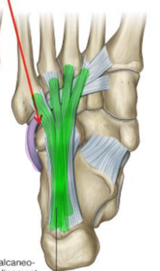
long plantar ligament
calcaneal tuberosity to base of MTs 2-5
strong ligament, supports the lateral longitudinal arch
helps protect peroneus longus tendon
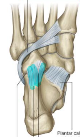
short plantar ligament
AKA calcaneocuboid ligament
calcaneal tubercle to cuboid
plantar ligaments
long plantar, short plantar, spring
very deep, remove 4 layers of muscle
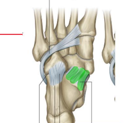
spring ligament
AKA plantar calcaneonavicular ligament
broad, thick ligament
sustentaculum tali to plantar surface of navicular
connection from calcaneus to navicular
function
helps maintain medial longitudinal arch
dorsal ligaments
talonavicular, bifurcate
talonavicular ligament
superior/anterior talus to superior navicular
connects bones and prevents anterior/inferior displacement of navicular
bifurcate ligament
Y-shaped ligament
superior/lateral calcaneus to cuboid and navicular
collateral ligaments
MTPs and IPs
resists abduction/adduction
plantar ligament (plate)
resists hyperextension
turf toe: first MTP injured (excessive hyperextension)
deep transverse ligament
between MT heads horizontally
resists separation of MTs
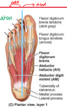
1st layer of intrinsic foot muscles
abductor hallucis
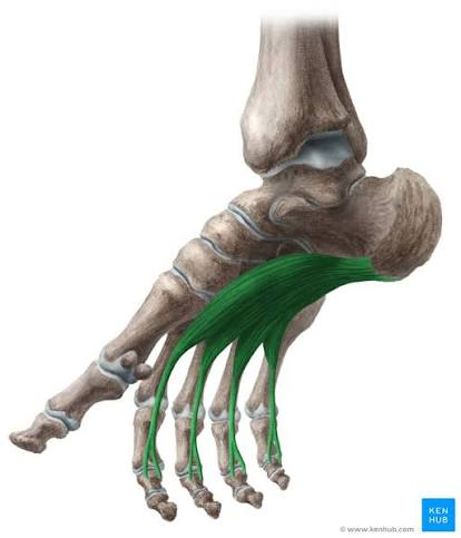
flexor digitorum brevis
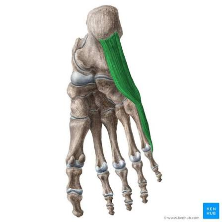
abductor digiti minimi
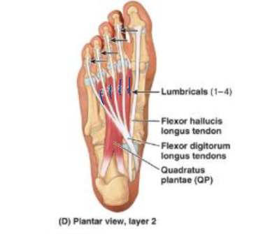
2nd layer of intrinsic foot muscles
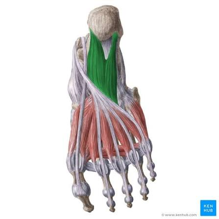
quadratus plantae
tendon for FDL
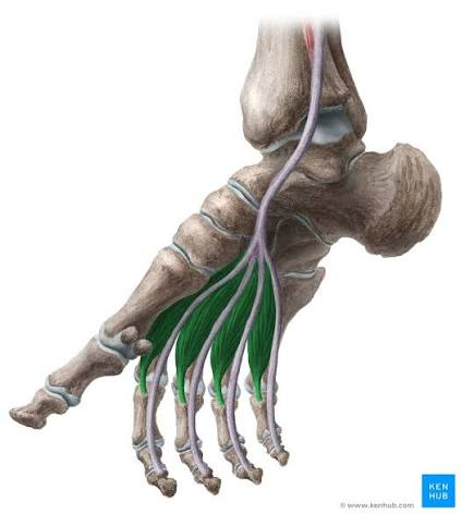
lumbricals (1-4)
tendon for FHL
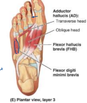
3rd layer of intrinsic foot muscles
flexor hallucis brevis (FHB)
adductor hallucis (transverse and oblique heads)
flexor digiti minimi brevis
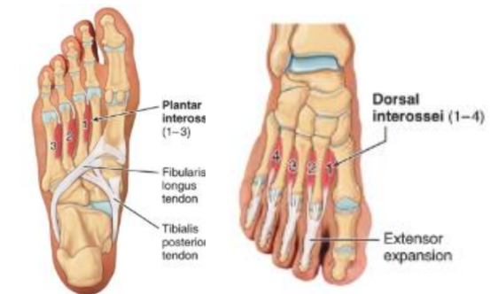
4th layer of intrinsic foot muscles
plantar interossei
dorsal interossei
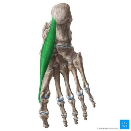
abductor hallucis
O: medial tubercle calcaneal tuberosity
I: medial side of base of 1st proximal phalanx
A: abducts 1st digit at MTP
N: medial plantar n (L5-S1)
B: medial plantar a.
flexor digitorum brevis
O: medial tubercle calcaneal tuberosity
I: both sides middle phalanges of digits 2-5
split allows for tendon FDL to pass through and distal
A: flex digits 2-5 at MTP and PIP
N: medial plantar n. (L5-S1)
B: medial and lateral plantar a.
abductor digiti minimi
O: medial and lateral tubercles calcaneal tuberosity
I: lateral side of base of 5th proximal phalanx
A: abducts 5th digit at MTP
N: lateral plantar n. (S1-S3)
B: lateral plantar a.
quadratus plantae
O: medial and lateral plantar calcaneus
I: lateral side of FDL tendon
A: assist FDL in flexion of digits 2-5 at DIP, PIP, and MTP
adjusts the line of pull for FDL
N: lateral plantar n. (S1-S3)
B: lateral plantar a.
lumbricals
O: tendon of FDL
I: extensor hood/expansion digits 2-5
A: flexion MTP digits 2-5, extension PIP and DIP digits 2-5
N:
medial 1: medial plantar n (L5-S1)
lateral 3: lateral plantar n. (S1-S3)
B: medial and lateral plantar a.