test 5 skin integrity and nutrition
 Call Kai
 Learn
Learn Practice Test
 Spaced Repetition
 Match
Match Flashcards
 Knowt Play
1/89

There's no tags or description
Looks like no tags are added yet.
Name | Mastery | Learn | Test | Matching | Spaced | Call with Kai | Chat |
|---|
No analytics yet
Send a link to your students to track their progress
90 Terms
dermatitis
inflammation of the skin
familial/hereditary
not contagious
is dermatitis an infection
no it is not an infection however it can become infected
atopic dermatitis
most common chronic, usually start at young age
tx. cortisone creams and light therapy
what do steroid creams do to the skin overtime
stain, thin, interrupt inflammatory process. can cause paperlike skin.
tx is usually 14 days
coricosteroid creams and where you use them
triamicinolone - face
clobetasol - body
h2 blockers
pepcid (famotidine)
can be used off-label in combination with H1 antihistamines (like Benadryl or Zyrtec) to treat allergic reactions.
It works by blocking H2 receptors on skin vessels, which can enhance relief from itching and hives
psoriasis
an autoimmune disorder
mostly plaque
usually effects trunk, elbows, knees and scalp
is a buildup of skin cells, the body attacking itself producing more rather than shedding
inflammatory response 3 parts
sequential
intensity of response
inflammatory response
sequential response to cell injury in inflammation process
neutralizes (destroying agent making it harmless) and dilutes (adds fluid to harmful agent to make it weaker) inflammatory agent
-removes necrotic material
-establishes environment for healing
"first we surround it, then we clean it."
intensity of inflammatory response (what it depends on)
-extent and severity of injury
-reactive capacity of injured person (the body's overall ability to initiate, manage, and resolve an inflammatory response following tissue damage)
what processes can the inflammatory response be divided into
-vascular response
-cellular response
-formation of exudate
-healing
vascular response to cell injury
1. injury occurs
2. brief vasoconstriction (to reduce blood loss)
3. chemical mediators are released (inflammatory chemicals- histamine, kinins, prostaglandins)
4. vasodilation (increase blood flow-hyperemia) causes redness and heat
5. capillary permeability increases (leaky) (allows fluid, proteins, immune cells to move out of bloodstream and into injured tissue)
6. local edema due to fluid leaking out of capillaries causing swelling
7. inflammatory exudate forms (fluid, WBCs, proteins- helps fight infection and start healing
Cellular response to cell injury
1. chemotaxis- WBCs chemically pulled to injury site
2. margination and diapedesis of leukocytes (WBCs move out of bloodstream) margination means move to edge of blood vessel wall, diapedesis means the WBCs squeeze through capillary wall into injured tissue. (essentially: blood -> vessel wall -> tissue)
3. migration of leuks to site of injury through chemotaxis signals
- neutrophils (first responders- arrive in mins to hrs, engulf bacteria (phagocytosis)
- monocytes- arrive next and transform into macrophages (macrophages live longer (long life span and can multiply) than neutrophils, continue phagocytosis, clean up dead cells and pathogens and help start healing)
- lymphocytes- immune response (recognize specific pathogens, coordinate adaptive immunity (identifies and destroys particular pathogens, creating long-lasting immunological memory), produce antibodies. arrive later; primary roles are cell mediated immunity, humoral immunity and chemical mediators like histamine, serotonin, and kinin
5. phagocytosis- neutrophils and macrophages engulfing and digesting pathogens and debris.
6. inflammatory exudate- produces pus in infected wounds, fights infection starts healing
pus components
dead neutrophils accumulated at site of injury, digested bacteria, other cell debris
what releases more neutrophils in response to infection, resulting in elevated WBC
bone marrow
complement system of cellular response
major mediator of inflammatory response, they destroy invading pathogens
- enhanced phagocytosis, increased vascular permeability (allowance of nutrients to move between bloodstream and tissues), chemotaxis (attracts and repels cells), cellular lysis (cell death)
final components of system create holes in cell membrane, causing targeted cell death by membrane rupture
pathway of generation
1st cell membrane injury, signals arachidonic acid (signals molecules that regulate inflammation, pain, and immunity). steroids inhibits this.
cyclooxygenase pathway = prostaglandins (pain fever inflammation) and thromboxane (clotting) - NSAIDS & Aspirin block these.
lipoxygenase pathway is leukotrienes (asthma, allergies)
steroids inhibits these pathways and weaken ability to fight things off
what meds inhibit the various steps in the pathway of generation
corticosteroids, nonsteroidal antiinflammatory drugs and acetylsalicylic acid (ASA)
left shift of inflammation
increased immature neutrophils, decreased grown neutrophils. because the body is trying to regenerate what's lost.
usually an indication of acute, active inflammation
acute inflammation
2-3 weeks, usually no residual damage.
neutrophils predominant cell type at site of inflammation
subacute inflammation
Same features as acute inflammation but persists longer (weeks or months)
chronic inflammation
-may last for years.
-injurious agent persists or repeats injury to site
-predominant cell types; lymphocytes and macrophages
-may result from immune system changes like an autoimmune disease
RICE
rest
ice
compression
elevate
primary intention of wound healing
wounds have little tissue loss like a surgical incision
edges can be approximated
low risk of infection and minimal scarring
secondary intention of wound healing
-occurs from trauma, burns, ulceration, infection
-large amounts of exudate, wide irregular wound margins with extensive tissue loss
-edges cannot be approximated
-high risk of infection, scarring and poss loss of tissue function
tertiary intention wound healing
when a wound is allowed to remain open for a time and then closed, allowing for debridement and other wound care - to reduce bacterial counts prior to closure
stages of wound healin
hemostasis (stop bleeding, initiating repair)
inflammation,
proliferation (repairing damaged tissue, preparing to close),
remodeling (tissue integrity restored)
what are two things wounds need to heal
oxygen and good blood flow
shearing
when skin sticks to a surface while muscles slide in the direction the body is moving
or bones in one direction, tissue in another
high fowlers is highest risk
blunt force wound
body hits a surface or is struck by a dull object, such as in assaults, falls, or car accidents
friction wound
caused when the skin is rubbed against another object
dehiscence
Bursting open of a wound, especially a surgical abdominal wound
evisceration
opening of wound, organs outside body cavity.
cover in moist gauze and call for help
hypertrophic scarring
too much collagen causes raised scarring, develop within the original boundary of a skin injury
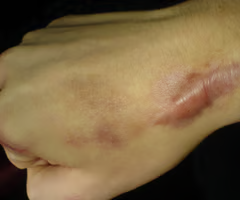
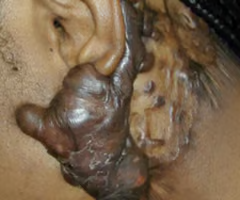
keloid scar
firm, raised, and often itchy skin growths that extend beyond the boundary of an original injury, caused by excessive collagen production during healing
wound class;
superficial
partial thickness
full thickness
superficial- affects only the topmost layer of skin (epidermis) and sometimes the upper dermis, typically caused by scrapes, minor cuts, or light burns
partial thickness- involves damage to the epidermis and superficial dermis, appearing as a shallow, red/pink, and moist wound. Common causes include abrasions, skin tears, and second-degree burns
full thickness- extends through the epidermis and dermis into the subcutaneous tissue, often exposing adipose tissue, muscle, tendon, or bone
how do you measure a wound
treat like a clock.
head to toe, right to left.
measure in CM
fistula
an abnormal passage, usually between two internal organs or leading from an organ to the surface of the body
(ex. tunnel from buttocks to vagina)
What is undermining?
wound has more involvement under the skin than we can see on the surface
if you can't measure dead tissue, what do you do?
say where it is (ex, on clock measurement, at 3pm)
hyperbaric oxygen therapy for wounds
accelerates the healing of chronic, non-healing wounds—such as diabetic foot ulcers, radiation injuries, and, crush injuries—by having patients breathe 100% oxygen in a pressurized chamber. This increases blood oxygen levels, enhancing the body's natural healing, reducing swelling, and fighting infections
negative pressure wound therapy
activity that promotes wound healing and wound closure through the application of uniform negative pressure on the wound bed, reduction in bacteria in the wound, and the removal of excess wound fluid.
(black open pore polyurethane foam applied directly to wound, dressing applied and wound vac applied to wound, sucks in to srhink and pull wound together)
nursing implementation of wounds;
drug therapy
becaplermin (regranex) prescription topical medication used to treat diabetic foot and leg ulcers by stimulating healing
nursing implementation wounds;
nutritional therapy
high in proteins, carbs, vitamins, with moderate fat
vitamins needed include c, b complex and a
infection prevention nursing implementation of wound
don't touch recent injured area
keep free from contaminated items
antibiotics given prophylactically
infection control
nursing implementation of wound
culture
concurrent swab specimens obtained using levine's technique (by rotating a swab over a area of viable tissue for 5 seconds with sufficient pressure to express tissue fluid)
psychological implications of nursing implementation; wounds
fear of scar or disfigurement
drainage or odor concerns
be aware of facial expressions w dressing changes
PAD:
A- absence of pulse/hair
R - red round sores
T - toes and feet pale or black
S - sharp calf pain (intermittent claudication)
TX PAD
antiplatelets, walking exercise, bypass/stents
DO; inspect feet daily, encourage walking, hang legs
avoid cold exposure, no tight clothing
PVD
V - VOLUPTUOUS pulses
E -edema
I - irregular shaped sores (pools)
N - no sharp pain
Y -yellow and brown ankles
TX PVD
anticoagulants
compression stockings
elevate legs
hydrate
DO NOT massage if DVT is suspected
avoid long immobility
ulcer location of PAD and PVD
PAD; toes, heels, pressure points (dry, round, punched out)
PVD; ankles, lower legs (wet, irregular edges)
FRUITS OF WOUNDS
stages
stage 1; tomato (can push on it, doesn't turn white)
stage 2; potato (1st layer skin gone, skin broken)
stage 3; apple (1st layer and some tissue gone)
stage 4; peach (deep, down to bone)
unstageable; down through tissue, can't tell about skin. icky. deep but not how deep. under dead tissue.
deep tissue injury; eggplant; skin intact, deep purple
pyrosis
heartburn
indicators of hemorrhagic gastritis
hematemesis, tachycardia, hypotension
electrolyte imbalances and hypoglycemia
acute; several hours to few days, caused from NSAIDS, aspirin, corticosteroids, alcohol, radiation therapy
chronic; repeated exposure to irritating agents/recurring episodes of acute gastritis. caused by H pylori, autoimmune disorders, long term drug therapy, duodenal content reflux
what vitamins can chronic gastritis interfere with and what causes it
vitamin b12, diminished instrinsic factor production (reduced secretion of a protein by stomach parietal cells necessary for Vitamin absorption in the small intestine) leads to pernicious anemia
DX for gastritis
endoscopy, biopsy
CBC for anemia
H pylori tests
H pylori TX
proton pump inhibitor, abx, bismuth salts sometimes
what electrolyte should you monitor for gastric disorders
sodium
potassium
chloride
every 24 hours
peptic ulcers and what causes them
gastric, duodenal, esophageal erosion of mucosa due to HCL and pepsin, h pylori infection, NSAID use
zollinger-Ellison syndrome
hypersecretion of gastrin, leading to severe peptic ulcers
associated with MEN1 syndrome
symptoms of peptic ulcer
dul gnawing pain or burning sensation in mid epigastrium (upper middle abdomen) or back
pyrosis, vomiting, constipation or diarrhea, bleeding
hematemesis or melena if bleeding ulcer
difference between gastric and duodenal
gastric- immediate after food
duodenal 23 hr after means (most common)
meds of peptic ulcers
abx combination, proton pump inhibitors (-prazole), bismuth salts (pepto bismol), H2 blockers
how does smoking increase peptic ulcer risk
decreases bicarbonate secretion increasing duodenal activity
symptoms of hemorrhage, perforation, obstruction
hemorrhage; hematemesis, tachycardia, hypotension, melena
perforation; abdominal tension, tachycardia, signs of shock (fainting, dizziness, sweating)
obstruction; vomiting of undigested food, feeling full quickly
achalasia
absent or ineffective peristalsis of the
distal esophagus and failure of lower esophageal sphincter (LES) to relax
distal esophageal spasm
affects esophageal motility, making swallowing difficult
high resolution manometry is the standard for diagnosing esophageal motility disorders. what does it measure
peristalsis, contraction amplitude, esophageal pressure
hiatal hernia
opening in the diaphragm becomes
enlarged, part of the upper stomach moves up into
the thoraxopening in the diaphragm becomes
enlarged, part of the upper stomach moves up into
the thorax
sliding hernia
paraesophageal hernia
sliding hernia- upper stomach and gastroesophageal junction displaced upward
paraoesophageal hernia- stomach pushes through the diaphragm beside the esophagus
diagnostic of hiatal hernia
endoscopy, esophageal, manometry, pH monitoring, or esophagography (barium swallow)
clinical manifestation of perforation
excruciating retrosternal pain (behind breastbone) followed by dysphagia
infection, fever, leukocytosis, severe hypotension
mediastinal sepsis with Boerhaave syndrome (rupture of distal esophagus), pneumothorax, subcutaneous emphysema
second line drugs GERD h2 receptor antagonists prokinetic drug
h2; famotidine, cimetidine; decrease gastric acid
prokinetic; metoclopramide; accelerate gastric emptying
proton pump inhibitors
prazole
decrease gastric acid production
baclofen
tx of GERD
reducing transient lower esophageal sphincter relaxations (muscle relaxant)
bethanechol chloride
reflux inhibitor
Barrett esophagus
altered esophageal mucosa lining predominantly white makes over 50. associated with family history, esophageal adenocarcinoma, GERD, smoking, obesity
symptoms of GERD
peptic ulcer disease
gastric acid injury causing inflammation and ulceration most common cause is H pylori
h pylori caused by
contaminated water or person to person
labs/dx peptic ulcer
urea breath test to test for h pylori upper GI h pylori and h pylori testing
nursing and medical care of peptic ulcer
nursing care; decrease gastric acid, determine cause of inflammation
medical care; h pylori tx antibiotics, ng tube decompression
two classifications of GERD
physiological (functional) and pathological (GERD)
GERD caused by
LES- lower esophageal sphincter tone
infants and GERD
peaks around 4 mos, and then declines
most common in premies and neurological deficit
functional GERD
painless effortless vomiting
normal nutrition criteria
factors increasing risk; small stomach size, short esophagus, liquid diet, horizontal positioning, frequent large feedings
pathological GERD
physical dysfunction
reflux greater than 18 mos
apnea
esophagitis
aspiration pneumonia
diagnosis GERD
history and physical
upper GI
barrium swallow
24 hour intraesophageally pH monitoring study (small catheter through nose to monitor pH of acid
normal pH of stomach acid
1-3, yellow or green in color for stomach contents