pathology lab pt1
1/30
There's no tags or description
Looks like no tags are added yet.
Name | Mastery | Learn | Test | Matching | Spaced |
|---|
No study sessions yet.
31 Terms
Diagnosis: Brown Heart Atrophy
organ: heart
Gross:
heart appears small
coronary blood vessles prominant & tortuous
Cut section:
ventricular wall thin
brown myocardium
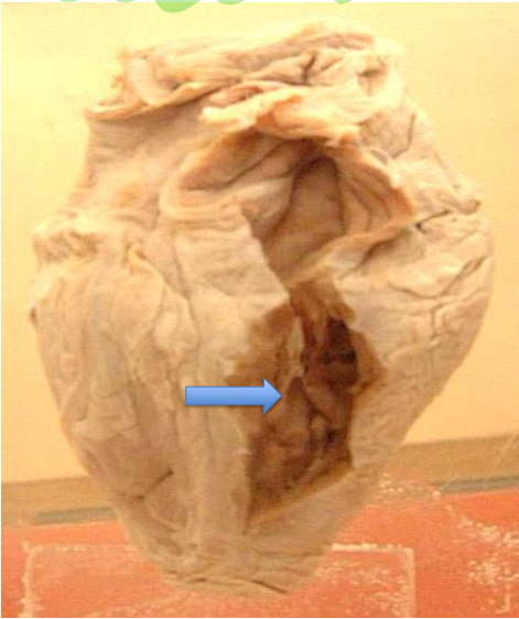
Diagnosis: urinary bladder hypertrophy due to benign prostatic hyperplasia
Organ: Urinary bladder and Prostate
Bladder:
urinary bladder englarged dialated
thick trabiculated smooth muscle wall
Prostate:
enlarged, grayish pink, firm leading to blader neck obstruction.

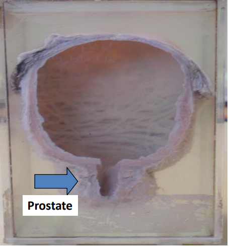
Diagnosis: benign prostatic hyperplasia
Outer surface: enlarged nodular prostate
Cut section:
firm, greyish pink, multiple nodules varies size & shape surrounded by fibrous tissue bands
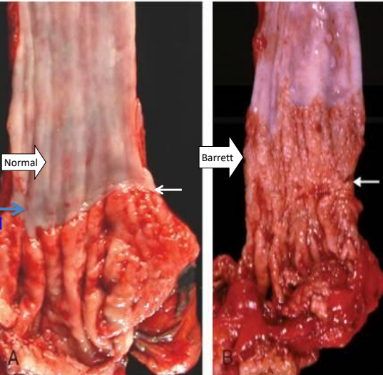
Diagnosis: Barrett esophagus
Organ: Esophagus
Esophageal mucosa above gastroesophageal junction show red velvety patches
Diagnosis: Fatty change liver
Organ: Liver
enlarged, smooth surface with rounded edges
Yellow in color, soft in consistency and greasy to touch.
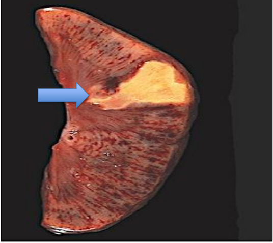
Diagnosis: Renal infarction (Coagulative necrosis)
Organ: Kidney
A wedge-shaped pale yellow area
Base towards Periphery
Apex towards Hilium
Surrouned by zone of hypermia
Capsule raised due to edema
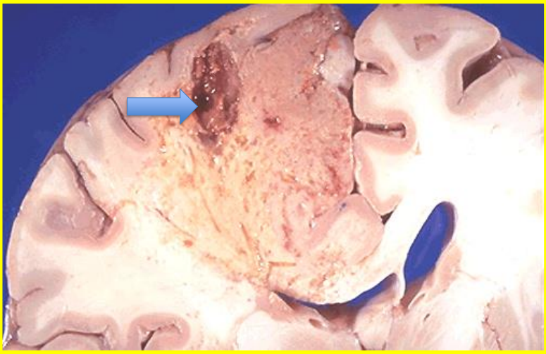
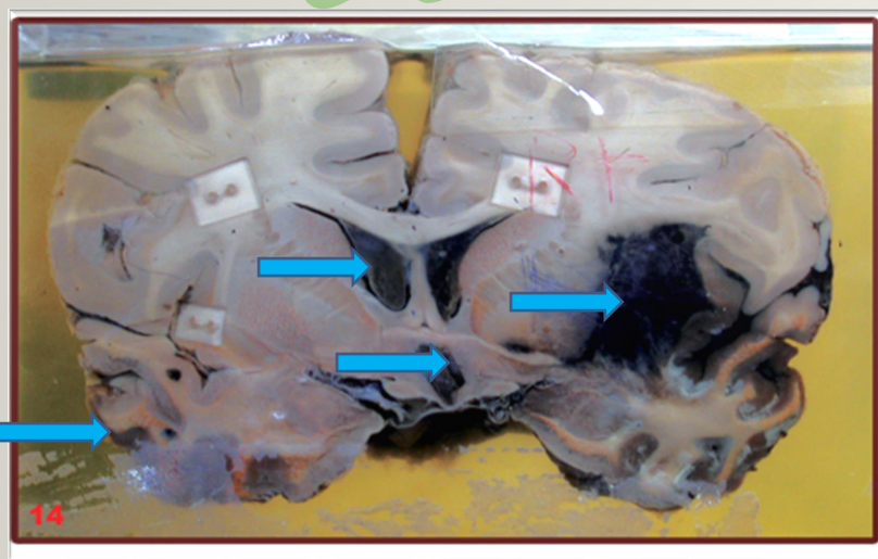
Diagnosis: Cerebral infarction (Liquefactive necrosis)
Organ: Brain
oval cyst, 2×1cm
filled with nectrotic tissue
surrounded by zone of edema and hypermia
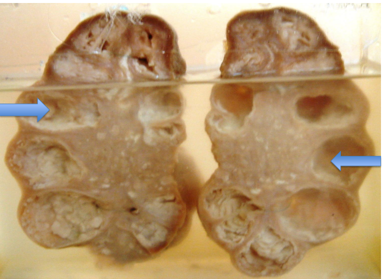
Diagnosis: Tuberculosis kidney (Caseous necrosis)
Organ: Kidney
Outer surface:
kidney enlarged
surface is nodular
capsule is thick and opaque
Cut section:
Cut section:
Shows multiple cavities of variable sizes (0.5 x 0.5 to 3x 3 cms) and shapes (oval and round)
Cavities contain yellow friable, cheese like material (caseous necrosis).
The renal parenchyma is atrophied and compressed by these cavities.
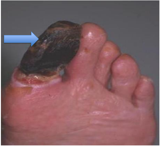
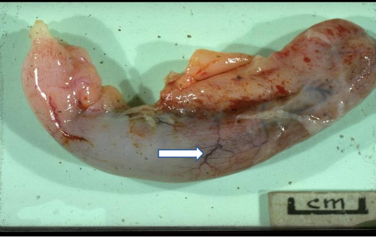
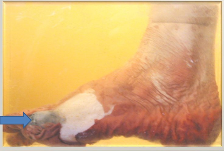
Diagnosis: Dry gangrene of big toe (Gangrenous necrosis)
Organ: Foot
Description:
The big toe is dry, mummified & black
Show line of demarcation and line of separation.
The surrounding skin is pale.
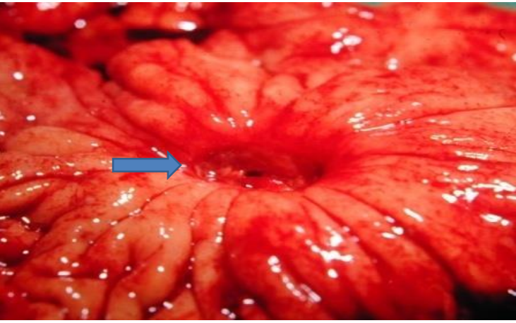
Diagnosis: Pseudomembranous colitis
Organ: Segment of colon
Wall appear edematous and hypermic. - The mucosa is covered partially by thin yellowish pseudomembrane.
Diagnosis: Acute diffuse suppurative appendicitis
Organ: Appendix
Appendix is elongated and edematous.
Serosa is hypermic show focal grey-yellow exudates (pus).

Diagnosis: Chronic cholecystitis
Organ: Gall bladder
Enlarged, thick wall & grey white.
Mucosa is hypermic focally
Contain multiple brown stones.
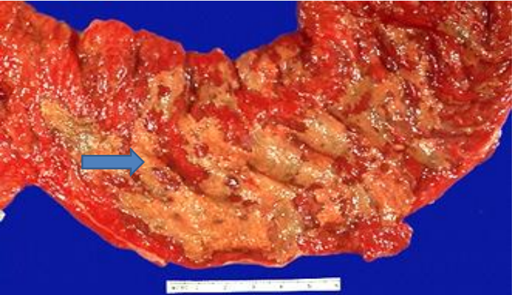
Diagnosis: Chronic peptic gastric ulcer
Organ: Stomach
Single oval ulcer measuring 2x1.5 cms
Punched out with clean floor and indurated base.
The margins are hyperaemic.
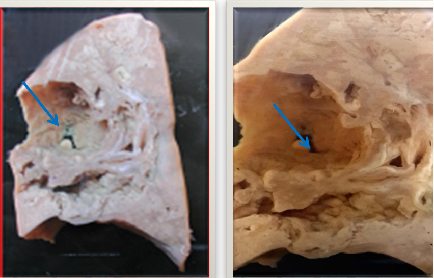
Diagnosis: Tuberculous bronchopneumonia with cavitation
Organ: Lung of a child
Description:
Outer surface: Covering pleura thick and opaque.
Cut section:
lung is heavy & shows areas of consolidation and caseation.
Many irregular cavities largest at mid lung,
oval in shape, 2X4 cms, was full of caseous cheesy material.
The wall is thick, irregular and ruptured into pleura.
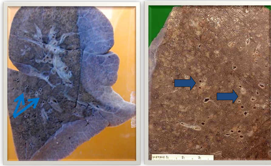
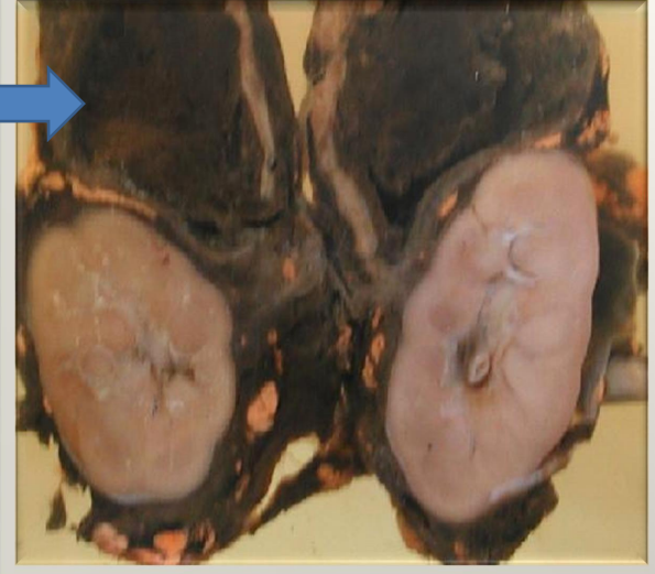
Diagnosis: Miliary tuberculosis lung
Organ: Lung
Description:
Outer surface: Pleura is thick, gery-white and adherent.
Cut section: shows multiple greyish-yellow tubercles
1-4 mms in diameter scattered all over the surface.
They contain yellow caseous necrotic material
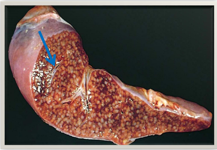
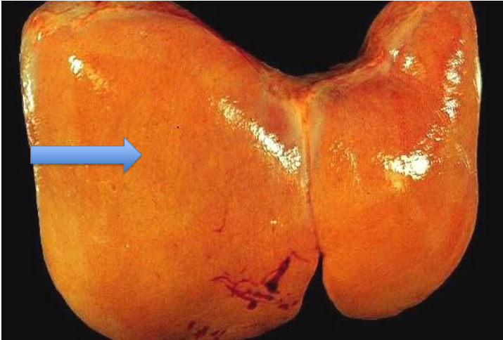
Diagnosis: Miliary tuberculosis spleen
Organ: Spleen of a child
Description:
Outer surface: Spleen is enlarged. Capsule is thickened, grey-white and adherent.
Cut section: bshows multiple greyish-yellow tubercles 1-4 mms in diameter scattered over surface.
They contain grey-yellow caseous necrotic material.
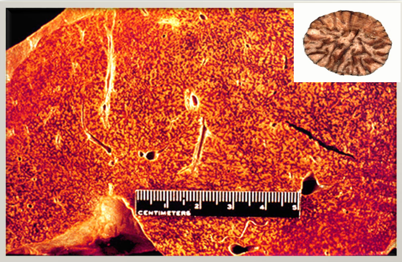
Diagnosis: Chronic venous congestion liver (CVC) (nutmeg liver)
Outer surface:
Enlarged with tense capsule and smooth surface.
Cut section: Two alternate areas seen throughout liver tissue consist of: Pale yellow (due to fatty change).
Dark-brown (due to congestion).
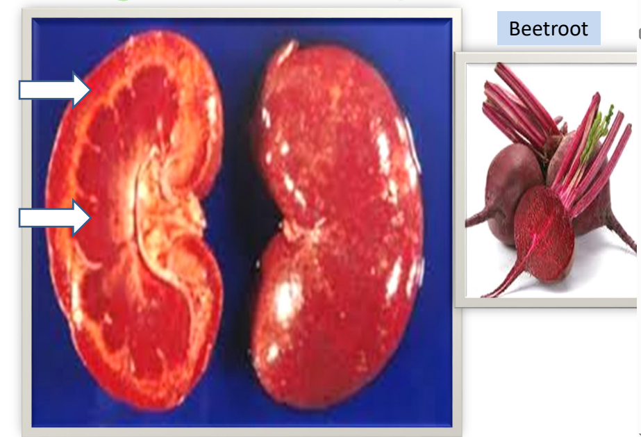
Diagnosis: Chronic venous congestion of kidney (Beetroot kidney)
Organ: Kidney
Enlarged with tense capsule and smooth surface.
Red-brown in colour especially medulla (as medulla is more vascular).
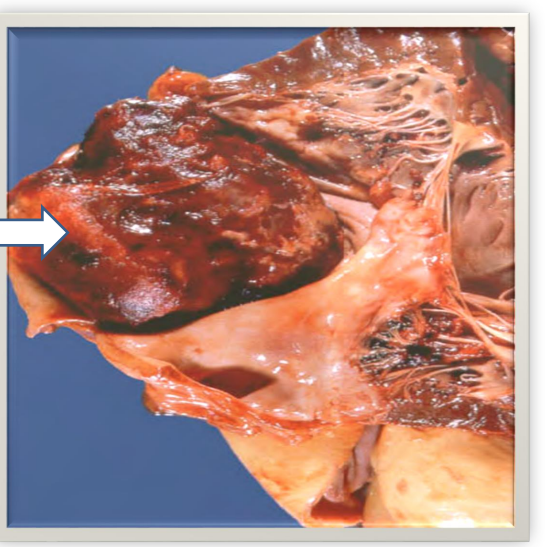
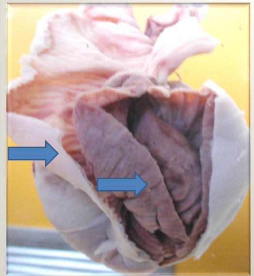
Diagnosis: Ball thrombus in the left atrium
Organ: Heart
Oval brown loose mass filling the left atrium 3x5 cms.
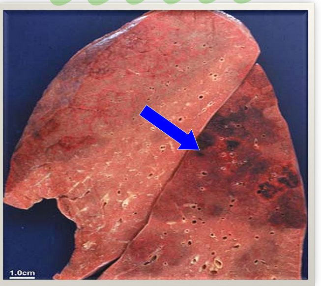
Diagnosis: Hemorrhagic (red) infarct of lung
Organ: Lung
Lung shows a wedge shaped infarct, well defined, red-brown in colour.
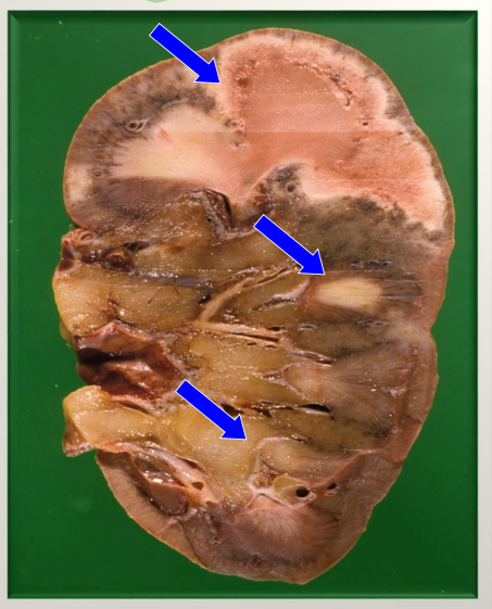
Diagnosis: Multiple old pale infarct of kidney
Organ: Kidney
Kidney show multiple grey depressed firm areas of variable size and shape.
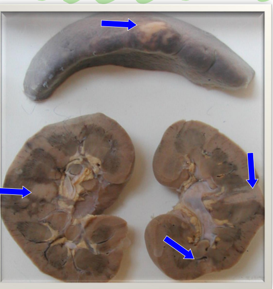
Diagnosis: Multiple old pale infarct of kidney and spleen
Organ: Kidney and spleen
Kidney shows multiple grey depressed firm areas of variable size and shape.
Splenic surface show thick capsule with central grey depressed firm area.
Diagnosis: Cerebral hemorrhage
Organ: Brain
Dark brown oval area of blood 3x2 cms seen in right cerebrum.
Small dark brown blood is also seen in third, lateral ventricles and subarachnoid space.
Diagnosis: Adrenal hemorrhage
Organ: Adrenal gland and kidney
Enlarged gland with marked dark brown hemorrhage.
Peripheral grey rim of remnant adrenal gland.
Diagnosis: Dry gangrene (senile)
Organ: Foot
The little toe is blackish in color.
The rest of the foot is pale with wrinkled skin.
Sharp line of demarcation.
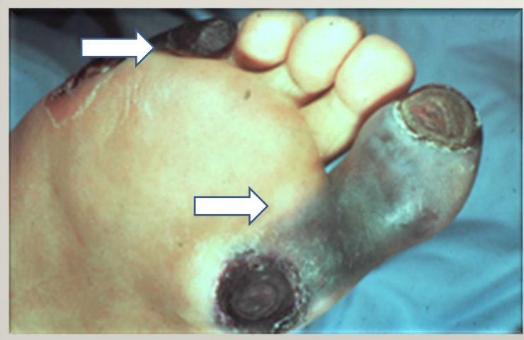
Diagnosis: Dry gangrene (senile)
Organ: Foot
The little toe and big toe are blackish in color.
The rest of the foot is pale.
Sharp line of demarcation.
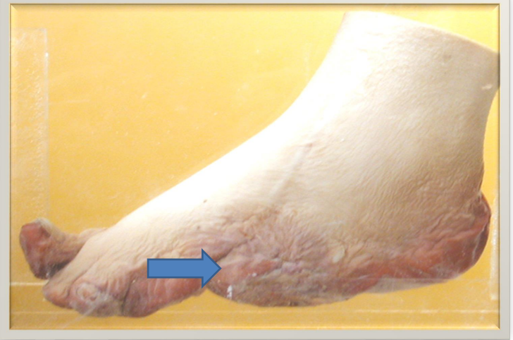
Diagnosis: Wet gangrene (Diabetic)
Organ: Foot
Lateral part of the foot are swollen and dark red.
Shrunken necrotic little toe.
No line of separation.
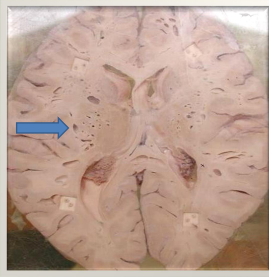
Diagnosis: Gas gangrene
Organ: Brain Description:
Multiple cavities of different shapes and sizes (1mm to 1cm).
Empty cavities as they were contain gas and necrotic material.
Diagnosis: Volvulus of small intestine (moist gangrene )
Organ: Small intestine
Small intestine is twisted, swollen and dark-brown in color.
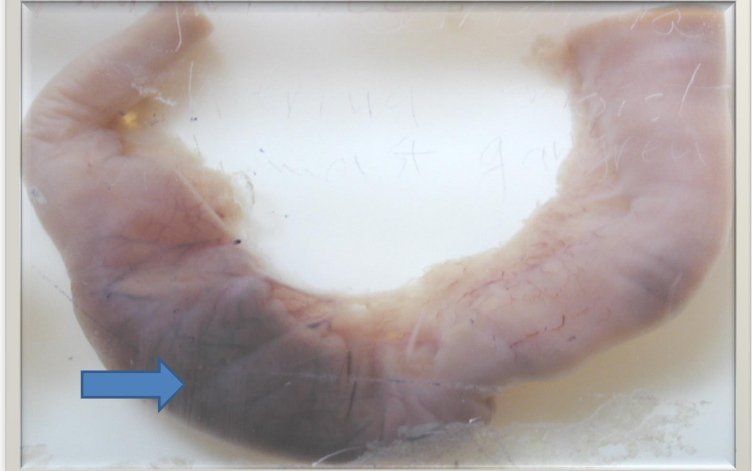
Diagnosis: Strangulated ileum with moist gangrene
Organ: Ileum
Dark brown swollen area, 3 cm in length.
No line of demarcation.
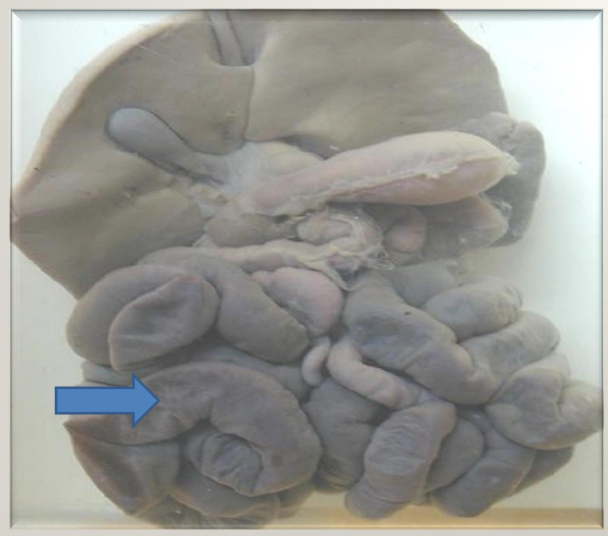
Diagnosis: Ileoileal intussusception
Organ: Small intestine
One segment of intestine is seen enter into another segment.
Inner segment is dark brown swollen with narrow lumen.
Outer segment is pale dilated with thin wall.