integumentary problems
 Call Kai
 Learn
Learn Practice Test
 Spaced Repetition
 Match
Match Flashcards
 Knowt Play
1/42

There's no tags or description
Looks like no tags are added yet.
Name | Mastery | Learn | Test | Matching | Spaced | Call with Kai | Chat |
|---|
No analytics yet
Send a link to your students to track their progress
43 Terms
sunscreen
UVA: tanning
UVB: sunburn
broad spectrum blocks UVA and UVB, must be at least SPF 15
daily protection: SPF 15
hx of skin cancer or photosensitivity: minimum SPF 30
apply 20-30 min before going outside, reapply every 2 hours
not waterproof
precancerous lesion
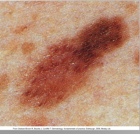
atypical or dysplastic nevus are atypical moles
irregular boarders, various shades of color, some ABCDE characteristics but less pronounced
increased risk of developing melanoma
skin cancer etiology and risk factors
most commonly diagnosed type of cancer, nonmelanoma or melanoma, most lesions are easily visible leading to early detection and diagnosis→ visible cancer
risk factors:
fair skin, blonde/red hair, blue eyes, outdoor sunbathing, living near the equator or high altitudes, hx of skin cancer, outdoor occupation/recreation, indoor tanning
nonmelanoma skin cancer
basal cell and squamous cell cancers most common from of skin cancer
do not develop from melanocytes
most common cause fro nonmelanoma skin is sun exposure
commonly found on the face, neck, back of the hands and arms
actinic keratosis
most common precancerous skin lesions
affect most of the older white population
sun exposure is the key factor
impossible to tell from squamous cell cancer→ aggressive treatment
characteristics: irregularly shaped, flat, pinkish/red papule with indistinct borders with an overlaying hard scale or horn
treatment: excision, topical meds (fluorouracil), photodynamic therapy, biopsy

basal cell carcinoma
basal cell layer of the epidermis
most common type of skin cancer and least deadly
UV exposure is most common cause
middle aged to older adults
found on head, neck, extremities, and trunk
characteristics: small, slowly enlarging papule, borders semitranslucent or pearly with telangiectasia red, erosion, ulceration, depressed center
treatment: surgical excision, curettage, cryosurgery, radiation, various targeted therapy meds

squamous cell carcinoma (SCC)
keratinizing epidural cells
aggressive and potential to metastasize
risk factors: sun exposure and immunosuppression post organ transplant
pipe, cigar and cigarette smoking contribute to SCC on mouth and lips
characteristics: thin, scaly red plaques with indistinct borders and ulcerations
treatment: surgical excision, radiation, curettage, medications, immunotherapy (metastatic lesions)

melanoma
arising from melanocytes
causes most skin cancer deaths
sun exposure is the greatest risk factor
may start as a benign mole with changes, itching/bleeding
⭐️ highly metastatic; survival depends on early diagnosis and treatment
characteristics: irregular color, surface, border, flat or elevated, men-back and chest, women-legs and back
treatment: excision, lymph. node dissection, immunotherapy and targeted therapy, chemotherapy
malignant melanoma
risk factors:
red/blond hair
blue or light colored eyes
light colored skin that freckles
chronic UV exposure
family Hx of melanoma

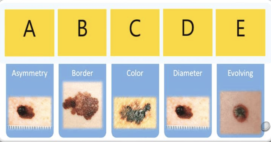
ABCDE rule
Asymmetry
Border irregularity
Color change
Diameter greater than 6 mm
Evolving in appearance
health promotion
most effective way to prevent skin cancer is avoiding or reducing exposure to the sun or tanning beds
wear sunscreen as appropriate
protect skin with hats, long sleeve clothing, umbrella, canopy
stay out og the sun during hottest part of the day 11am-3pm
educate about self screenings and when to contact provider
⭐️ if you notice a new or changed lesion, have it looked at ASAP
skin infections and infestations
bacterial infections: primary or secondary infection, staph and group a b-hemolytic strep are most common
viral infections: virus infects a cell or from an inflammatory response, create lesions that can be hard to treat
infestation and insect bites: allergy to venom, reaction to infestation of eggs, feces, parasite
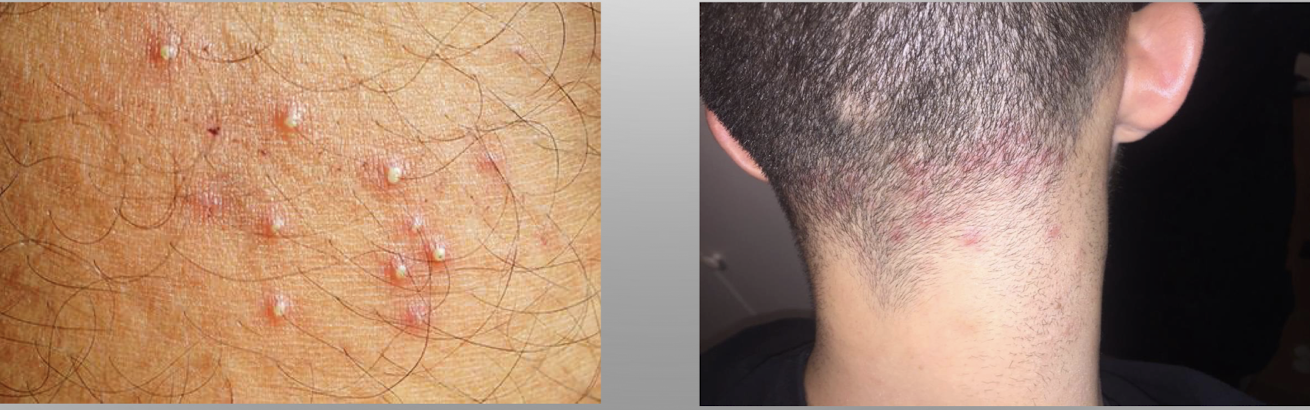
folliculitis
bacterial: inflammation of the hair follicles
cause: staph bacteria; found in areas exposed to friction, moisture, rubbing, oil
symptoms: small pustule at hair follicle, mild redness, tender
treatment: topical antibiotic, chlorhexidine, warm saline compresses
no scarring, if extensive may have hair loss and systemic antibiotics, proper hygiene methods teaching
furuncle
bacterial: deep infection of the hair follicle
cause: staph, often occurs with severe acne
symptoms: tender, red, painful area around hair follicle, drainage of pus upon rupture; can be found on face, back of neck, axillae, breastes, buttocks, perineum, thighs
treatment: warm, moist compress; if abscess develops, will need incision and drainage and antibiotics, resolves slowly with scaring, teach proper hygiene

carbuncle
bacterial: collection of infected hair follicles (furuncles)
cause: staph bacteria
symptoms: pustules forming in reddened area firm lesion that is red, painful, and swollen, drainage, abscess may be present; most common on nape of neck, also on upper back, lateral thighs
treatment: warm, moist compress, if abscess develops will need incision and drainage, antibiotics, slowly with scarring, proper hygiene teaching

cellulitis
bacterial: deep inflammation of subcutaneous tissues from enzymes produced by bacteria; primary or secondary
cause: staph or group b strep
symptoms: hot, tender, red, edematous area with diffuse borders; gangrene is left untreated
treatment: topical→ moist heat, elevation, immobilization; systemic→ antibiotics
may need to be hospitalized for IV antibiotics

impetigo
bacterial: skin infection; primary or secondary
cause: staph, group B strep or both
symptoms: vesiculopustular lesions that develope thick, honey-colored crust surrounded by redness and itchiness; contagious, most commonly on face
treatment: topical→wound care with special solution or warm, soapy water, topical antibiotic cream, good personal hygiene; systemic→ oral antibiotics: doxycycline, cephalexin, clindamycin

preventing the spread of MRSA
Methicillin Resistant Staphylococcus Aureus (MRSA)
avoid close contact with others
keep areas covered
change dressing prn and dispose of soiled dressings appropriately
wash hands often, clean surfaces with bleach
private room, maintain contact precautions
take antibiotics as prescribed
shower rather than bathe, antibacterial soap
wash soiled linen separately
avoud sharing personal care items
herpes simplex virus
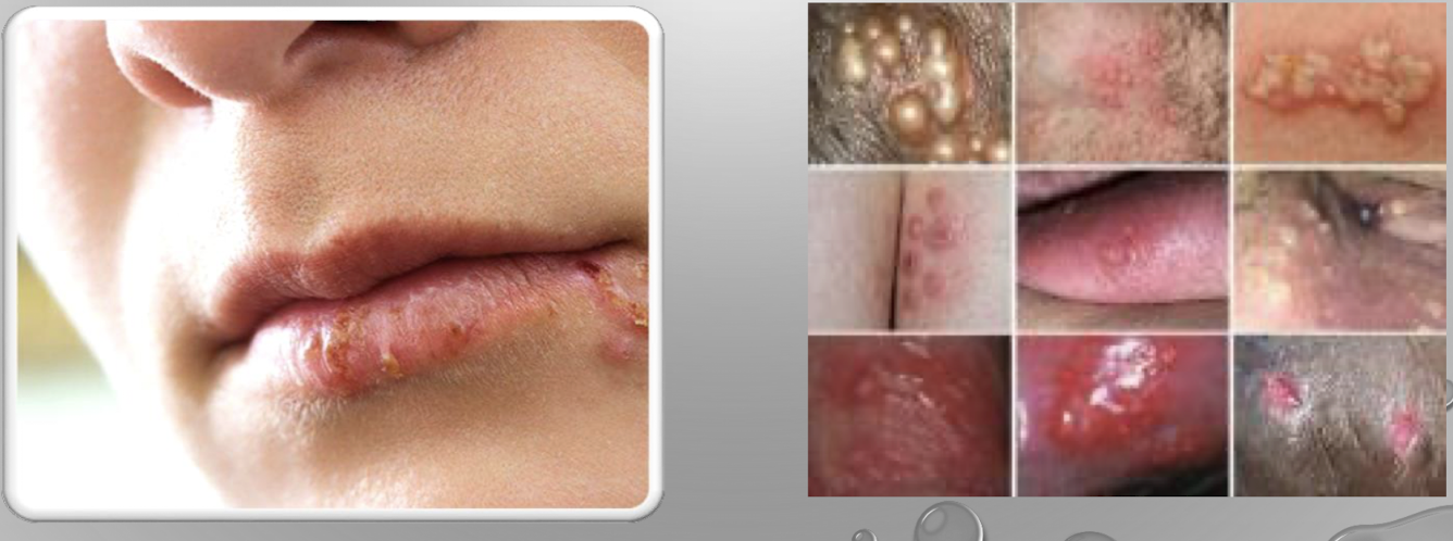
recurrent, lifelong viral infection; worsened by sunlight, trauma, menses, stress, systemic infection; contagious, can be transmitted from one area to another by contact.
HSV1: primarily on mouth (cold sore, fever blisters)
HSV2: primarily found on the genitalia (genital warts)
symptoms: first→ develop 2days-2weeks after contact; itchy, painful vesicles may form and become pustules and burst, weep, then crust, lesions last 2-6 weeks, contagious; recurrent→ similar spot, same symptoms.
treatment: soothing, moist compresses, topical agents (abreva); antiviral oral agents: acyclovir (Zovirax), valacyclovir (Valtrex)
herpes zoster virus
activated by the varicella-zoster virus
virus causes shingles→incidence increases with age
risk for infection increases if no previous varicella infection or immunocompromised
symptoms: painful lesions are unilateral and follow nerve root; commonly found on anterior/posterior trunk, face, eye; burning, painful and neuralgia before outbreak, mild to severe pain during outbreak
treatment: topical→ wet compresses, silver sulfadiazine (Silvadene) to ruptured vesicles; systemic→antivirals (Acyclovir) should be given within 72 hours of eruption; pain relief→ analgesia, gabapentin may be prescribed for neuralgia
prevention: CDC reccomends single dose of Zostavax vaccine for adults 50 years and older

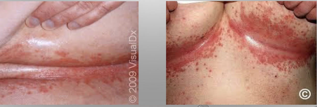
candidiasis
fungal infection of the skin
cause: associated with candida albicans
symptoms: oral mucosa→ white, cheesy plaques on roof of mouth, tongue, can be irritating; skin→ diffuse, red rash, odor usually present
treatment: topical→ Nystatin powder or ointment, keep skin clean, cool and dry; systemic→oral antifungal therapy, Fluconazole (Diflucan)
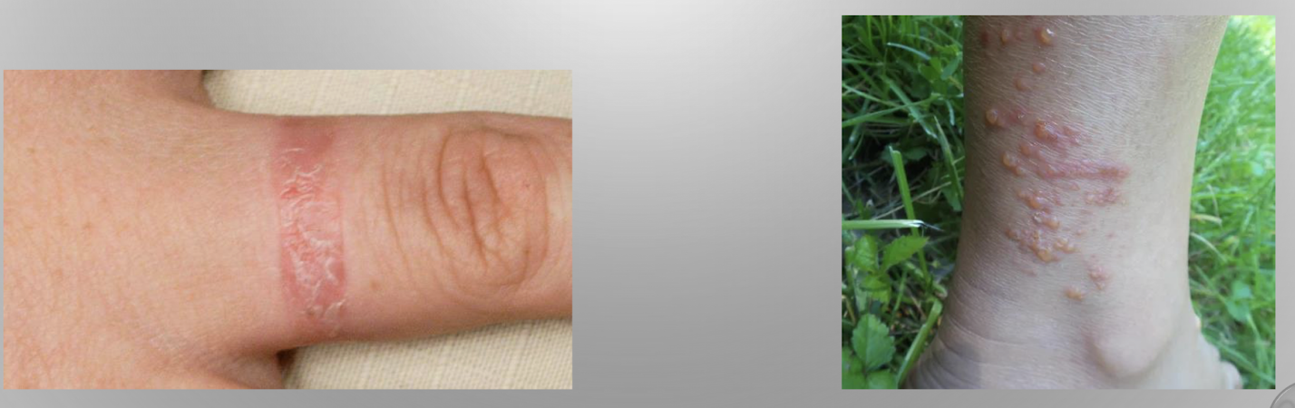
tinea corporis (ringworm)
caused by various dermatophytes
manifestations: red, annular (ringlike) scaly appearance, well-defined borders
management: contagious, keep skin covered, don’t share personal care items, cool compresses, topical antifungals (creams ending in -zole) usually OTC

tinea cruris (jock itch)
caused by various dermatophytes
manifestations: well-defined, scaly plaque in groin area
management: keep skin clean and dry, no sharing of personal care items, topical antifungal creams or solutions

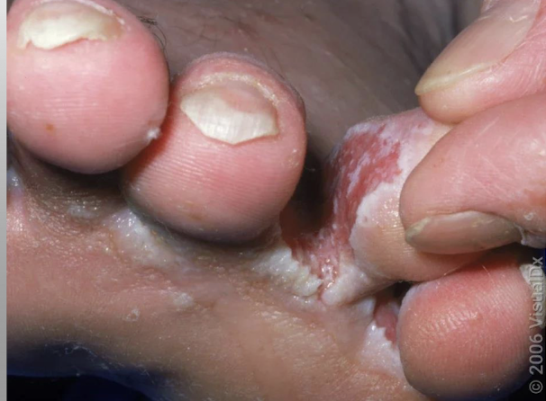
tinea pedis (athlete’s foot)
caused by various dermatophytes
manifestations: interdigital scaling and maceration; scaly skin, may be peeling
treatment: topical antifungal agents, keeping feet clean and dry, allow feet to have air, wear shoes in public showers
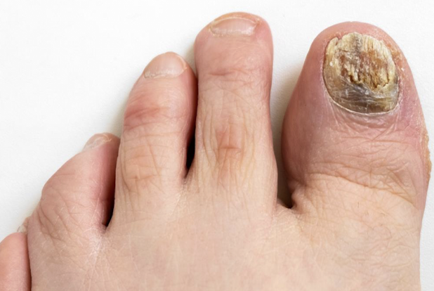
tinea unguium (toenail fungus)
caused by various dermatophytes, incidence increases with age
manifestations: affects toenails most often, may affect fingernails on one hand; brittle, thickened, broken, or crumbling nails with yellowish discoloration
management: oral/topical antifungals, podiatry consult, thinning of toenails or nail removal
pediculosis (lice)
lice infestation of either head, body, or pubic area
most common symptom is itching, observe hair for nits, pubic lice cause severe itching of the vulvar or perirectal area→ can be contracted through bed linens or during sexual intercourse with infected person
treatment: topical sprays, creams, and shampoos; otic lice kits, topical permethrin cream or malathion lotion, oral ivermectin, wash contaminated linens and personal items in hot water with detergent

scabies
contagious skin infection caused by mite infestation
characteristics of linear or curved ridge, intense itching, worse at night; transmitted through close contact with an infected individual or bedding/clothing; common in nursing homes and other crowded living areas, those with poor hygiene; infestation is confirmed with scraping of a lesion
treatment: scabisides→ topical permethrin or oral/topical ivermectin, launder all clothing and personal items with hot water and detergent

bedbugs
common parasite with infestations increasing due to travel and resistance to pesticides
live in mattresses, fabrics, cracks/crevices of furniture
feed on human blood at night, do not live on humans, bites resemble mosquito bites, resolve on own, hydrocortisone cream or oral antihistamine to help with itching
prevention: examine hotel room bedding especially box springs, place luggage away from bed when traveling, soiled clothing goes in sealed plastic bag when traveling, observe and clean items bought at garage sales, thrift stores, other resales before bringing into home
eradication: usually requires exterminator
allergic contact dermatitis
reaction of the skin most often r/t allergic immune response
cause: contact with allergen, specific cause not always known
symptoms: red papules and plaques, itching, area of irritation may take shape of causative agent
treatment: topical or oral corticosteroids, antihistamines, skin lubrication, cool, moist compresses, lukewarm colloidal oatmeal baths, elimination of contact allergen, avoid irritating affected area, if severe symptoms IV steroids may be needed
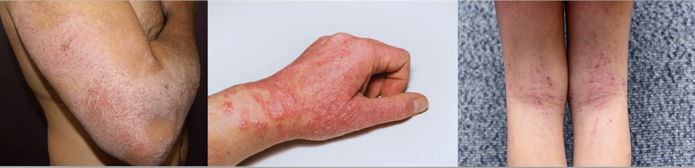
atopic dermatitis
reaction to the skin most often r/t type 1 hypersensitivity immune response→ chronic, relapse disease, skin response to environmental allergens, associated with asthma and allergic rhinitis
symptoms: acute→redness, oozing vesicles, extreme itching; subacute→scaly, light red to red-brown plaques, itching; chronic→thickened, dry skin with lichenification, possible hypo/hyper-pigmentation
treatment: topical immunodulators, moisturize dry skin, stress reduction; antibiotics for secondary; severe inflammation and itching→ steroids, phototherapy
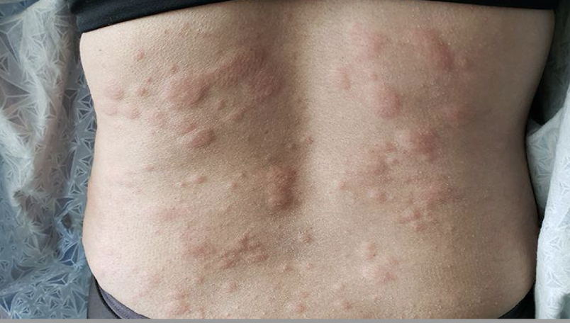
urticaria (hives)
allergic reaction to the skin
cause: histamine release in response to allergic reaction
symptoms: rash of white or red raised areas (wheals) of various sizes, can occur anywhere on the body
treatment: removal of triggering agent, oral antihistamine, possible need for systemic corticosteroids
skin drug allergies
rare, life-threatening immune-mediated hypersensitivity reaction, most commonly caused by certain medications.
results in acute destruction and detachment of the epidermis and mucous membrane epithelium, leading to extensive skin loss and impaired barrier function.
common drugs: allopurinol, carbamazepine, lamotrigine, phenobarbital, phenytoin, NSAIDs, sulfonamides
symptoms: 4-21 days after medication start; initial→proceed skin reaction by 1-3 days, fever, cough, headache, anorexia, myalgia, and nausea; skin→ red, macular rash with purpuric centers, over hours to days, blisters form with sheets of epdermal detachment
management: stop med immediately, airway management, kidney function, fluid/electrolyte balance, pain control, wound care, parenteral/enteral nutrition, supportive care
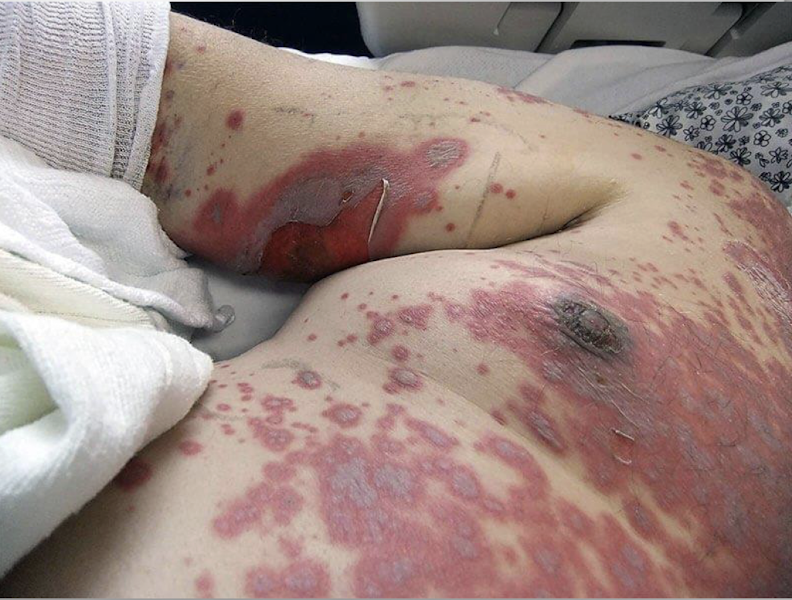
Stevens-Johnson syndrome
skin drug reaction
skin detachment of less than 10% of body surface
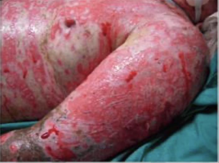
toxic epidermal necrolysis
skin drug reaction
skin detachment of more than 30% of the body surface
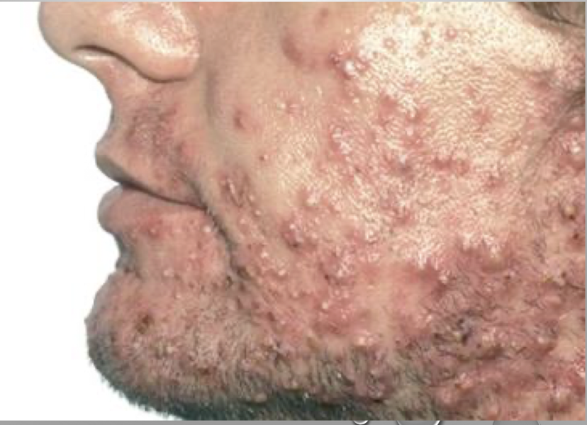
acne vulgaris
benign: inflammatory disorder of the sebaceous glands; more common in teenagers, may worsen with hormone fluctuations
symptoms: various sizes of blackheads and whiteheads, may include papules and pustules, most common of face, neck and upper back
treatment: topical→mechanical removal of multiple lesions with extractor, topical solutions (benzoyl peroxide, retinoids, antimicrobials); systemic→antibiotics (Isotrerinoin)
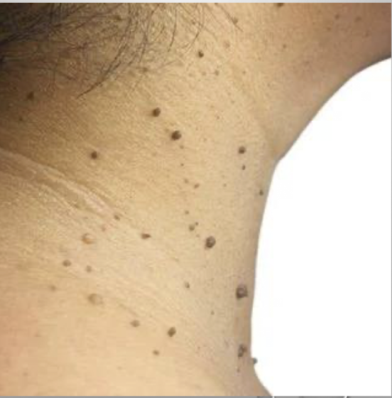
acrochordons (skin tags)
benign growths
cause: unknown
symptoms: small, skin colored, soft, pedunculated papules; can become irritated in friction areas; common on neck, axillae and upper trunk
treatment: none necessary; surgical removal when needed, snipping without anesthesia
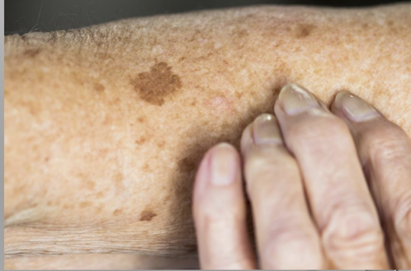
lentigo
benign: increased number of normal melanocytes in the basal layer of the epidermis; aka liver spots or age spots
cause: sun exposure and aging
symptoms: hyper pigmented, brown to black macule or patch on the sun-exposed skin
treatment: evaluated regularly for changes (biopsy if needed), may want removed for cosmetic purposes
psoriasis
benign: autoimmune chronic skin disorder marked by rapid turnover of epidermal cells; periods of exacerbations and remissions; family predisposition
symptoms: silver scaling plaques on reddish skin; often found on scalp, elbows, palms, soles, and fingernails; itching, burning, pain; mild-severe symptoms
treatment: reduce inflammation and suppress rapid turnover of epidermal cells; no cure; topical→corticosteroids, various other topical meds, phototherapy; systemic→methotrexate, Otezla; biologic therapies (Enbrel, Skyrizi, Humira)

rosacea
benign; common disorder of the central face; unknown cause
manifestations: redness, flushing of the face
management: avoid triggers; sun, extreme heat/cold, spicy food, alcohol; oral and topical agents

seborrheic keratoses
benign; common, noncancerous skin growth that often looks waxy, scaly, or like it was ‘stuck on’ the skin
management: biopsy if necessary, remove by curettage or cryosurgery

punch biopsy
no sutures, cover with dressing

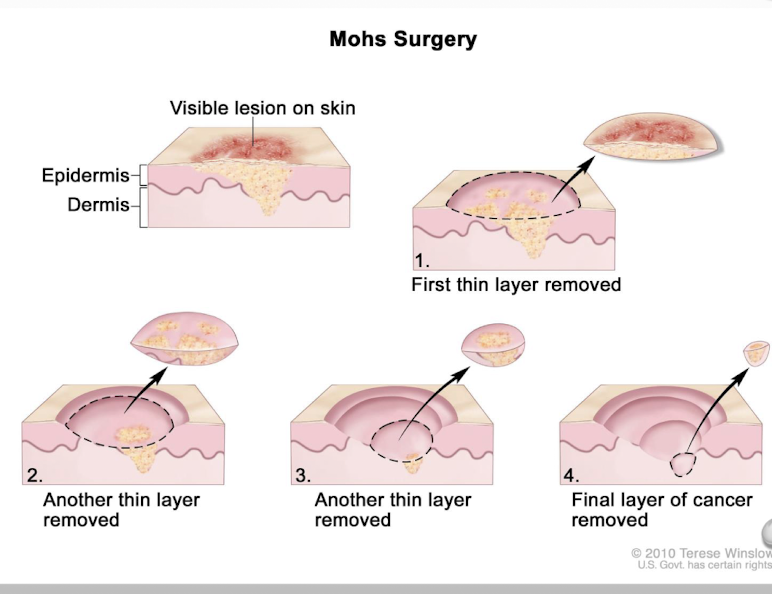
Mohs surgery
layers of the tissue are shaved away and placed under a microscope
shaving continues until there id no longer presence of the lesion
Nursing management of skin problems
wet compresses
colloidal oatmeal baths
good hygiene practices
correct application of topical medications
educate on ways to control itching
prevent spread of skin infections
prevention of secondary infections
educate about postprocedure care
offer patient support