Sect 2 - Cell Morphologies & Inclusions
 Call Kai
 Learn
Learn Practice Test
 Spaced Repetition
 Match
Match Flashcards
 Knowt Play
1/92

There's no tags or description
Looks like no tags are added yet.
Name | Mastery | Learn | Test | Matching | Spaced | Call with Kai | Chat |
|---|
No analytics yet
Send a link to your students to track their progress
93 Terms
importance of reporting morphologies
1. helps with diagnosis
2. gives provider into about progression/severity of disease
3. confirms finding/flags from analyzer
why peripheral smears are ordered
Provider ordered it
Specific manual testing by hematology (LAP/FMH testing)
Met criteria from the analyzer flags, needs verified
Pediatric patients (esp. fingerstick draws)
Specifically asked for by pathologist for confirmation of abnormal cells
LAP stain
stain used for bacteria or cancer; always look for neutrophils (specifically stained granules)
Fetal-Maternal hemorrhaging
What does FMH stand for?
fresh blood samples & copy of analyzer printout
make several stained & unstained slides
When a peripheral smear is specifically ordered to be sent to the pathologist for confirmation or abnormal cells, what are the 2 requirements neede?
1. patient's full name (first and last)
2. sample ID number or DOB
patient identifiers needed on slides when labeled (2):
1 month
How long are slides kept?
push (wedge)/cover slip method
technique used when making a peripheral smear

1. using right size blood drop
2. speed at which you spread
3. angle at which the drop is spread
3 factors that help determine a good technique is being used for a peripheral blood smear:
optimal smear
min length 2.5 cm
transitional from thick to thin
straight feathered edge
no streaks, waves, or troughs

true
T or F:
Always mix the specimen before making a peripheral smear
Wright's stain
stain used for performing manual differentials
Romanowsky stain
another name for a Wright's stain
Methylene Blue
Eosin
Glycerin
three components of a Wright's stain
Supravital stain
Stain used for Retic counts and identification of other inclusions in suspected disease processes for confirmation
true
T or F:
The Supravital Stain does NOT kill the cells, so we can observe them living
New Methylene Blue
Brilliant Cresyl Blue
two types of Supravital stains
RNA
DNA
Heinz bodies (denatured hgb(
Siderotic granules of RBC
What cell components are stained by the Supravital stain?
stain is orange
buffer pH
stain is purple
buffer pH >6.0 or too alkaline
Caused by excess stain to buffer and not adequately rinsed
CFU in cell maturation
G = granulocytes
E = erythroid cells
M = macrophage/monocyte cells
M = megakaryocyte cells (platelets)
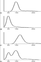
Normocytic
Red Cell Distribution Width
What is A?

Microcytic
Red Cell Distribution Width
What is B?

Macrocytic
Red Cell Distribution Width
What is C?

Dimorphic
Red Cell Distribution Width
What is D?
**hint: seen in blood transfusion pts (2 pops present)
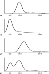
the patient (microcytic)
In D, the left curve represent which population?

donor (normocytic)
In D, the right curve represents which population?
Anisocytosis
Variation in sizes of RBCs
(RDW is indicator of this: ↑ RDW = ↑ anisocytosis = more variation is sizes)
Conditions associated: blood transfusion

Microcytes
RBCs that are abnormally small
- MCV < 80fl
- oval & big central pallor
Conditions Assoc: Iron deficiency anemia, thalassemia minor, chronic inflammatory, lead poisoning, hemoglobinopathies, and sideroblastic anemia

Macrocytes
RBCs that are abnormally large
- MCV >100fl
- NO central pallor
Conditions Assoc: Liver disease, vitamin B12 deficiency, folate deficiency, neonates, and reticulocytes

Poikilocytosis
variation in RBC shape
- reporting is specific to lab
Conditions Assoc: Severe anemia, Hemolytic states
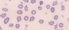
Polychromasia
indication of immature erythrocyte
- basophilic color = blue
Conditions Assoc: Increased erythropoiesis, Hemolysis, Acute & Chronic hemorrhage, Regenerative RBC process

Spherocytes
created by defect in membrane proteins in RBCs (decreased surface area:volume ratio)
- MCHC >36%
- NO central pallor, dense
Conditions Assoc: Hereditary, Hemolytic anemias (Immune/Autoimmune), Post-transfusion, Severe burns

Microspherocytes
like spherocytes, but smaller
- CRITICAL FINDING & SIGNIFICANT
Conditions Assoc: Autoimmune hemolytic anemia, Post transfusion

Codocytes (Target Cells)
caused by a membrane defect
- excess membrane cholesterol and phospholipids; decreased cellular hgb
- red/salmon color
- "bullseye"
Conditions Assoc:
Hemoglobinopathies (sickle cell (HgbS), HgbC)
Thalassemia's (decreased production in alpha & beta chains)
Iron-Deficiency Anemia (IDA)
other: Splenectomy, Obstructive liver disease

Dacryocytes (Tear Drop)
RBC squeezing and passing through spleen/fibers
- "tear drop" shape facing different directions
Conditions Assoc: Primary myelofibrosis, Ineffective erythropoiesis, Megaloblastic anemia, Thalassemia
artifacts if all tear drops face same direction

Elliptocytes (Ovalocytes)
abnormal cytoskeletal proteins
- cannot change back to normal shape, but still able to carry O2 like regular RBCs
- irregular, elliptically shaped RBCs
Conditions Assoc: Hereditary elliptocytosis, Iron Deficiency Anemia (IDA)
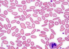
Sickle Cell (Drepanocyte)
abnormal presence of Hgb S (cell inflexible)
- polymerization of deoxygenated hgb (taking O2 out - polymerizes & changes shape)
- NO central pallor
- crescent shaped
- normal MCV
Conditions Assoc: Sickle-cell anemia

<120 days
life span of Sickle cell
Degmacyte (Bite/Helmet cell)
spleen pits out the Heinz body
- cell body is denatured hgb due to oxidative injury
- seen in Wright's stain
- RBC has "bite" taken out of it
Conditions Assoc: G6PD deficiency, Drug induced hemolysis

Heinz body
Which degmacyte cell is seen in the supravital stain?

Echinocytes (Burr Cells)
regular contracted cells with short, evenly spikey projections & preserved central pallor
Conditions Assoc: KIDNEY PROBLEMS: Uremia, Secondary ATP deficiency, altered membrane lipid
artifact due to hypertonicity/alkalinity of stain solution

Acanthocytes
increased cholesterol/phospholipid ratio (increase surface area); decreased lecithin
- spiny projections on the surface of RBC
- slight loss of central pallor
- hint: ACANT = I CANT make normal shape :(
Conditions Assoc: Hemolytic anemia, Alcoholic cirrhosis (liver issue), Vitamin E deficiency, Post-splenectomy, Severe liver disease, Pyruvate kinase (PK) deficiency, Congenital abetalipoprotienemia

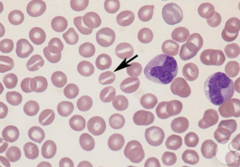
Stomatocytes
increased permeability to sodium
- bowl-shaped with slit like area in central pallor
- looks like a screwhead for flat screwdriver
Conditions Assoc: LIVER PROBLEMS: Acute alcoholism (screwdrivers), Hereditary Stomatocytosis, Liver disease, Cirrhosis, Hemolytic anemia
artifacts due to staining solution is too acidic
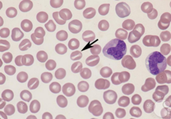
Schistocytes
fragmentation of RBCs due to membrane damage
- CLINICALLY SIGNIFICANT (life or death)
- should not be seen in peripheral blood
Conditions Assoc:
Disseminated Intravascular Coagulation (DIC)
Pulmonary Emboli (clot in lungs)
Microangiopathic hemolytic anemia (small clots in vessles)
Other: Thrombotic thrombocytopenia purpura (TTP), G6PD deficiency, Hemolytic uremic syndrome (HUS), Burns, Heart valves

HbC Crystals
lysine replaces glutamic acid in beta chain
- rectangular and dense
- looks like "bar of gold"
Conditions Assoc: Hgb C; Hgb S (glutamic acid replaced with valine)

too alkaline stain
Causes RBCs to become crenated (look like Echinocytes/Burr cells)
Abnormality in peripheral smear that is caused by slide making techniques

too acidic stain
Causes RBCs to become Stomatocytes
water artifacts
What type of artifact will you have if the slide is not allowed to dry properly?
makes identification of morphologies harder to determine than normal
true
T or F:
Abnormal cells should be distributed EVENLY through the slide for them to be considered notable or significant
Basophilic stippling
Cabot Ring
Howell-Jolly body
Pappenheimer body
RBC inclusions:
Basophilic stippling
Components: RNA and mitochondrial segments (precipitated ribosomes)
Wright stain used

lead posining
Coarse Basophilic stippling is seen in:
increased polychromasia
Fine Basophilic stippling in seen in:
Cabot Rings
Components: Nuclear membrane remnants, part of mitotic spindle, remnant of microtubules
Conditions Assoc: post-splenectomy, hemolytic anemia, megaloblastic anemia
Wright stain used

Howell-Jolly Body
Components: DNA nuclear fragments, fragment of nucleus that was extruded or fragments of chromosome separated by the mitotic spindle
Conditions Assoc: post-splenectomy, hemolytic anemia, megaloblastic anemia
Wright stain used
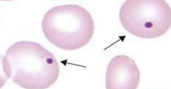
Pappenheimer body
Unused iron (non-heme) that was in excess
Conditions Assoc: sideroblastic anemia, post-splenectomy
Wright stain used

Heinz body
Body found in periphery of the RBC that is precipitated due to denatured Hgb due to oxidative injury
Conditions Assoc: G6PD deficiency
Supravital stain used

false; see Bite/Helmet cells
T or F:
Heinz bodies can be seen in the Wright Stain
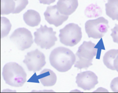
parasitic infection only found within RBCs
What type of infection is Malaria?
protozoan transmitted by the bite of a female Anopheles mosquito
How is Malaria transmitted?
Plasmodium falciparum
most common type of Malaria seen in lab
produces delicate rings; do not enlarge the cell
Plasmodium falciparum
Which type of Malaria?

Rouleaux
appears as a "stack of coins"
clumping induced by circulating paraproteins
Condition associated: paraproteinemia (plastic cell myeloma) & Multiple myeloma

Cold Agglutinin
random clumps of red cells (warming sample will break them up) that are caused by cold temperature of the sample
very high MCV (>130)
Conditions Assoc: Associated with EBV infection:
Mono in kids
Mycoplasma infections in adults

IgM anti-I antibodies
Cold agglutinins are most commonly associated with which anitbodies?
Dohle bodies
Toxic granulation
Vaculization
Hypersegmentation
Ehrlichiosis
WBC inclusions:
Dohle bodies
round-oval pale grayish-blue inclusions composed of RNA that are found in the periphery of the cytoplasm of neutrophils
Conditions Assoc: burns, infections, physical trauma, neoplastic disease, may also indicate May-Hegglin anomaly

neutrophils
Which type of WBC do you ONLY see Dohle bodies?
Vacuolization
formation of vacuoles within or adjacent to cells that is only seen in neutrophils
related to bacterial infection and caused by increased activity of phagocytosis in the cell
observed in pts that are septic
neutrophils
Which type of WBC do you ONLY see
Vacuolization?
Toxic Granulation
observed in bacterial infection where granules are darker in the neutrophils
seen in vacuolization and Dohle bodies
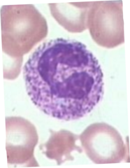
>5 lobes
How many lobes must a neutrophil have in order to be considered hypersegmented?
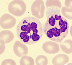
megaloblastic anemia
What type of anemias would hypersegmentation be seen in?
Ehrlichiosis
infectious disease caused by bacteria that belongs to the Rickettsia group that is transmitted by bite of infected tic
pale purple circle in cell of granulocytes

decreased WBCs and platelets
increased liver enzymes
other lab findings associated with Ehrlichiosis
Analyzer flags for clumping
Pt results do not match previous results (check for micro clots, verify analyzer results)
What situation would call for a platelet estimation to be done?
equation of platelet estimation

150,000-400,000/ul
normal range for platelets
adequate, increased, decreased
terms used when resulting out platelet estimations
Agranular platelet
platelet without granulation
- giant
- associated with rapid platelet production

giant platelets
platelets the size of WBCs & RBCs (looks like WBCs without nucleus)
Conditions Assoc: thrombocytopenia, myelofibrosis, CML, Bernard-Soulier Syndrome

bizzare platelets
irregularly shaped
large sized platelets
pseudopods
associated with Myelofibrosis

neutrophils
Which WBC does platelet satellitism occur with?

more than 4 platelets
How many platelets are involved in order to call satellitism?
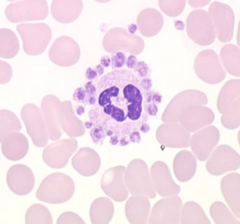
in vitro artifact in EDTA anticoagulant blood
platelet satellitism is associated with:
inadequate mixing
over-filling anticoagulant tubes
"sewing machine" method
factors that cause platelet clumping
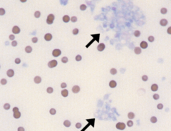
water
What type of artifact?

Pyknotic cell
What type of artifact?
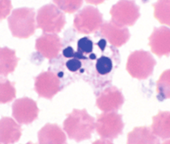
platelet superimposed
What type of artifact?
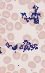
stain precipirate
What type of artifact?
same direction
Dacryocytes that face __________ are considered artifact
dead cells
What are pyknotic cells?