Cell Death
1/19
There's no tags or description
Looks like no tags are added yet.
Name | Mastery | Learn | Test | Matching | Spaced |
|---|
No study sessions yet.
20 Terms
4 examples of pathological causes of apoptosis
Organ not receiving stimulus (portosystemic shunt)
Cells containing infectious agent
Cell and/or DNA irreparably damaged
Cell is cancerous
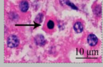
Histological morphology of apoptosis (5)
shrunken cell
loss of adhesions to nearby cells and basement membrane
pyknotic, with chromatin condensed around periphery
no/minimal inflammation
cytoplasmic blebs = apoptotic bodies
3 main causes of necrosis
Loss of blood supply = hypoxia/anoxia
Living agents (pathogens) that damage the cell
Non - living agents (chemicals, ROS, physical injuries)
Histological changes seen with necrosis
Nuclear changes:
Karyorrhexis (nucleus broken up into pieces)
Karyolysis (faint nucleus due to break down)
Pyknosis
Inflammation
white blood cell influx seen grossly and histologically
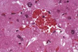
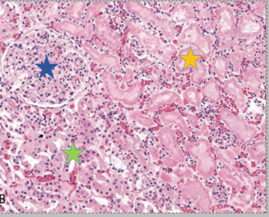
What nuclear change is seen in this necrotic image?
Karyolysis
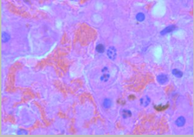
What nuclear change is seen in this necrotic image?
Karyorrhexis
How does anoxia/hypoxia lead to necrosis
Reduction of ATP production = active pumps stop working
Na+/K+ pumps
Calcium efflux pumps = intracellular calcium accumulation causes cell damage = hypercalcaemia (poor prognostic indicator)
= oncotic necrosis - cells swelling due to water following sodium
= hyperkalaemia
Increase in anaerobic glycolysis
= increased lactic acid and decreased pH = damaged cellular enzymes and DNA
4 ways cell membranes can be directly damaged
Pore-forming infectious agents/toxins
ROS
Phospholipase activation
Protease activation
Name a popular pore-forming bacteria
Clostridium perfringens - many types, produce different toxins
In what ways can viruses cause membrane damage?
Enveloped virus takes host membrane. Some can leave host cell intact whilst others cause cell lysus
Non-enveloped virus can only leave cell by lysis of the cell
Cell lysis caused by disruption to the cytocavitary network and other homeostatic mechanisms
Can induce apoptosis
What are free radicals and their causes? How are they controlled?
Any molecule with a free electron. Can be reactive oxygen species or reactive nitrogen species
Produced by oxidative metabolism (mitochondria usually) but can damage mitochondria if cannot be removed
Constantly produced by all cells
Free radicles neutralised by Vitamin E and selenium
What are the 4 gross morphologies of necrosis
Lytic/liquefactive
Coagulative
Caseous
Gangrenous
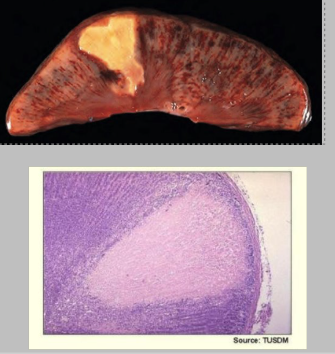
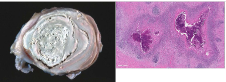
What is this type of necrosis, its causes, and describe the gross and histo features
Coagulative necrosis - basic outline of necrotic cells is preserved
hypoxic injury - infarction
Gross:
well demarcated (infarction), rim of inflammation
Firmer and dryer
Histo:
preserved tissue architecture - basement membrane often intact
necrotic cells
inflammation
early attempts at healing
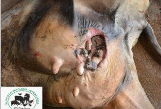
What type of necrosis is this? Causes. Describe the gross and histological appearance. Give example causative agents
Chronic coagulation necrosis
Body unable to remove agent e.g. intracellular bacteria or fungi
Birds and reptiles more common due to heterophils producing solid pus (reduced myeloperoxidase)
Gross:
friable, granular, white appearance
usually encapsulated
Histo:
loss of architecture
central accumulation of remnants of lysed leukocytes
may have granulomatous inflammation and outer fibrous tissue
usually have dystrophic calcification centrally
Causative agents:
= Corynebacterium pseudotuberculosis (lymphadenitis in ruminant and horse)
= mycobacterium tuberculosis
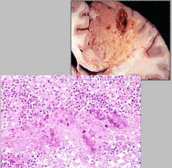
What is this type of necrosis? What is the cause? Describe the gross and histological appearance.
Liquefactive/lytic necrosis
cells lysed and necrotic tissue converted to a fluid phase
Caused by bacteria, fungi (cryptococcus neoformans), thiamine deficiency (CNN in ruminants)
mostly affects CNS
Gross:
soft, viscous
usually has cavity containing pus
poorly demarcated boundaries
Histo:
eosinophilic fluid, cell debris
messy, hard to orientate in high magnification
no background supportive tissues
What is gangrenous necrosis and cause?
Sequel of coagulative necrosis
Associated with loss of blood flow to a tissue
Type of gangrenous necrosis. What are its causes (4) and features?
Dry
Caused by coagulation necrosis followed by mummification (no water)
Usually affects the lower portion of extremities
No bacterial proliferation
Features: dry, shrivelled, brown/black. May slough
Cause:
Ingested toxins
Frostbite
Peripheral arteriolar constriction and damage to capillaries
Thrombosis and infarction
Type of gangrenous necrosis and what are the features?
Wet/moist
Areas of necrotic tissue are further degraded by liquefaction action of saprophytic bacteria = sloughing of tissue
Death can occur from toxaemia (E.coli/lipopolysaccharide)
Gross appearance: soft, moist, red/brown/black, may have gas, putrid odour (hydrogen sulphide)
Type of gangrenous necrosis and explain how it occurs and its gross features.
Gaseous
Bacteria, usually anaerobic, proliferate and produce toxins in necrotic tissue
Bacteria introduced by penetrating wounds
Gross = dark red/black, gas bubbles, fluid and haemorrhagic exudate.
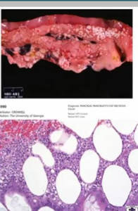
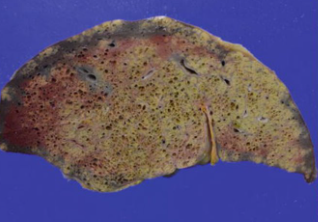
What type of necrosis is this? 4 possible causes. Describe the gross and histological features.
Fat necrosis
nutritional (yellow fat disease) from high unsaturated fats and low vitamin E
enzymatic (pancreatitis- enzyme release causes liquefaction of adipocytes)
traumatic (crushing)
idiopathic e.g. Jersey and Guernsey cattle can cause stricture and stenosis