Skeletal System Notes
1/39
There's no tags or description
Looks like no tags are added yet.
Name | Mastery | Learn | Test | Matching | Spaced | Call with Kai | Chat |
|---|
No analytics yet
Send a link to your students to track their progress
40 Terms
How many bones do the adult human consists of?
206 bones (cartilage, ligaments, and joints included)
What are the 5 Functions of the Skeletal Systems
Support
Protection
Storage
Blood Cell Production
Movement
Functions of the Skeletal System: Support
The skeleton provides a rigid framework that supports the body and bears its weight
Functions of the Skeletal System: Protection
Bones protect vital organs; for example, the skull protects the brain, the rib cage protects the heart and lungs, and the vertebrae protect the spinal cord.
Functions of the Skeletal System: Storage
Bones store essential minerals such as calcium and phosphate, which can be released into the bloodstream as needed. Fat is also stored in yellow bone marrow.
Functions of the Skeletal System: Blood Cell Production
Red bone marrow produces red blood cells, white blood cells, and platelets through a process called hematopoiesis.
Functions of the Skeletal System: Movement
Bones act as levers, and joints allow movement when skeletal muscles contract
Bones Composition
Solid matrix of living cells and collagen fibers
Bones are classified by their shape:
Long Bones
Short bones: roughly cubed shape
Flat bones: thin and flattened
Irregular bones: complex shapes
Sesamoid bones: Small bones embedded in tendons
Anatomy of a Long Bone
Diaphysis: The shaft of the bone
Epiphyses: The expanded ends of the bone
Epiphyseal plate (growth plate): A layer of hyaline cartilage between the diaphysis and epiphysis that allows for bone growth during childhood and adolescence
Articular cartilage: Covers the epiphyses, reducing friction and absorbing shock at joints
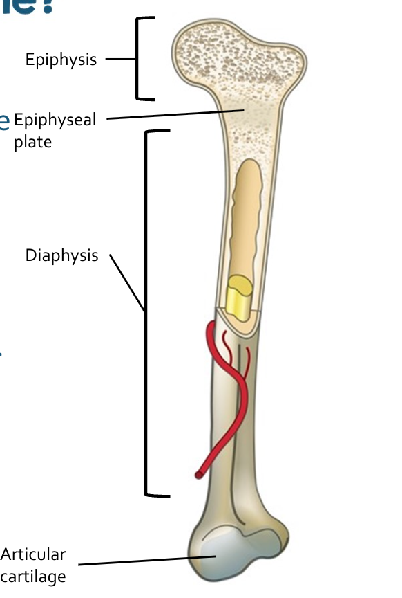
The Diaphysis
Contains a medullary cavity
Holds red bone marrow in children (stays in hematopoiesis)
Replaced by yellow bone marrow (stores fat)

Compact Bone
Dense and Strong, found beneath the periosteum
Forming the outer layer of bones
Cylindrical units called osteons
Consists of lamellae (concentric circles)
Lamellae surround the Haversian canal (containing blood vessels and nerves)
Haversian canals are connected by Volkmann’s canals
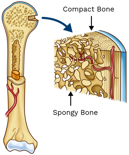
Spongy Bone
Found at ends of long bones and inside flat bones
Consists of a lattice of trabeculae (little beams)
Found along lines of stress for perfect resistance to compression
Between the trabeculae, space contains marrow and blood vessels
Periosteum
Tough connective tissue membrane covering the outer surface of bones. It is essential for bone growth, repair, and muscle attachm
Osteocytes
Mature bone cells make up the majority of the bone structure
Canaliculi connects all bone cells (receive nutrients and remove wastes)
Osteoblasts
Bone-forming cells that produce new bone matrix
Osteoclasts
Bone-resorbing cells that break down bone tissue
Bone Formation
Embryo’s skeleton is made of cartilage
Osteoblasts start at third month (ossification)
Cartilage is replaced by bone (secretes mineral)
Osteoblasts mature into osetocytes
Ossificiation
Calcium and minerals is incorporated into cartilage to become bone
Two hormones control
Bone remodeling is a continuous process regulated by calcium levels in blood
Parathyroid Hormone (PTH): Increases blood calcium by stimulating bone resorption
Calcitnonin: Lowers blood calcium by promoting bone formation
Bone Composition: Organic
35% Osteoid (made of ground substance & collagen)
Provides the flexibility & tensile strength required to keep bones from constantly breaking.
Lack of collagen causes “Brittle Bone Disease”
Bone Composition: Inorganic
65% mineral salts
Crystalline salts made of hydroxyapatites
Ca10(PO4)(OH)2
Provides bone strength & hardness
Lack of hydroxyapatite causes “Rickets”
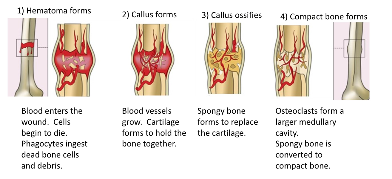
Four Stages of Bone Repair
Hematoma formation: Blood vessels break, forming a clot and initiating inflammation.
Fibrocartilaginous callus formation: New blood vessels and cartilage stabilize the fracture.
Bony callus formation: Spongy bone replaces cartilage.
Bone remodeling: Compact bone replaces spongy bone, restoring the medullary cavity.
What are the 2 major sections of the skeleton?
Axial Skeleton and Appendicular Skeleton
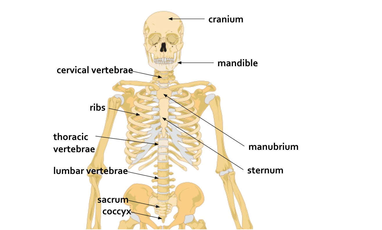
Axial Skeleton
80 bones
Central axis of the body
Skull, ribs, sternum, and vertebrae
Appendicular Skeleton
126 bones
Pectoral and Pelvic girdles
Bones of the arms, legs, pelvis, and shoulders

The Skull
22 bones
Cranial Bones and Facial Bones
Protect the brain, support the facial structure
House the special sense organs
Most skull bones are joined together by immovable joints called sutures
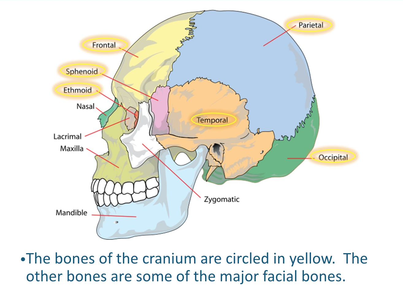
Cranial bones
The cranium encloses and protects the brain. It is composed of eight cranial bones
Facial Bones
The facial skeleton consists of 14 bones that form the framework of the face, support the teeth, and shape the nasal and oral cavities.
Sutures
Fibrous joints that tightly connect the bones of the skull
coronal, sagittal, lambdoid, and squamous structures
Fontanels
In infants, sutures are separated by flexible connective tissues
Allow skull growth and deformation during childbirth
Fontanels gradually ossify as skull develops
Paranasal Sinuses
Skull bones containing air-filled spaces
Frontal, maxillary, sphenoid, and ethmoid bones
Reduce weight of skull, help warm and humidify inhaled air
Contribute to voice resonance
Vertebral Column
Vertebral column extends from skull to pelvis
Protects spinal cord
consists of 33 vertebrae at birth
33 Vertebrae’s
Cervical (7)
Thoracic (12)
Lumbar (5)
Sacral (5 fused)
Coccygeal (4 fused)
Fibrous Joints
Immovable or slightly movable
Held together by fibrous connective tissue
Cartilaginous Joints
Immovable or slightly movable
Held together by cartilage
Synovial Joints
Highly movable
Contain synovial fluid for frictionless movement
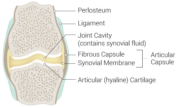
What is the structure of synovial joints?
Joint capsule filled with synovial fluid surrounds the end of the bones
A synovial membrane and articular cartilage line the joint cavity
Ligaments attach the bones of the joint
Ligaments
connect bone to bone
What are the types of Synovial Joints?
Pivot Join (between C1 and C2 vertebrae)
Hinge Joint (elbow)
Ball & Socket Joint (hip)
Saddle Joint (between trapezium carpal bone & first metacarpal bone)
Condylar joint (between radius and carpal bone of wrist)
Gliding joint or plane joint (between tarsal bones)
Bone Fractures
Disruption in the continuity of bone tissue that occurs when a bone is subjected to forces greater than it can withstand. May result from acute trauma, repetitive stress, or underlying disease processes that weaken bone structure