205 exam 1 (cells, tissue, bones)
 Call Kai
 Learn
Learn Practice Test
 Spaced Repetition
 Match
Match Flashcards
 Knowt Play
1/132

There's no tags or description
Looks like no tags are added yet.
Name | Mastery | Learn | Test | Matching | Spaced | Call with Kai | Chat |
|---|
No analytics yet
Send a link to your students to track their progress
133 Terms
Four key functions of the cell membrane are:
physical isolation
regulation of exchange in cell environment
communication between cell and environment
structural support
describe physical isolation
integral proteins are the key
channels
proteins bind to change shape of channels
keep ECF out and ICF in
describe regulation of exchange in the cell environment
channels (ion channels, water channels)
describe communication between the cell and its environment
communication through integral proteins and immune system
alerts system about foreign cells and disease
describe structural support
shape is maintained by membrane, integral proteins, and internal & external fluctuations
What are the main structures of the cell membrane?
phospholipids, integral proteins, and cholesterol (lipid rafts)
what are phospholipids?
phosphate head and two lipid tails
PO4 likes water, hydrophilic
two lipid tails hate water, hydrophobic
the PO4 heads face the ECF and the tails are facing the ICF
what are integral proteins and how are they imbedded?
Imbedded three ways:
transmembrane
facing outside
facing inside
They can move across the membrane when needed (moved by the cholesterol lipid raft)
they can be lipid-anchored
hat is cholesterol and why is it needed?
It is all within the ICF
Form lipid rafts to move integral proteins throughout the cell membrane tor
What is the fluid mosaic model?
phospholipids arranged w/ their polar (hydrophilic heads) face protein layer and non-polar (hydrophobic tails) face each other
what are modified proteins?
modifications can be made with molecules such as glycolipds
How do proteins interact
covalent bonds
Specific functions of phospholipids
structural stability, cell recognition, and barrier formation
specific functions of integral proteins
transport of substances across cell membrane
provision of binding site
formation of cell:cell junctions by contact adhesions
provision of enzymes
cell recognition
specific functions of cholesterol
barrier formation and lipid rafts
integral proteins: active vs passive transport
active: requires energy, uses ATP
passive: no energy spent, no ATP
integral proteins: passive transport (what does it transport and how? water channels vs ion channels vs glucose?)
diffusion: moves to lower concentration (down gradient)
osmosis
Transports nutrients, water, and ions
no net gain & no net loss (ie. 5 molecules go in, 5 must come out)
Water channels:
uses aquaporins
Ion channels:
moves Na+, K+, or both (dual ion transport)
Glucose moved with facilitated transport (glucose transport receptor, GTR)

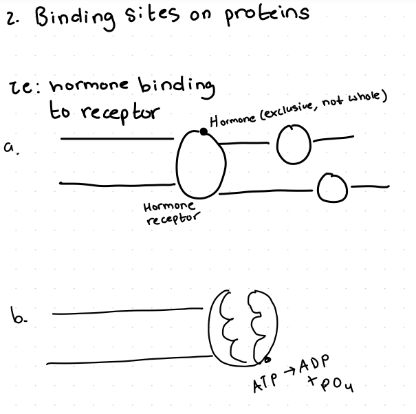
Integral proteins: provision of binding sites
Becomes a receptor (only a portion, not the entire protein, exclusive)
Ie. hormones binding to receptor
or dual ion channel that binds with ATP and turns it into ADP to change shape to bind to many things
Integral proteins: cell:cell junction
Two types: non-permissive and permissive
Non-permissive: integral proteins form a zipper-like binding of cells w/ strong covalent bonds
Permissive: integral proteins still join the cells, but there is a gap for hormones or ions to pass from one cell to the other

Integral proteins: provision of enzymes
portion of the integral protein is enzymatic
(*all enzymes are proteins, but not all proteins are enzymes!)

Integral proteins: immunosurvalence
tests for self vs non-self
checks w/ short, weak covalent bonds
then signals to terminate non-self

Diffusion (& describe simple vs facilitated)
Passive bc DOWN gradient (from high to low conc.)
distribution of a substance in a solvent such that it gets equally concentrated
Simple: DOWN conc. gradient (no help, ions, gas, & alcohol)
Facilitated: DOWN a conc. gradient but requires a CARRIER system (transmembrane protein) (help, ie. glucose transport receptor)
5 main points of osmosis
(integral proteins: transport)
Passive transport
water moves (substances are “stuck”)
across the membrane
via aquaporins
from low to high solute/particles concentration (dilutes high conc.)
What is ATP?
Adenosine triphosphate
What is the normal concentration of NaCl (saline) in the body?
0.9%
Isotonic solution
iso means “same”
Equal conc. of solute in the ICF and ECF
water moves equally between the ECF and ICF so there is no net gain and no net loss

Hypotonic solution
Higher conc. of solute in ICF, so water from the ECF rushes in to make the conc. equal again
Cells will swell and can lyse

Hypertonic solution
Higher conc. of solute in the ECF than the ICF
Water rushes out of the cell into the ECF
cells with shrivel (crenation)

What tissues does active transport happen in?
excitable cells: muscle cells and neurons
3 types of stimuli for excitable cells
chemical (ie. smell)
physical (ie. touch)
electrical (ie. changes)
When excited, channels open
At rest, channels closed
breakage of ATP term
hydrolysis
(becomes adenosine diphosphate, then monophosphate)
Describe & draw how the Na+, K+, and dual ion channels work in cell membranes (& polarizations, depolarization)
More Na+ on outside & more K+ on inside, + outside & - inside
Stimulus
Na+ travels through Na+ ion channel
Inside of membrane depolarizes (excited)
Na+ diffuses and K+ leaves through the K+ ion channel in response, repolarization occurs (relaxation)
These are all PASSIVE traspnsport, no ATP used bc the particle are going from high to low concentration
Then, Na+ & K+ can travel through the dual ion channel transmembrane integral protein pump
3 Na+ ions bind in the native state
pump cleaves a PO4 from an ATP (phosphorylation) and changes shape to expel 3 Na+ ions into the ECF
Two K+ bind as PO4 is still attached
Dephosphorylation occurs and pump returns to native state & expels 2 K+ ions into the ICF
Repeat

Specifically describe the dual ion channel
Integral protein

Depolarization = ?
Depolarization excitation
Polarization = ?
Polarization = relaxation
Repolarization & RMP
The initial gain in Na+ in the ICF makes the RMP less negative, repolarization makes it negative again with a loss of K+
The K+ leaves in response to the Na+ arriving into the ICF
Na+-K+ ATPase Pump

Identify this cell:cell junction
Gap junctions (communicating junctions)
AKA cell adhesion molecules (CAMs)
Permissive
Direct cell:cell communication (ex: smooth & cardiac muscle)

Identify this cell:cell junction
Tight junctions (occluding junctions)(“buttons”)
Non-permissive
Blocks movement between cells (ie. Epithelial cells)

Identify this cell:cell junction
Desmosome (a cell:cell anchoring junction)
Holds cells together & to ECM
Non-permissive
(Ie. Skin cells) (ex: protein ares heat labile, so when friction melts proteins on the skin, fluid fills the space & forms a blister. The desmosomes are what dissolve when blisters are formed)

What is in the cytoplasm?
Cytosol (ICF) (liquid component)
Organelles
mitochondria (powerhouse, ATP)(excitable cells hv a lot)
endoplasmic reticulum (rough & smooth)
ribosomes (site of protein synthesis)
lysosomes (holds enzymes, acids, H2O, etc. & kills & breaks down, aggressive)
Cytoskeleton (filaments/microtubules)
Describe plasma & its role & ratio to cells
55% plasma & 45% cell
Forms bodily fluids
(ECF derived from plasma)

Mitochondria
Excitable cells hv the most (muscle & neurons)

Endoplasmic reticulum (specify rough vs smooth)
Rough: ribosomes on surface
in immune cells (bc they make a lot of proteins)
Smooth: no ribosomes, site for lipid synthesis
(ie. Steroidal glandular cells, muscle cells bc sarcoplasmic reticulum is the storehouse of Ca+ in muscles)

Lysosomes
Vesicles that contain powerful enzymes
Ie. White blood cell macrophage and osteoclasts

Four basic types of tissue
Epithelial (most abundant)
Connective
Muscle (excitable)
Nervous/neural (neurons excitable)
Epithelial classification by layers: simple
One layer

Epithelial classification by layers: stratified
Many layers

Epithelial classification by shape: squamous
Flat

Epithelial classification by shape: Cuboidal
Square

Epithelial classification by shape: columnar
Tall


Identify this tissue
Simple squamous epithelial (simplest)
Function: Allows passage of materials by diffusion where protection is not important, flat for gaseous exchange
Found in parts of kidney, alveolar cells (lung air sacs), lining of heart, blood vessels, and lymphatic vessels

What is the simple squamous cell in capillaries called?
Endothelial cell epithelial

Identify this tissue
Simple cuboidal epithelial
Function: secretion & absorption
Found in kidney tubules, ducts, & small glands


Identify this tissue
Simple columnar epithelial
Function: absorption; secretion of mucus, enzymes, etc
Non-ciliated: in digestive tract, gallbladder
Ciliated: bronchi, uterine tubes, and parts of uterus


Identify this tissue
Pseudostratified epithelial
“Falsely” stratified, single layer
Varied height in cells due to nucleus changing position is why it appears stratified
Functions: secretion of mucus, entraps pathogens before it enters the body
Found in sperm ducts and parts of trachea and upper respiratory tract
The goblet cells hv a lumen, provides thin layer of sticky mucous to trap foreign particles, then macrophages destroy them with lysosomes


Identify this tissue
Stratified squamous epithelial
Function: text underlying tissues in areas subjected to abrasions
Nonkeratinized type is found in moist linings of the esophagus mouth and vagina
Keratinized variety forms, the epidermidis of the skin, which is a dry membrane (impervious)


Identify this tissue
Stratified cuboidal epithelial
Function: protection
Found in large ducts of sweat, glands, mammary glands, and salivary glands
If they’re in a duct: it is the stratified cuboidal cells of the exocrine gland
If they are n a cluster (and directly putting their hormone into the blood supply): they are endocrine stratified cuboidal cells


Identify this tissue
Stratified columnar epithelial, RARE
Function: protection, secretion, conveys urine and semen
Found in male urethra
Variation in height is just due to the inner cells having less space to stretch out

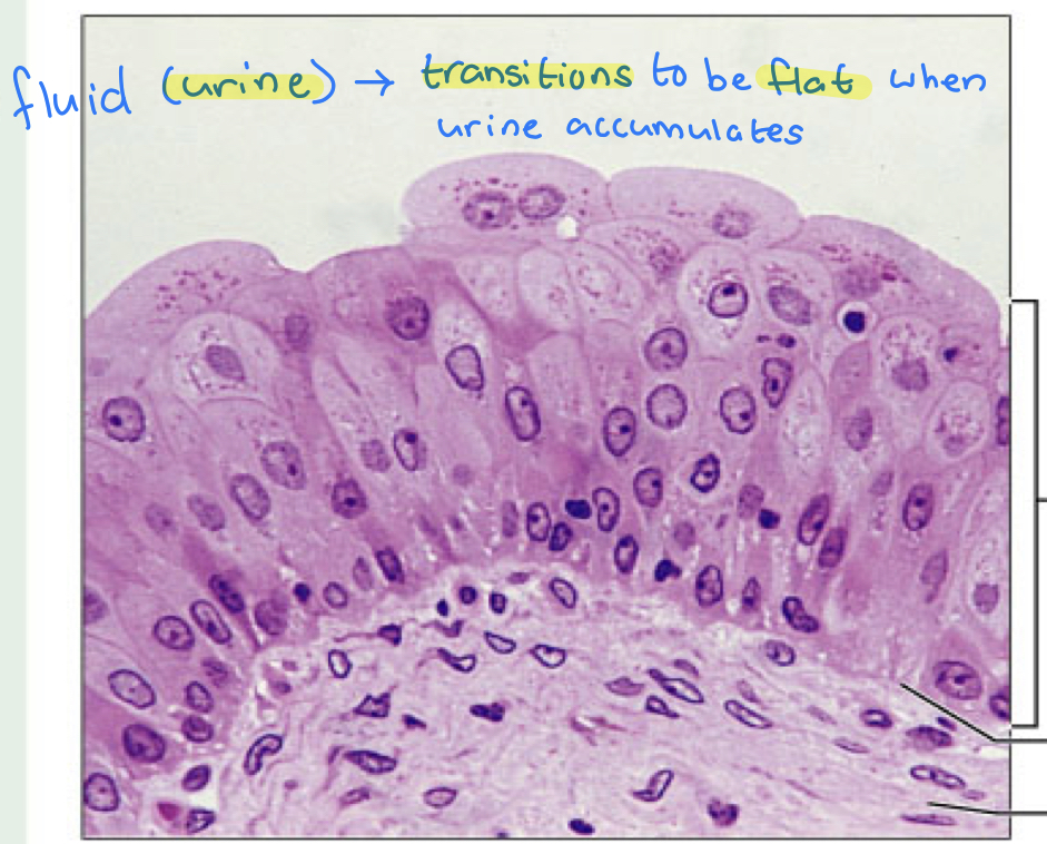
identify this tissue
Transitional epithelial
Function: stretches and expels urine in the kidney pelvis (compresses when a lot of urine is present)
Found in the kidney, ureters, and part of urethra

Connective tissue (what does it do, what are the two categories, and what are the specific examples?)
Connects other tissues
Gives form and strength to organs
Two types: Loose and dense
cartilage
Bone
Blood
Extracellular matrix (ECM)
Matrix is being produced by the cells, and it is gooey and proteinaceous
It then becomes the environment in which the cell lives, communicates, etc.
It plays a large role in the skeletal system
Determines what kind of cartilage it will be
Mesenchymal (stem) cells

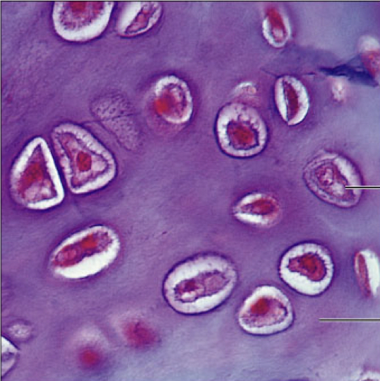
Identify this tissue
Hyaline cartilage (connective)
Function: supports and reinforces, cushioning
Found in costal cartilage, nose, trachea, larynx, embryonic skeletons, ends of long bones (plays a role in long bone synthesis)
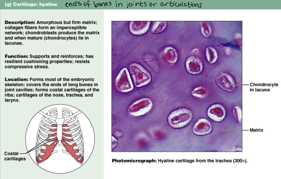

Identify this tissue
Fibrocartilage (connective)
Function: tensile strength that absorbs compressive shock
Found in intervertebral discs, pubic symphysis, discs of knee join


Identify this tissue
Elastic cartilage (connective)
Function: maintains the shape of a structure while allowing great flexibility
Found in the external ear (pinna)
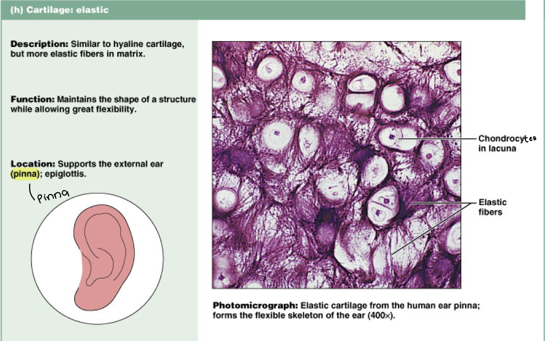

Identify this tissue
Compact bone (connective)
Rigid outer layer of bone, has central blood vessel
Most mineralized and inorganic, 80% CaPO4
Function: supports and protects; provides levers for muscles to act on, stores calcium and other minerals, marrow in the bone is the site for blood/hematopoiesis


Identify this tissue
Blood (connective)
Function: transports respiratory gases, nutrients, water, and other substances
Found in blood vessels
mature RBC hv no nuclei, only immature ones do

Muscle tissue traits and types
Contractile
Excitable- they conduct signals
Actin and myosin
Types:
Skeletal
Cardiac
Smooth

Identify this tissue
Skeletal muscle (connective)
Function: voluntary movement
Found in muscles attached to bones or skin


Identify this tissue
Cardiac muscle (connective)
Function: contracts to propel blood, involuntary
Found in the walls of heart


Identify this tissue
smooth muscle (connective)
Function: propels substances or objects along internal passageways; involuntary
Found in that walls of organs

Identify the tissue and cells
Neuron and glial cells (nervous tissue)
Neurons send signals
excitable
Electrical
Chemical
Glial cells (aka neuroglia) support neurons (more abundant)

Function of skeletal system

What is compact bone
Hard layer covers most bones, forms shaft of long bones
What is cancellous bone
Spongy bone
Spicules arranged in a porous network, has small open spaces filled with marrow
What is medullary cavity
Space surrounded by cortex of a long bone filled with red or yellow marrow
What is the epiphysis
Ends of the bone
What is the diaphysis
Shaft of the bone which surrounds the medullary cavity
What is articulate cartilage
Hyaline, cartilage in joints (glassy and smooth for movement)
Cushions ends of bones and allows smooth movement
What is epiphyseal plate
Areas made of cartilage allowing for growth of the bone; lengthens
Chondroblasts live in here, lengthen bone
What is periosteum and endosteum
Fibrous membrane covering compact bone and cancellous bone/medullary
Peri: outside, lining the compact bone
Endo: inside, lining the cancellous bone
What does the periosteum contain?
Mesenchymal (stem) cells and osteoblasts (immature)
For repair, ie. broken bone
What are mesenchymal cells and what do they create?


Label this long bone
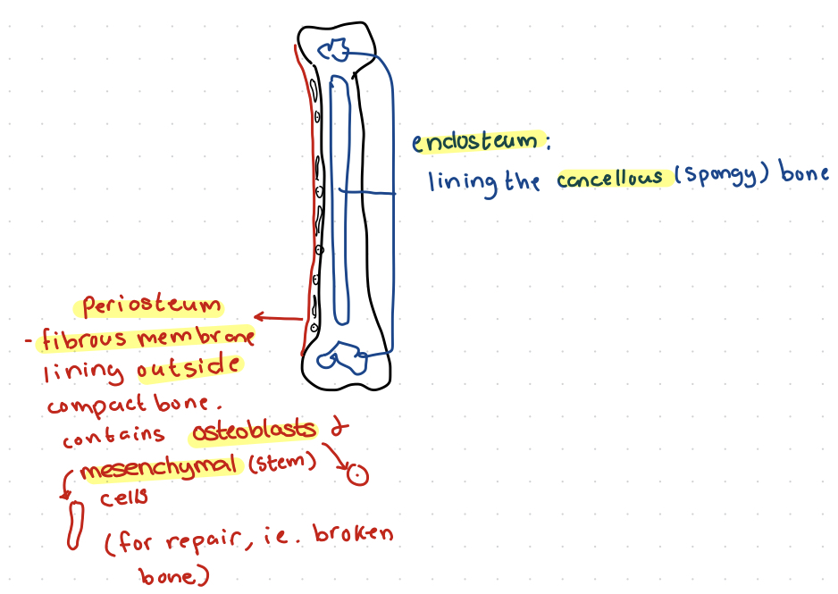

Label
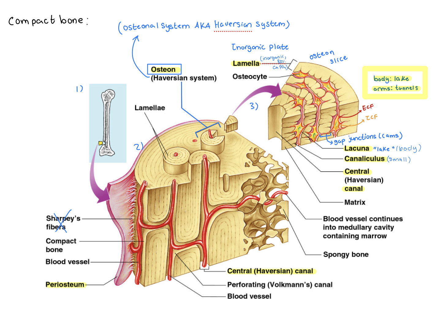
Label (mainly osteon)
Long bones
Levers, aid in support, locomotion, pretension
Ie. femur
Short bones
Gliding/sliding movements, absorb shock
Ie. carpals
Flat bones
Protect vital organs
Ie. skull
Irregular bones
Protection, support, muscular attachment
Ie. Vertebrae
Sesamoid bones
Reduce friction, increase leverage, change direction of pull
Ie. patella & digital joints
Name the parts of the axial and appendicular skeleton
Axial:
skull
Vert. column
Sternum
Ribs
Appendicular:
thoracic limb (forelimb)/pectoral
Pelvic limbs (hind limb)
Shoulder
Pelvis


Anatomy label

What bone cell is this and what does it do?
Osteogenic cell (Mesenchymal cell aka bone stem cell)
produce osteoblasts


What bone cell is this and what does it do?
Osteoblast: immature bone cell that secretes the organic part of matrix (osteoid)

What is osteoid?
Bone matrix on which Ca++ is deposited on from the blood
(Blood to bone, bone to blood is Ca++ flow)
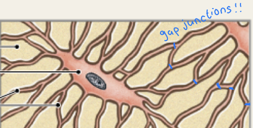
What bone cell is this and what does it do?
Osteocyte: mature bone cell that maintains the bone matrix


What bone cell is this and what does it do?
Osteoclast: multinucleate cell that secretes acids and enzymes to dissolve bone matrix
Has lysosomes that break down CaPO4 to make Ca++ available in the blood (shoots the lamellae in the compact bone with the lysosomes)(lamellae are the inorganic, CaPO4 containing part of the bone)

What is osteon?
Concentric circle, telescopic tube running the compact bone
and the atypical osteon running through the cancellous bone that does not hv a blood vessel running through it (blood supply on outside)