Chapter 5 The Integumentary System (Openstax)
1/97
There's no tags or description
Looks like no tags are added yet.
Name | Mastery | Learn | Test | Matching | Spaced | Call with Kai |
|---|
No analytics yet
Send a link to your students to track their progress
98 Terms
integumentary system
Consists of the skin and accessory organs; hair, nails, and cutaneous glands
provides the body with overall protection
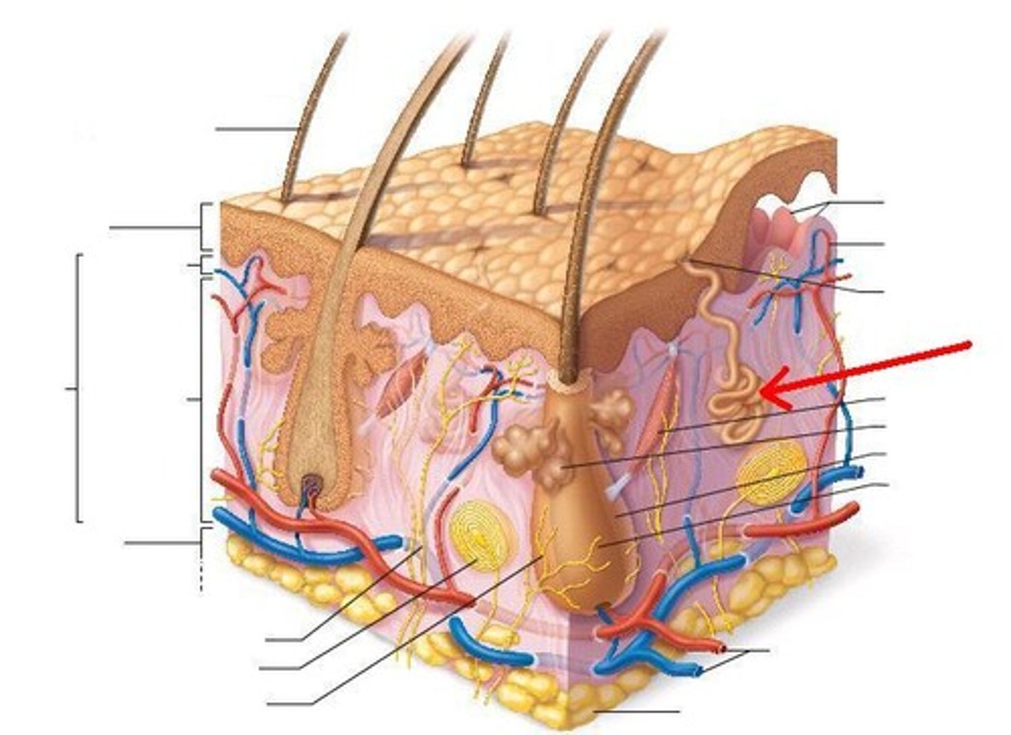
skin is made of multiple layers of cells and tissues, which are held to underlying structures by connective tissue
skin
made of two main layers: epidermis and dermis
epidermis
- composed of keratinized, stratified squamous epithelium
- made of four or five layers of epithelial cells, depending on its location in the body
- does not contain any blood vessels
thin skin
- epidermis skin with 4 layers of epithelial cells
- layers from deep to superficial are: stratum basale, stratum spinosum, stratum granulosum, and stratum corneum
thick skin
- epidermis tissue with 5 layers of epithelial cells
- located on palms of hands and soles of feet
- 5th layer of epithelial cells is stratum lucidum
- layers from deep to superficial are: stratum basale, stratum spinosum, stratum granulosum, stratum lucidum, and stratum corneum
keratinocyte
- is a squamous cell that manufactures and stores the protein keratin
- located in all layers of the epidermis EXCEPT the stratum basale
- these cells located in the stratum corneum are dead and are regularly sloughed away and replaced by cells in deeper layers
- are produced by the singular layer of basal cells in teh stratum basale
keratin
is an intracellular fibrous protein that gives hair, nails, and skin their hardness and water-resistant properties
stratum basale
- also called the stratum germinativum
- consists of a single layer of cuboidal, basal cells
- Merkel cells and melanocytes are also found in this layer dispersed in the layer of basal cells
- deepest layer of epidermis and attaches the epidermis to the basal lamina
- cells in this layer bond to the dermis via intertwining collagen fibers, referred to as the basement membrane
dermal papillae
- fingerlike projection, or fold found in the superficial portion of the dermis
- increase the strength of the connection between the epidermis and dermis; the greater the folding, the stronger the connections made
basal cell
is a cuboidal-shaped stem cell that is a precursor of the keratinocytes of the epidermis
merkel cell
- cell found in the stratum basale
- which functions as a receptor and is responsible for stimulating sensory nerves that the brain perceives as touch
- especially abundant on hands and feet
- touch receptors
melanocytes
- cell found in stratum basale
- a cell that produces the pigment melanin
melanin
- gives hair and skin its color, and also helps protect the living cells of the epidermis from ultraviolet (UV) radiation damage
- increased melanin accumulation protects the DNA of epidermal cells from UV ray damage and the breakdown of folic acid, a nutrient necessary for our health and well-being
fingerprints
- form where the cells of the stratum basale meet the papillae of the underlying dermal layer (papillary layer), resulting in the formation of the ridges on your fingers
- unique to each individual and do not change with age or growth
stratum spinosum
- is spiny in appearance due to the protruding cell processes that join the cells via a structure called a desmosome
- composed of 8-10 layers of keratinocytes, formed as a result of cell division in the stratum basale
- dispersed among the keratinocytes are a type of dendritic cells called the Langerhan cells
- superficial to the stratum basale
desmosome
- interlock with each other and strengthen the bond between the cells
- found in the stratum spinosum
Langerhan cells
- dendritic cells found dispersed in the keratinocytes in the stratum spinosum
- which functions as a macrophage by engulfing bacteria, foreign particles, and damaged cells that occur in this layer
keratinocytes in stratum spinosum
- begin the synthesis of keratin and release a water-repelling glycolipid that helps prevent water loss from the body, making the skin relatively waterproof
stratum granulosum
- has a grainy appearance due to further changes to the keratinocytes as they are pushed from the stratum spinosum
- cells (three to five layers deep) become flatter, their cell membranes thicken, and they generate large amounts of the proteins keratin (fibrous) and keratohyalin, which accumulates as lamellar granules within the cells
- These two proteins make up the bulk of the keratinocyte mass in the stratum granulosum and give the layer its grainy appearance
keratohyalin
- protein produced in the stratum granulosum that accumulates as lamellar granules within cells
stratum lucidum
- is a smooth, seemingly translucent layer of the epidermis located just above the stratum granulosum and below the stratum corneum
- found only in thick skin
- composed of dead and flattened keratinocytes that are densely packed with eleiden
eleiden
- a clear protein rich in lipids, derived from keratohyalin, which gives these cells their transparent (i.e., lucid) appearance and provides a barrier to water
- located in stratum lucidum
stratum corneum
- is the most superficial layer of the epidermis and is the layer exposed to the outside environment
- increased keratinization (also called cornification) of the cells in this layer gives it its name
- 15-30 layers of cells
- dry, dead layer helps prevent the penetration of microbes and the dehydration of underlying tissues, and provides a mechanical protection against abrasion for the more delicate, underlying layers.
- layer sheds periodically and is entirely replaced in the span of 4 weeks
dermis
- considered the "core" of the integumentary system
- It contains blood and lymph vessels, nerves, and other structures, such as hair follicles and sweat glands
- is made of two layers (papillary and reticular) of connective tissue that compose an interconnected mesh of elastin and collagenous fibers, produced by fibroblasts
papillary layer
- superficial layer of the dermis
- is made of loose, areolar connective tissue, which means the collagen and elastin fibers of this layer form a loose mesh
- projects into the stratum basale of the epidermis to form finger-like dermal papillae
- contains fibroblasts, a small number of fat cells (adipocytes), and an abundance of small blood vessels and phagocytes, lymphatic capillaries, nerve fibers, and touch receptors called the Meissner corpuscles
adipocytes
- fat cells
- located in papillary layer
phagocytes
defensive cells that help fight bacteria or other infections that have breached the skin
reticular layer
- thicker layer in dermis, deeper than papillary layer
- composed of dense, irregular connective tissue
- well vascularized and has a rich sensory and sympathetic nerve supply
- appears reticulated (net- like) due to a tight meshwork of fibers (elastin and collagen)
elastin fibers
- fibers located in the reticular layer of the dermis
- provide some elasticity to the skin, enabling movement
collagen fibers
- fibers located in the reticular layer of the dermis and extends into the papillary layer of the dermis and the hypodermis
- provide structure and tensile strength
- binds water to keep the skin hydrated
hypodermis
- also called the subcutaneous layer or superficial fascia
- is a layer directly below the dermis and serves to connect the skin to the underlying fascia (fibrous tissue) of the bones and muscles
- It is not strictly a part of the skin
- consists of well- vascularized, loose, areolar connective tissue and adipose tissue, which functions as a mode of fat storage and provides insulation and cushioning for the integument
melanosome
A cellular vesicle that transfers melanin into keratinocytes
- temporary structures eventually destroyed by lysosomes
moles
larger masses of melanocytes that should be monitored for changes that might indicate the presence of cancer
albinism
- is a genetic disorder that affects (completely or partially) the coloring of skin, hair, and eyes.
- The defect is primarily due to the inability of melanocytes to produce melanin
- remember melanin protects from UV damage--> typically people with this disorder limit sun exposure because they are more prone to sunburn and skin cancer
vitiligo
- the melanocytes in certain areas lose their ability to produce melanin, possibly due to an autoimmune reaction
Addison's disease
- disease that can stimulate the release of excess amounts of adrenocorticotropic hormone (ACTH), which can give the skin a deep bronze color
- darkening of skin
Accessory structures of the skin
hair, skin glands, and nails
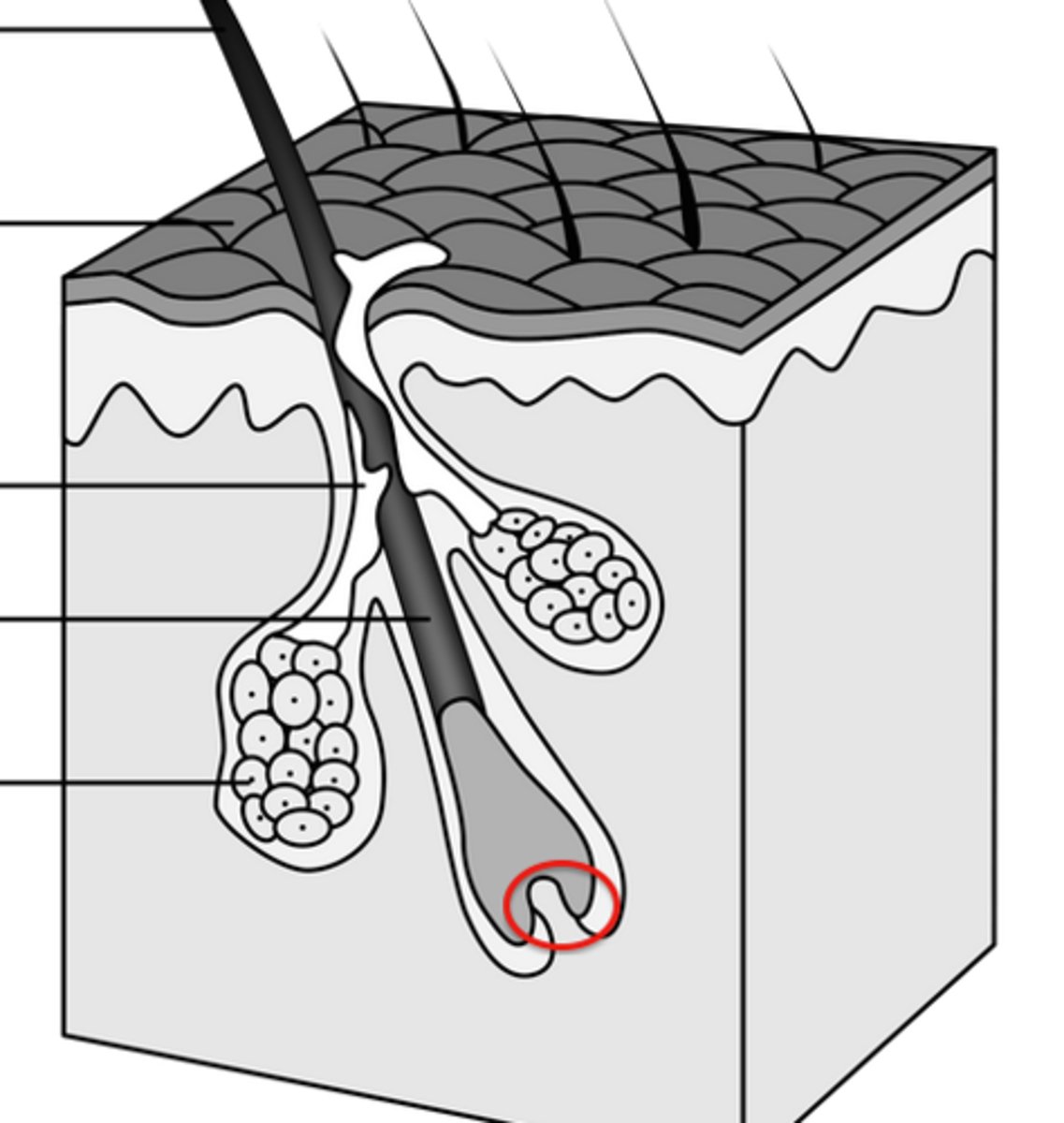
hair
- is a keratinous filament growing out of the epidermis
- primarily made of dead, keratinized cells
- originate from the hair follicle
- serves a variety of functions, including protection, sensory input, thermoregulation, and communication
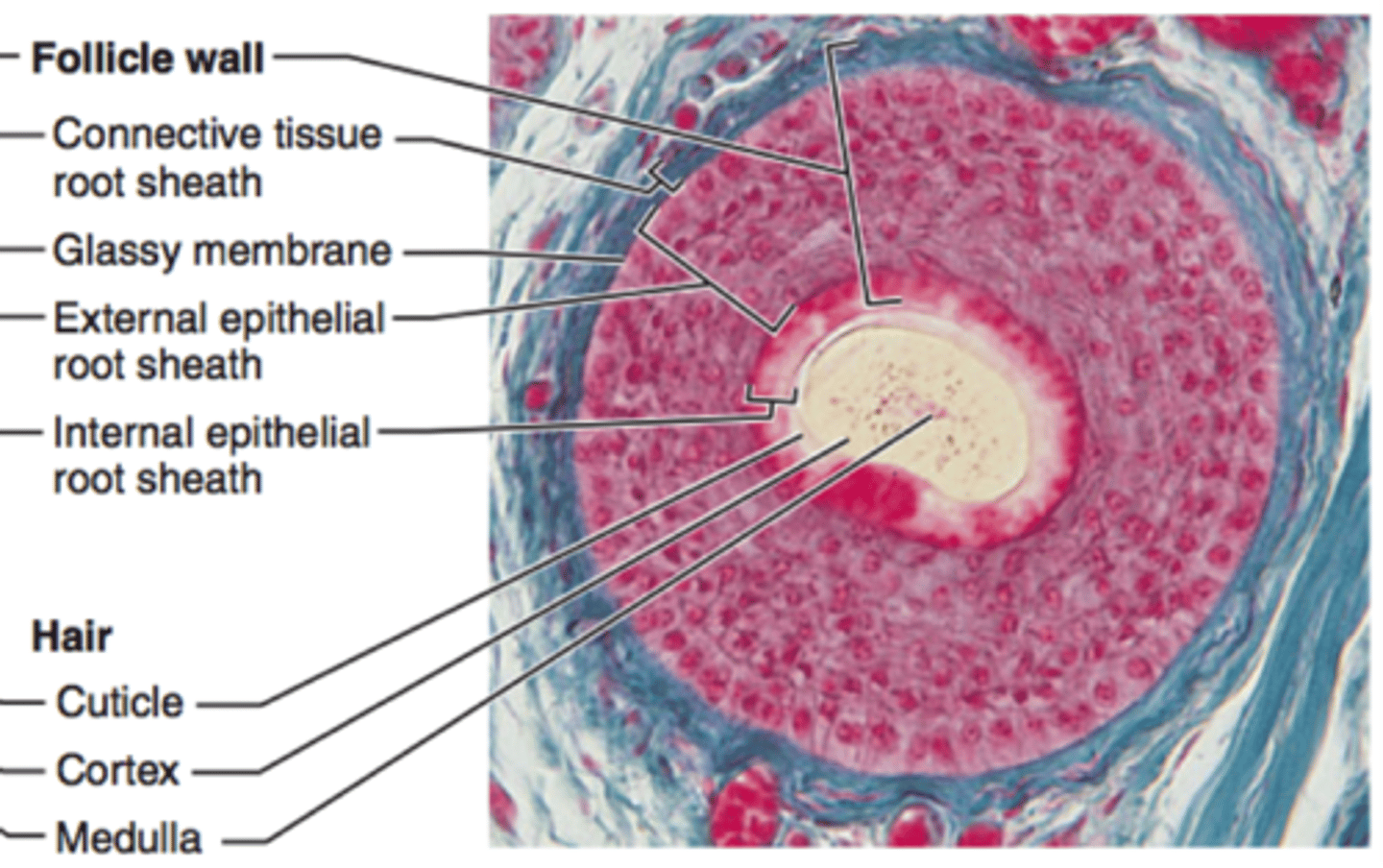
hair follicle
- where strands of hair originate in an epidermal penetration of the dermis
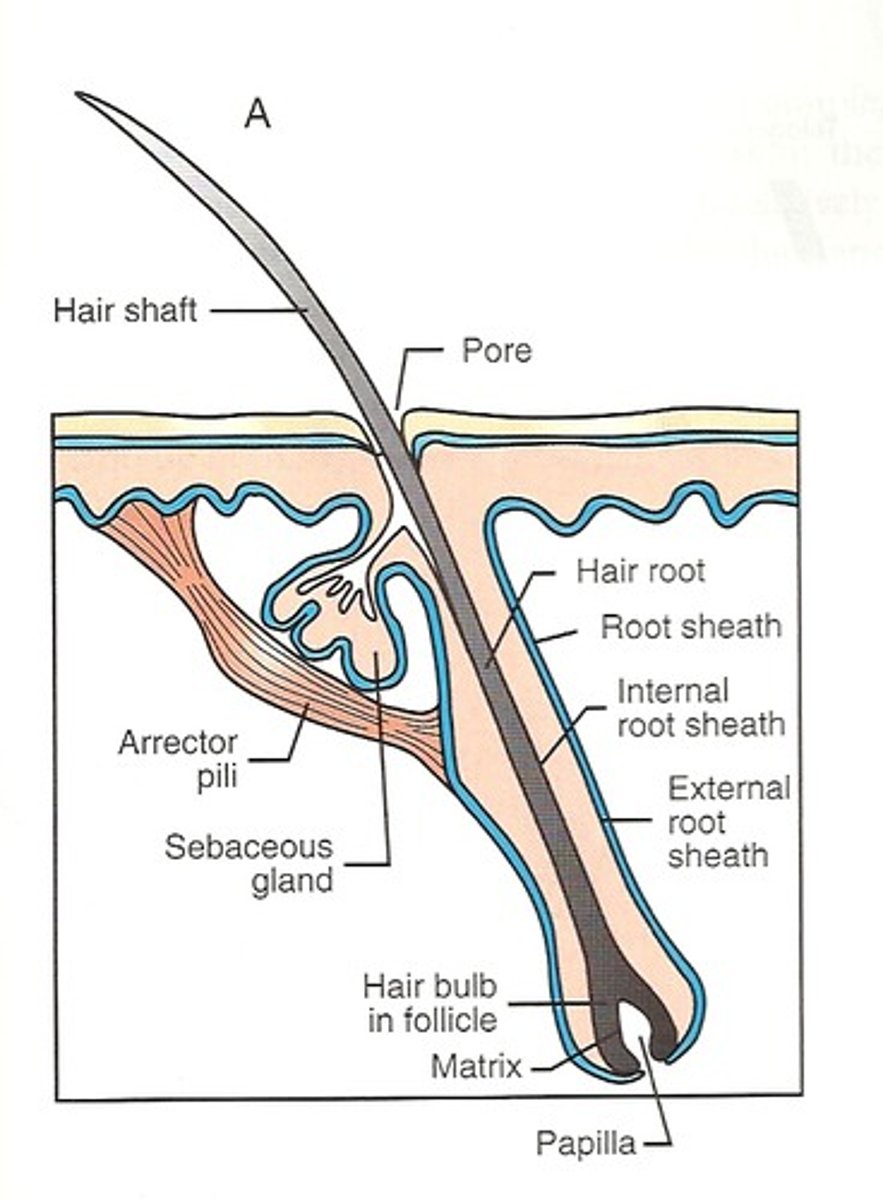
hair shaft
- is the part of the hair not anchored to the follicle, and much of this is exposed at the skin's surface
- hair exposed to the surface is dead and completely composed of keratin

hair root
- rest of the hair, which is anchored in the follicle, lies below the surface of the skin
- ends deep in the dermis at the hair bulb, and includes a layer of mitotically active basal cells called the hair matrix
hair bulb
- where the hair root ends deep in the dermis
- the basal cells of the hair bulb divide and push cells outward in the hair root and shaft as the hair grows

hair matrix
a layer of mitotically active basal cells in the hair bulb
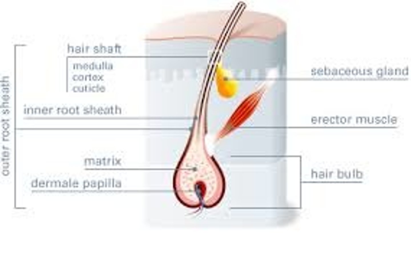
hair papilla
- is made of connective tissue and contains blood capillaries and nerve endings from the dermis
- surrounded by the hair bulb
medulla
Central core of hair shaft
cortex
- surrounds the medulla
- a layer of compressed, keratinized cells that is covered by an outer layer of very hard, keratinized cells, called the cuticle
cuticle
surrounds the cortex and is composed of very hard, keratinized cells
internal root sheath
- the cells of this sheath surround the root of the growing hair and extend just up to the hair shaft
- They are derived from the basal cells of the hair matrix
external root sheath
- which is an extension of the epidermis, encloses the hair root
- It is made of basal cells at the base of the hair root and tends to be more keratinous in the upper regions
glassy membrane
is a thick, clear connective tissue sheath covering the hair root, connecting it to the tissue of the dermis
arrector pili muscle
- smooth muscle connected to each hair root
- contracts in response to nerve signals from the sympathetic nervous system, making the external hair shaft "stand up."
Hair growth phases
(1) Anagen: growth
(2) Catagen: intermediate/regression (apoptosis)
(3) Telogen: rest/shedding
anagen
- The period of active hair growth
- during which cells divide rapidly at the root of the hair, pushing the hair shaft up and out.
- The length of this phase is measured in years, typically from 2 to 7 years
catagen
- The period of break down and change of hair growth
- phase lasts only 2 to 3 weeks, and marks a transition from the hair follicle's active growth
telogen
- Resting phase of hair growth
- typically lasts 2-4 months
- the hair follicle is at rest and no new growth occurs
- after this phase, anagen begins again
hair color
- hair gets it color from melanin in the hair papilla
nail bed
- is a specialized structure of the epidermis that is found at the tips of our fingers and toes.
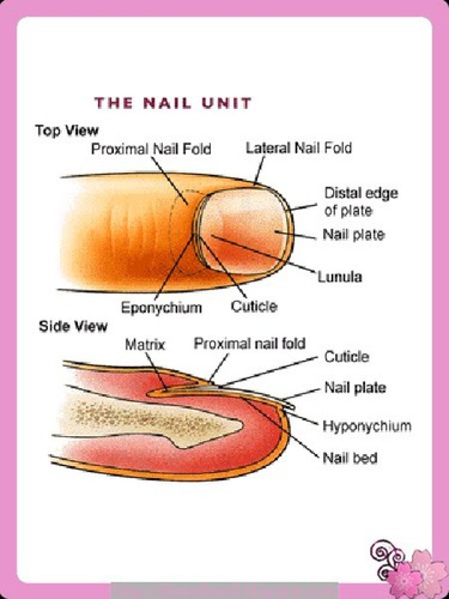
nail body
- is formed on the nail bed, and protects the tips of our fingers and toes as they are the farthest extremities and the parts of the body that experience the maximum mechanical stress
- forms a back-support for picking up small objects with the fingers
- is composed of densely packed dead keratinocytes
- is rich in blood vessels, making it appear pink
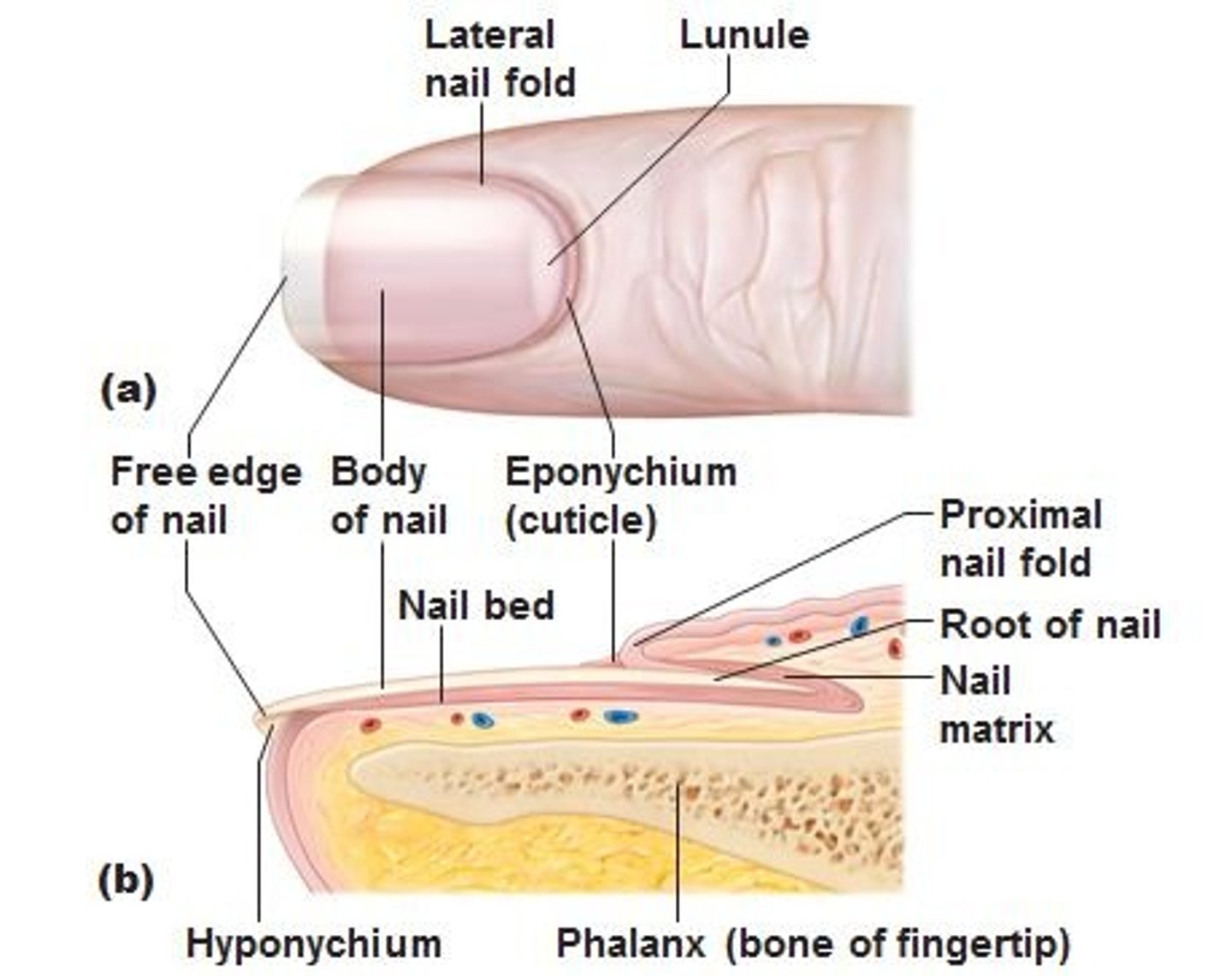
nail root
- a specialized structure upon which nail bodies can form
- which has a matrix of proliferating cells from the stratum basale that enables the nail to grow continuously

nail fold
- overlaps the nail on the sides, helping to anchor the nail body

nail cuticle
- forms when the nail fold that meets the proximal end of the nail body
- also called the eponychium

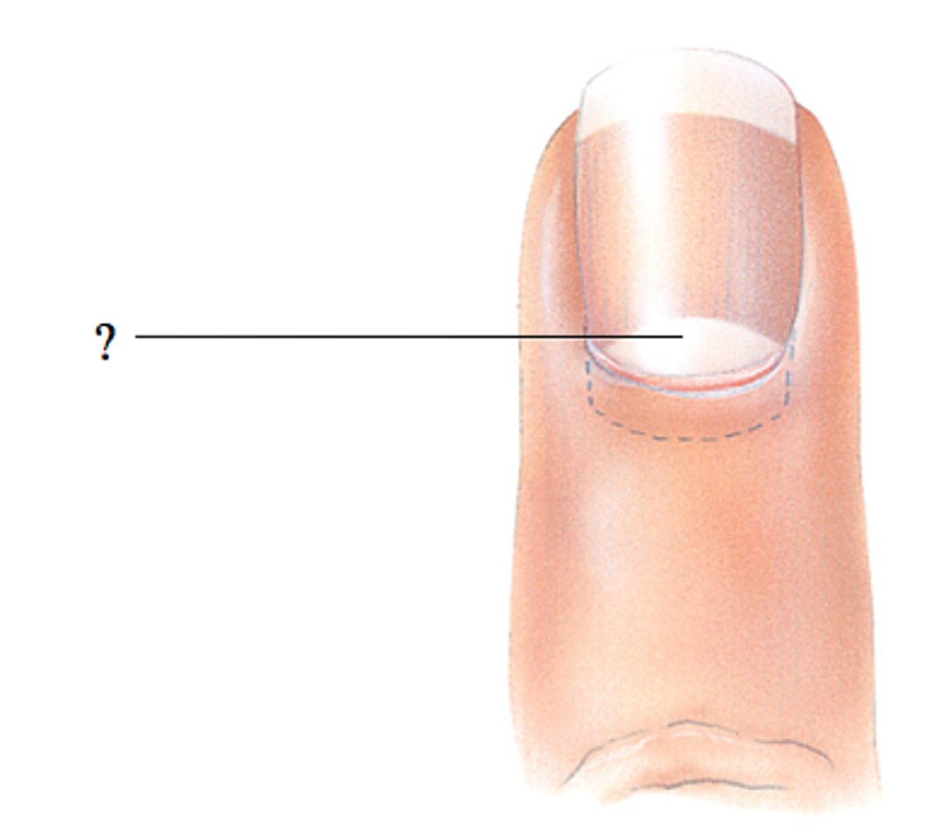
lunula (little moon)
- at the base of the nail body
- a crescent-shaped region on the nail body where a thick layer of epithelium covers the nail matrix
hyponychium
- The area beneath the free edge of the nail, furthest from the cuticle
- consists of a thickened layer of stratum corneum

sweat glands
- develop from epidermal projections into the dermis and are classified as merocrine glands
- sudoriferous glands
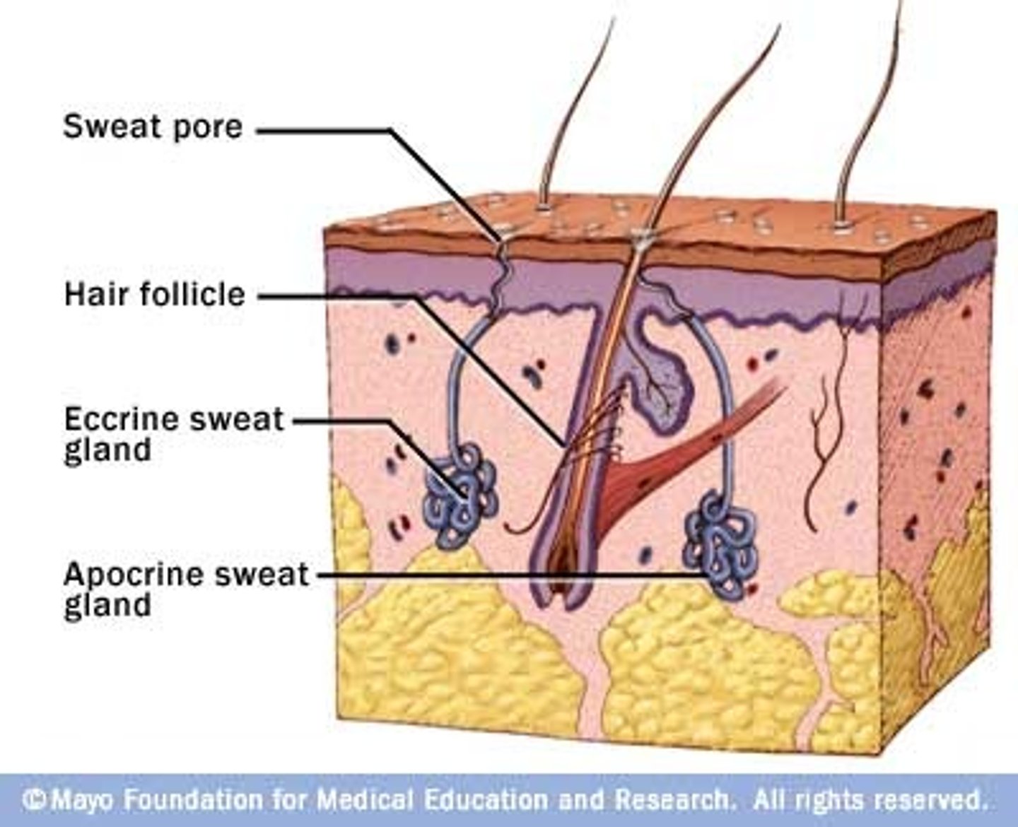
merocrine glands
the secretions are excreted by exocytosis through a duct without affecting the cells of the gland
eccrine sweat gland
- is type of gland that produces a hypotonic sweat for thermoregulation
- These glands are found all over the skin's surface, but are especially abundant on the palms of the hand, the soles of the feet, and the forehead
- They are coiled glands lying deep in the dermis, with the duct rising up to a pore on the skin surface, where the sweat is released.
- This type of sweat, released by exocytosis, is hypotonic and composed mostly of water, with some salt, antibodies, traces of metabolic waste, and dermicidin, an antimicrobial peptide.
Eccrine glands are a primary component of thermoregulation in humans and thus help to maintain homeostasis.
apocrine glands
- usually associated with hair follicles in densely hairy areas, such as armpits and genital regions
- larger than eccrine sweat glands and lie deeper in the dermis, sometimes even reaching the hypodermis, with the duct normally emptying into the hair follicle
- sweat includes water and salts, but also organic compounds that make the sweat thicker and subject to bacterial decomposition and subsequent smell
- release of this sweat is under both nervous and hormonal control, and plays a role in the poorly understood human pheromone response
sebaceous glands
- is a type of oil gland that is found all over the body and helps to lubricate and waterproof the skin and hair. - Most are associated with hair follicles.
- generate and excrete sebum onto the skin surface
sebum
- secreted by the sebaceous gland
- a mixture of lipids that are secreted onto the skin surface, naturally lubricating the dry and dead layer of keratinized cells of the stratum corneum, keeping it pliable
- the fatty acids have antibacterial properties, and prevent water loss from the skin in low-humidity environments
- secretion is stimulated by hormones
integumentary system functions
- protecting the body from invasion by microorganisms, chemicals, and other environmental factors
- preventing dehydration; acting as a sensory organ
- modulating body temperature and electrolyte balance
- synthesizing vitamin D
hypodermis functions
- fat storage
- forming a cushion over underlying structures
- providing insulation for colder temperatures
Meissner corpuscle
- tactile corpuscle
- responds to light touch
Pacinian corpuscle
- lamellated corpuscle
- responds to vibration
sympathetic nervous system
- the division of the nervous system involved in our fight-or-flight responses
- continuously monitoring body temperature and initiating appropriate motor responses
Vitamin D
- synthesized by the epidermal layer when exposed to UV radiation
- is essential for normal absorption of calcium and phosphorous, which are required for healthy bones
- absence of sun exposure can lead to a lack of vitamin D in the body, leading to a condition called rickets
- is essential for general immunity against bacterial, viral, and fungal infections.
rickets
- lack of vitamin D
- a painful condition in children where the bones are misshapen due to a lack of calcium, causing bowleggedness
osteomalacia
- condition when elderly individuals suffer from vitamin D deficiency
- softening of bones
metastasis
The spread of cancer cells to locations distant from their original site.
basal cell carcinoma
- is a form of cancer that affects the mitotically active stem cells in the stratum basale of the epidermis
- the most common of all cancers that occur in the United States and is frequently found on the head, neck, arms, and back, which are areas that are most susceptible to long-term sun exposure
- start in the stratum basale and usually spread along this boundary
squamous cell carcinoma
- is a cancer that affects the keratinocytes of the stratum spinosum and presents as lesions commonly found on the scalp, ears, and hands
- is the second most common skin cancer
melanoma
- is a cancer characterized by the uncontrolled growth of melanocytes, the pigment-producing cells in the epidermis
- typically, develops from a mole
- it is highly metastatic and can be difficult to detect before it has spread to other organs
- usually appear as asymmetrical brown and black patches with uneven borders and a raised surface
melanoma treatment
- Doctors often give their patients the following ABCDE mnemonic to help with the diagnosis of early-stage melanoma
- Asymmetry - the two sides are not symmetrical
- Borders - the edges are irregular in shape
- Color - the color is varied shades of brown or black
- Diameter - it is larger than 6 mm (0.24 in)
- Evolving - its shape has changed
- Elevated - it is raised on the skin surface
- Firm - it feels hard to the touch
- Growing - it is getting larger
eczema
- is an allergic reaction that manifests as dry, itchy patches of skin that resemble rashes
- It may be accompanied by swelling of the skin, flaking, and in severe cases, bleeding
- Symptoms are usually managed with moisturizers, corticosteroid creams, and immunosuppressants
acne
- is a skin disturbance that typically occurs on areas of the skin that are rich in sebaceous glands
- is most common along with the onset of puberty due to associated hormonal changes
- An overproduction and accumulation of sebum along with keratin can block hair follicles
skin healing process
- first step to repairing damaged skin is the formation of a blood clot that helps stop the flow of blood and scabs over with time
- fibroblasts mobilize and divide rapidly to repair the damaged tissue by collagen deposition, forming granulation tissue
- Blood capillaries follow the fibroblasts and help increase blood circulation and oxygen supply to the area
- Immune cells, such as macrophages, roam the area and engulf any foreign matter to reduce the chance of infection
burn
- results when the skin is damaged by intense heat, radiation, electricity, or chemicals
- damage results in the death of skin cells, which can lead to a massive loss of fluid
- Dehydration, electrolyte imbalance, and renal and circulatory failure follow, which can be fatal
first degree burn
- is a superficial burn that affects only the epidermis.
- Although the skin may be painful and swollen, these burns typically heal on their own within a few days
- ex: mild sunburn
second degree burn
- burn that goes deeper and affects both the epidermis and a portion of the dermis.
- These burns result in swelling and a painful blistering of the skin
- must keep wound site clean and sterile to prevent infection
third degree burn
- burn that fully extends into the epidermis and dermis, destroying the tissue and affecting the nerve endings and sensory function.
- These are serious burns that may appear white, red, or black; they require medical attention and will heal slowly without it
fourth degree burn
- burn affecting the underlying muscle and bone
scar
- is collagen-rich skin formed after the process of wound healing that differs from normal skin
- occurs in cases in which there is repair of skin damage, but the skin fails to regenerate the original skin structure
- the regenerated tissue is fibrous in nature and does not allow for the regeneration of accessory structures, such as hair follicles, sweat glands, or sebaceous glands
fibroblast
- generate scar tissue in the form of collagen, and the bulk of repair is due to the basket-weave pattern generated by collagen fibers and does not result in regeneration of the typical cellular structure of skin
keloid
- an overproduction of scar tissue, because the process of collagen formation does not stop when the wound is healed
- results in the formation of a raised or hypertrophic scar
atrophic scars
scars that result from acne and chickenpox have a sunken appearance
bed sores
- also called decubitis ulcers
- caused by constant, long-term, unrelieved pressure on certain body parts that are bony, reducing blood flow to the area and leading to necrosis (tissue death)
stretch marks
- results when the dermis is stretched beyond its limits of elasticity, as the skin stretches to accommodate the excess pressure
- usually accompany rapid weight gain during puberty and pregnancy
callus
- the basal stem cells in the stratum basale are triggered to divide more often to increase the thickness of the skin at the point of abrasion to protect the rest of the body from further damage
corn
- specialized form of callus
- form from abrasions on the skin that result from an elliptical-type motion