BIO 231 - Ch. 13 - Central Nervous System Review Questions
1/59
There's no tags or description
Looks like no tags are added yet.
Name | Mastery | Learn | Test | Matching | Spaced |
|---|
No study sessions yet.
60 Terms
What is the general function of the cerebrum?
It controls voluntary actions, thinking, memory, emotions, and sensory interpretation.

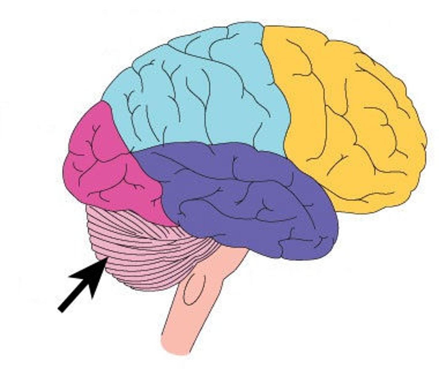
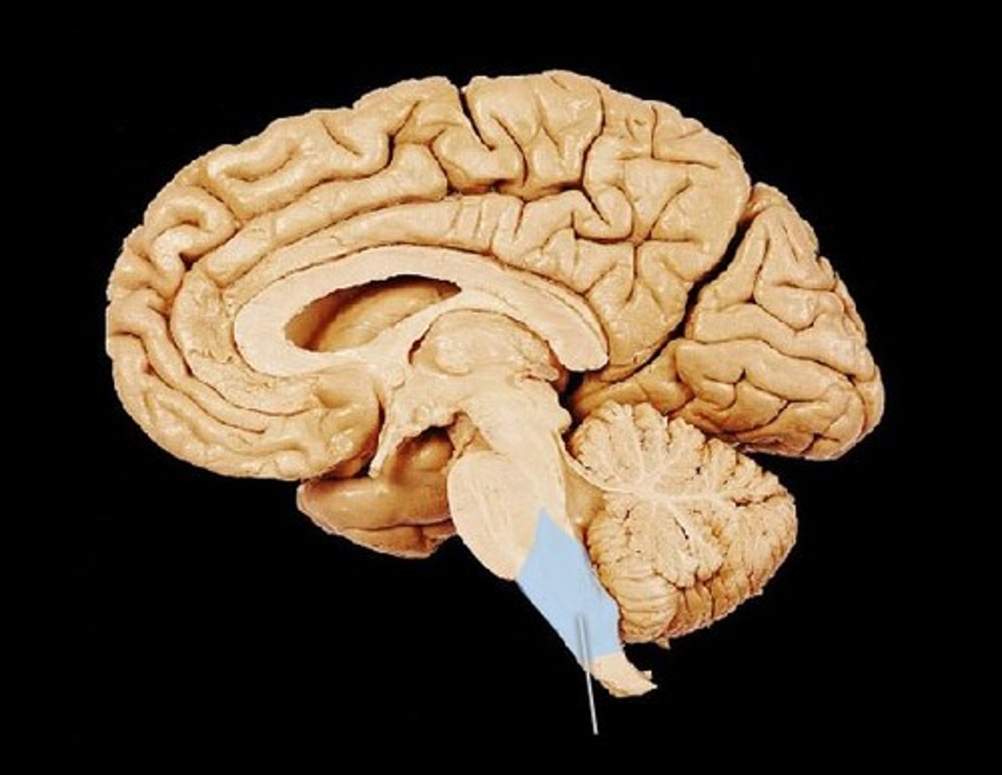
What is the general function of the cerebellum?
It coordinates balance, posture, and smooth body movements. Helps maintain equilibrium.
“Cerebellum = Balance Bell” — imagine a ballerina ringing a bell to keep her balance.
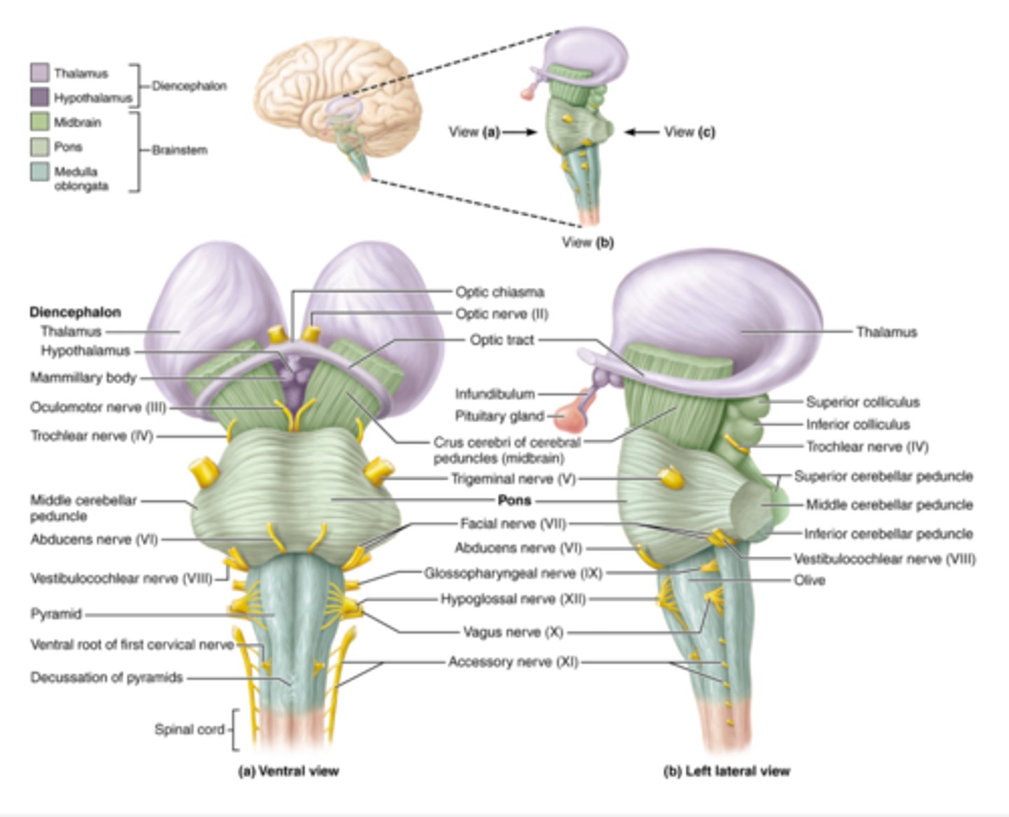
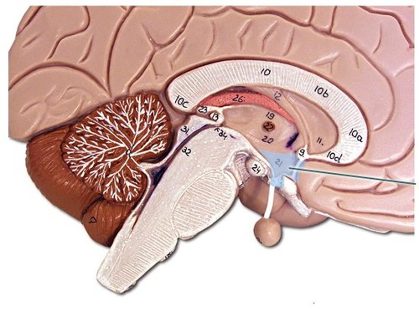
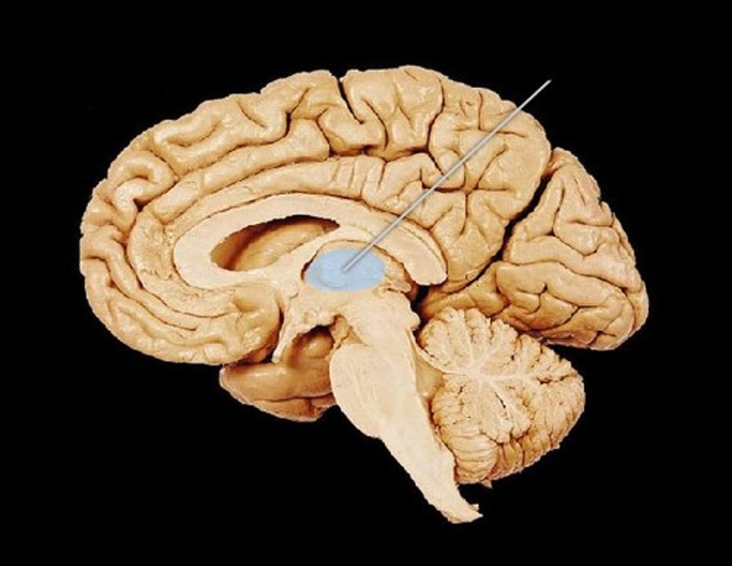
What is the general function of the diencephalon?
The diencephalon acts as a relay and control center.
It includes structures like the thalamus, hypothalamus, and pineal gland, which help regulate sensory information, homeostasis, emotions, and hormones.
Primarily composed of gray matter

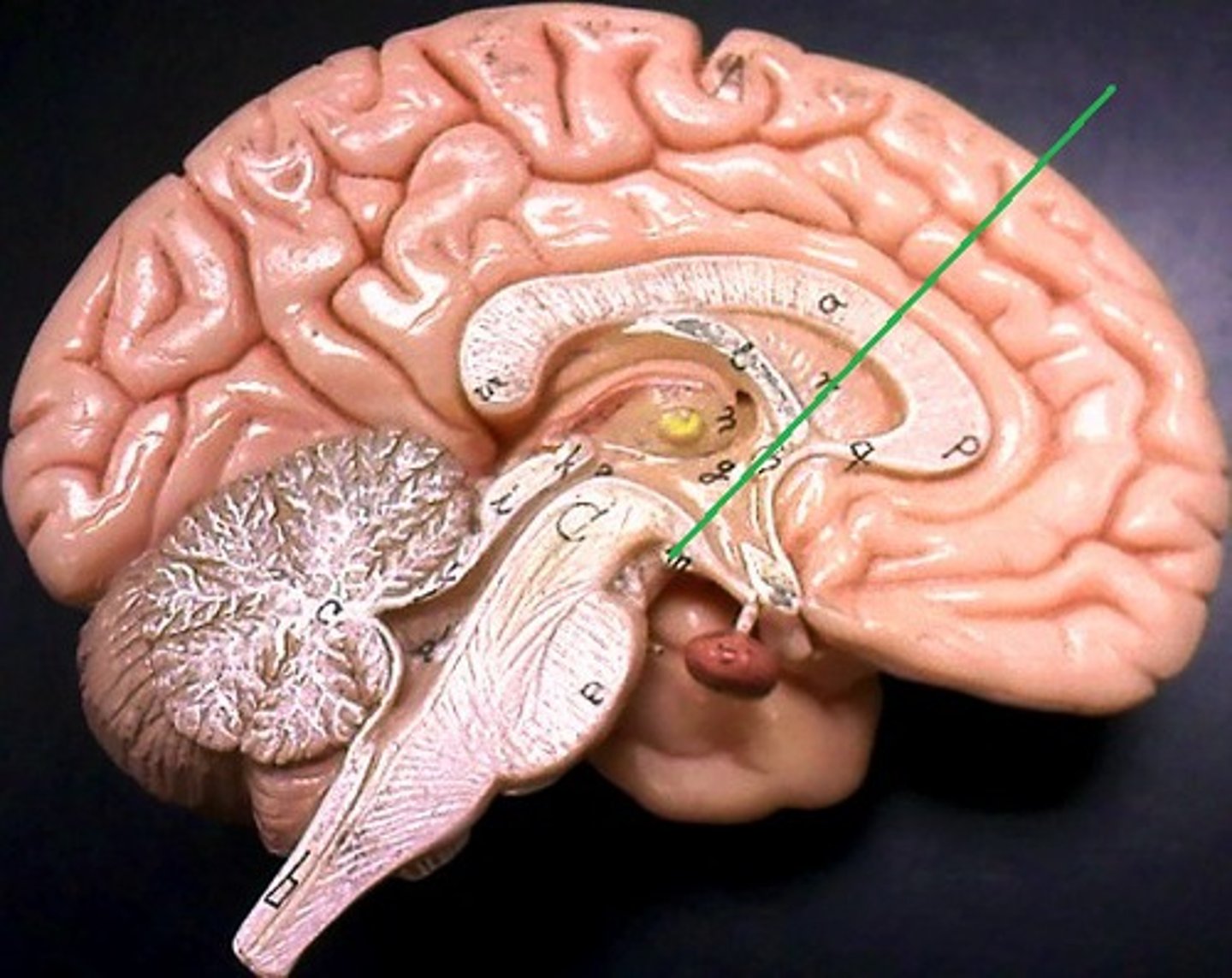
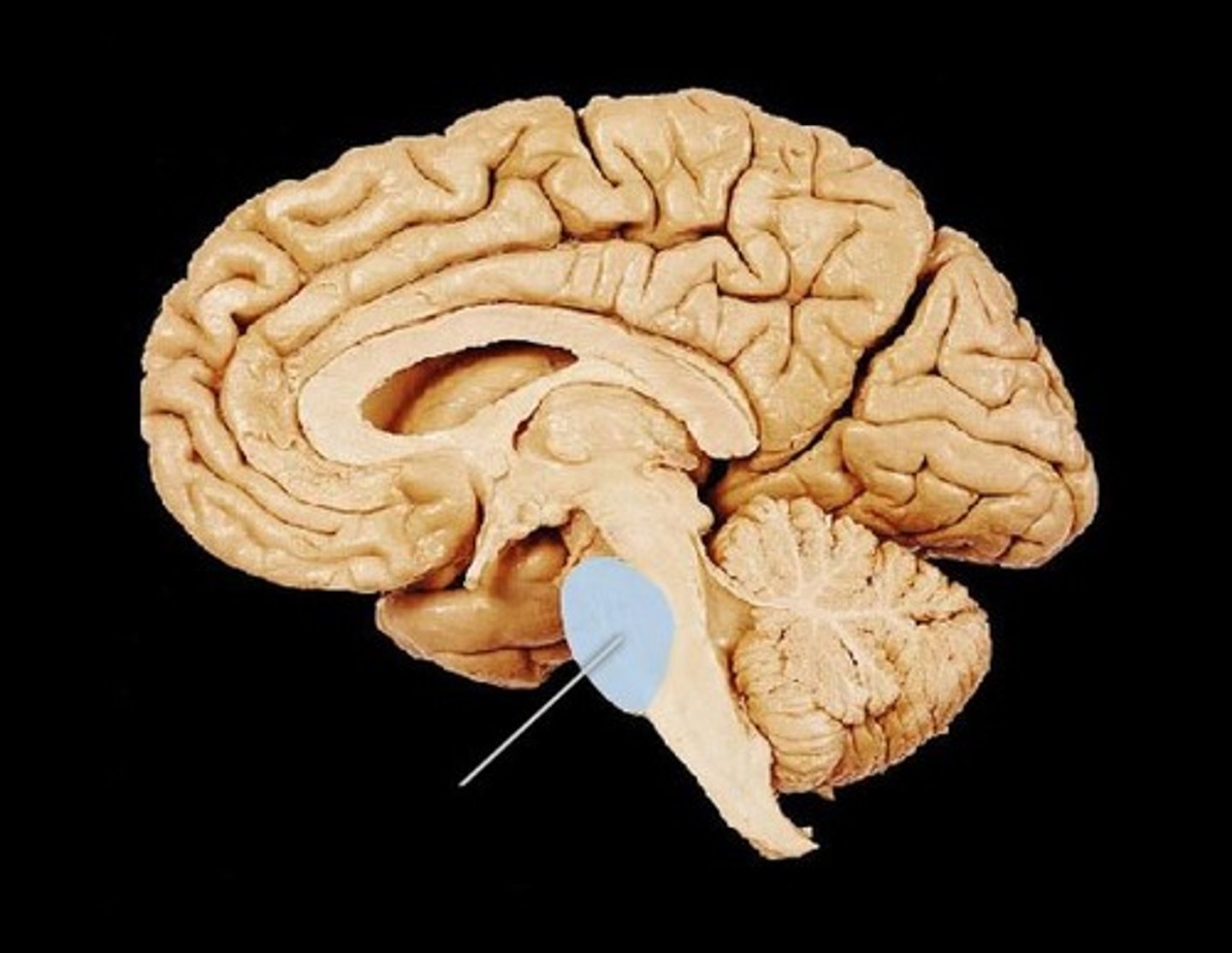
What is the general function of the brainstem?
It controls automatic functions like breathing, heart rate, and blood pressure.
“Brainstem = Life Stem” — it keeps you alive by managing vital functions.
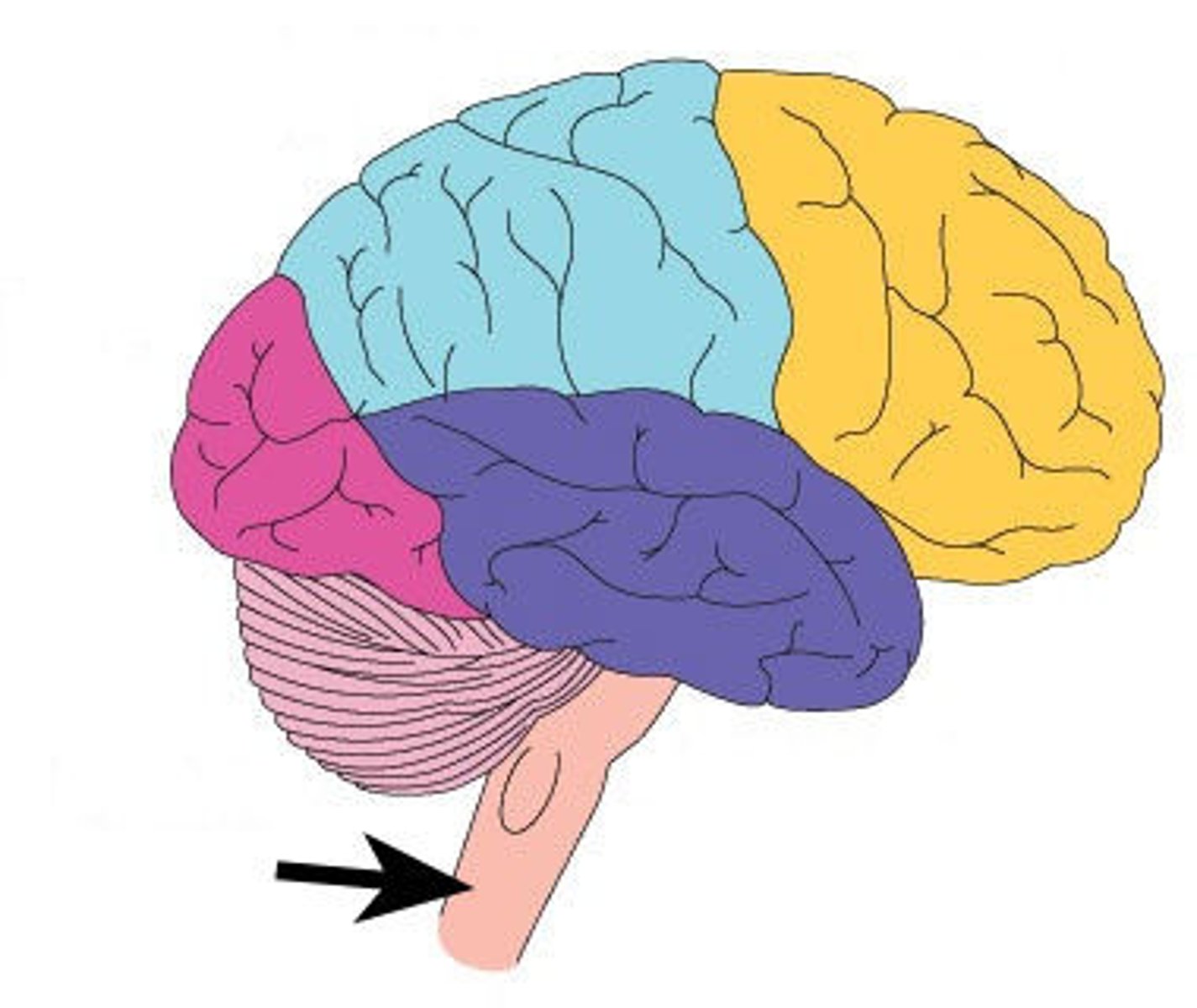
What structures make up the brainstem, what is the function of each?
1. Midbrain - Processes visual and auditory information and controls reflexive eye and head movements.
“Midbrain = Middle for Movement” — helps you automatically turn toward a sound or sight.
2. Pons - Acts as a bridge between different parts of the brain and helps regulate breathing and sleep.
“Pons = Bridge” — think of “Pons → Ponds have bridges.”
3. Medulla Oblongata - Controls involuntary life functions like heartbeat, breathing, swallowing, and blood pressure.
“Medulla = Must Do to Live” — without it, vital body functions stop.
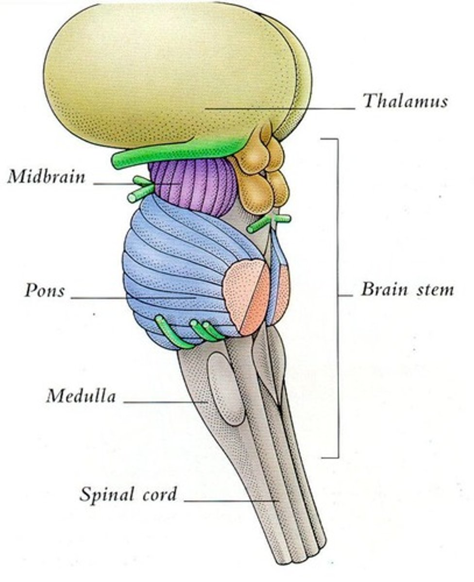
What structures make up the diencephalon, what is the function of each?
1. Thalamus - Serves as the main relay station for sensory information going to the cerebral cortex (except smell).
- Plays a role in alertness, awareness, and consciousness.
2. Hypothalamus - Control of ANS (visceral motor), endocrine system, and behavior. Regulates emotional responses, body temp, hunger & thirst sensations, and sleep-wake cycles. Formation of memory
- Controls the pituitary gland, linking the nervous and endocrine systems.
3. Epithalamus (Pineal Gland) - The epithalamus contains the pineal gland, which produces melatonin to regulate sleep-wake cycles.
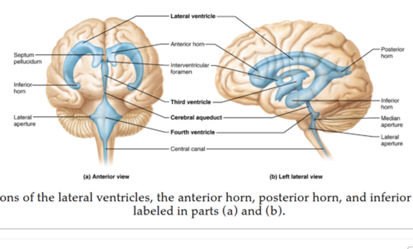
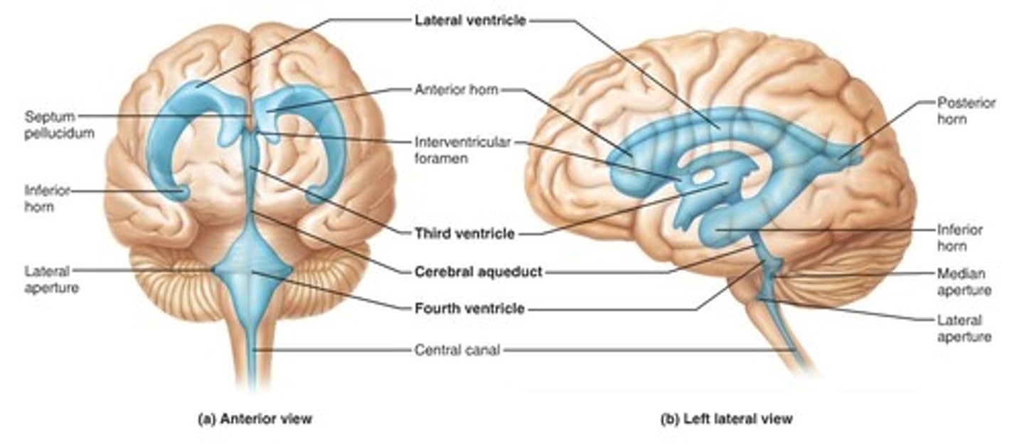
What are the four ventricles and what is their function?
1. Right Lateral Ventricle & Left Lateral Ventricle -
Produce and hold most of the cerebrospinal fluid (CSF).
Drain CSF into the third ventricle through the interventricular foramina (foramen of Monro).
3. Third Ventricle - Acts as a passageway for CSF from the lateral ventricles to the fourth ventricle. Surrounds parts of the thalamus and hypothalamus.
4. Fourth Ventricle - Receives CSF from the third ventricle via the cerebral aqueduct.
Allows CSF to flow into the central canal of the spinal cord and around the brain’s surface.
“Fourth = Final Flow” — it’s the last stop before CSF circulates around the brain and spinal cord.
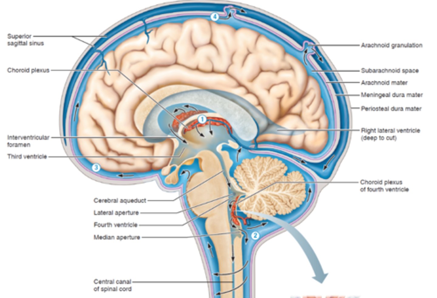
What are the functions of cerebrospinal fluid (CSF)?
CSF is produced in the ventricles of the brain, circulates through the hollow central cavities of the CNS and the subarachnoid space, and is reabsorbed back into the blood in the dural venous sinuses.
Protection (Cushioning) - Acts as a shock absorber that protects the brain and spinal cord from injury.
Buoyancy (Support) - Keeps the brain floating within the skull, reducing its effective weight and preventing it from pressing on the base of the skull.
Nutrient Transport - Delivers nutrients and oxygen to brain tissue and carries waste products away to the bloodstream.
Describe the following about the CSF
a. Where is it made.
b. How is it made CSF.
c. What is the path it takes through the brain and spinal cord.
Where it is made: In the choroid plexuses - a network of capillaries and ependymal cells located inside all four ventricles of the brain.
How it is made: The choroid plexus filters blood plasma and secretes CSF.
Ependymal cells (special brain cells) actively transport ions and nutrients, drawing water in to form CSF.
Pathway of CSF Flow:
1. Lateral ventricles →
2. Interventricular foramina (of Monro) →
3. Third ventricle →
4. Cerebral aqueduct (of Sylvius) →
5. Fourth ventricle →
6. Subarachnoid space (around brain and spinal cord) →
7. Reabsorbed into the dural venous sinuses through arachnoid villi (granulations).
“Lazy Tigers Chase Fast Spotted Rats”(Lateral → Third → Cerebral aqueduct → Fourth → Subarachnoid → Reabsorbed)
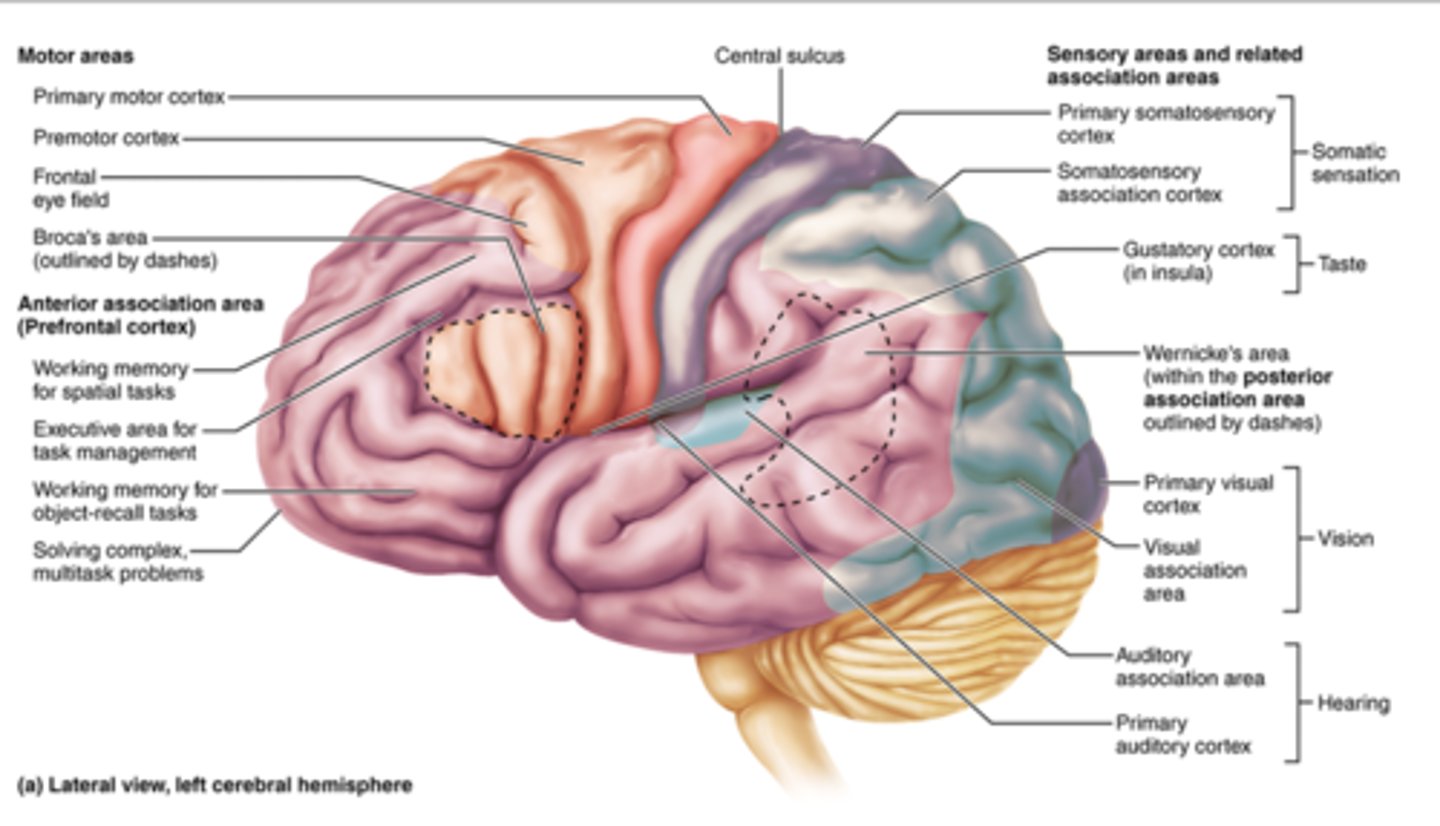
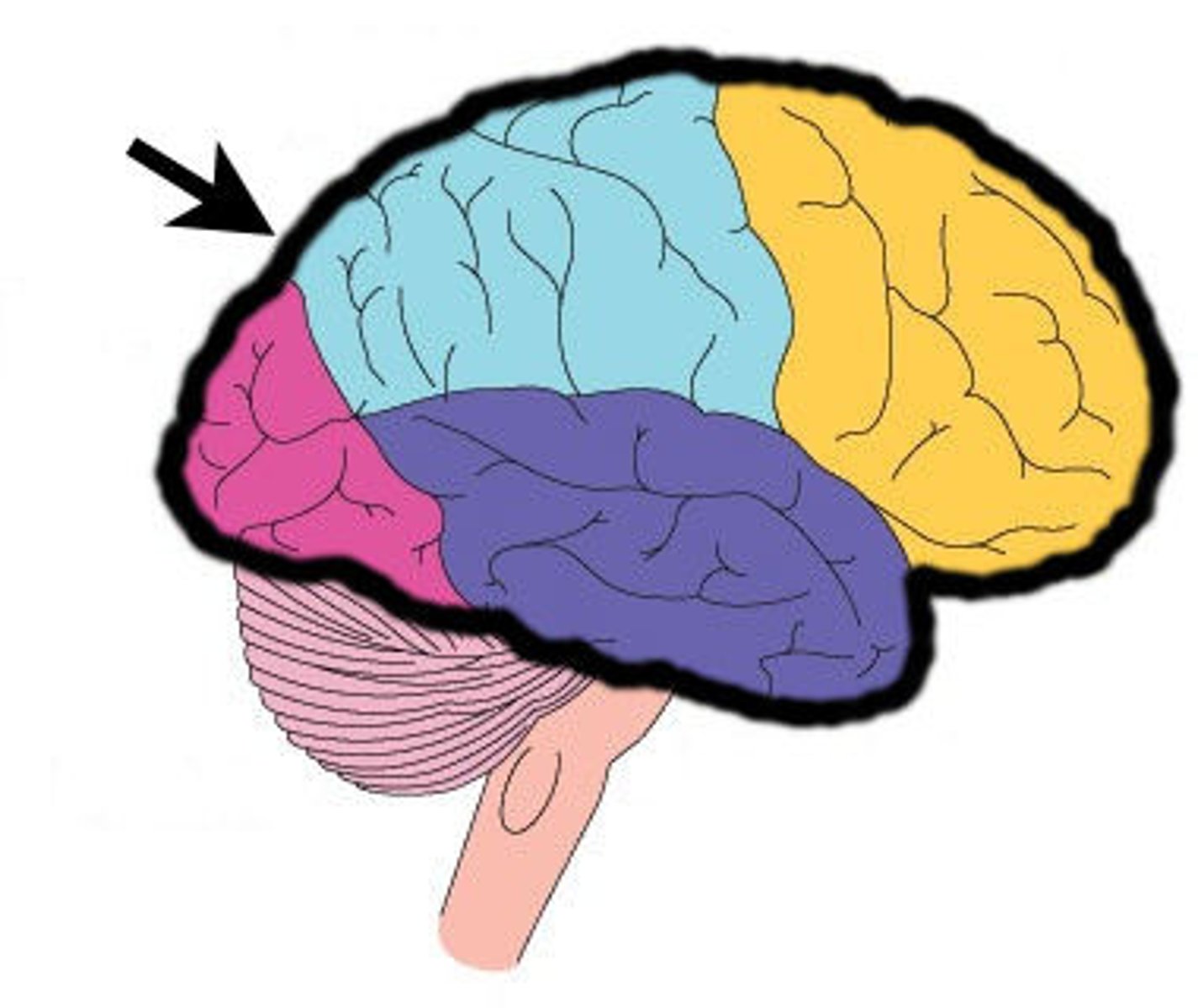
What are the 5 lobes of the brain, what are they named after, what functional regions are found in each?
Frontal:
- Voluntary movement (primary motor cortex)
- planning movement (premotor cortex)
- Eye movement (frontal eyelid field)
- Speech production (Broca's area)
- Executive cognitive functions (anterior association area)
- Emotional response (limbic association area)
Parietal:
- General somatic (somatosensory cortex & association area)
- Spatial awareness of objects, sounds, body parts (posterior association area)
- Understanding speech (Wernicke's area)
Occipital Lobe:
- Vision (visual cortex and association areas)
Temporal Lobe:
- Hearing (auditory cortex & association area)
- Object identification (posterior association area)
- Emotional response, memory (limbic association)
Insula:
Taste (gustatory cortex)
What is a sulcus?
A shallow groove or fold on the surface of the brain.
Short Groove

What is a gyrus?
A raised ridge or hill on the surface of the brain between sulci
Brain fold
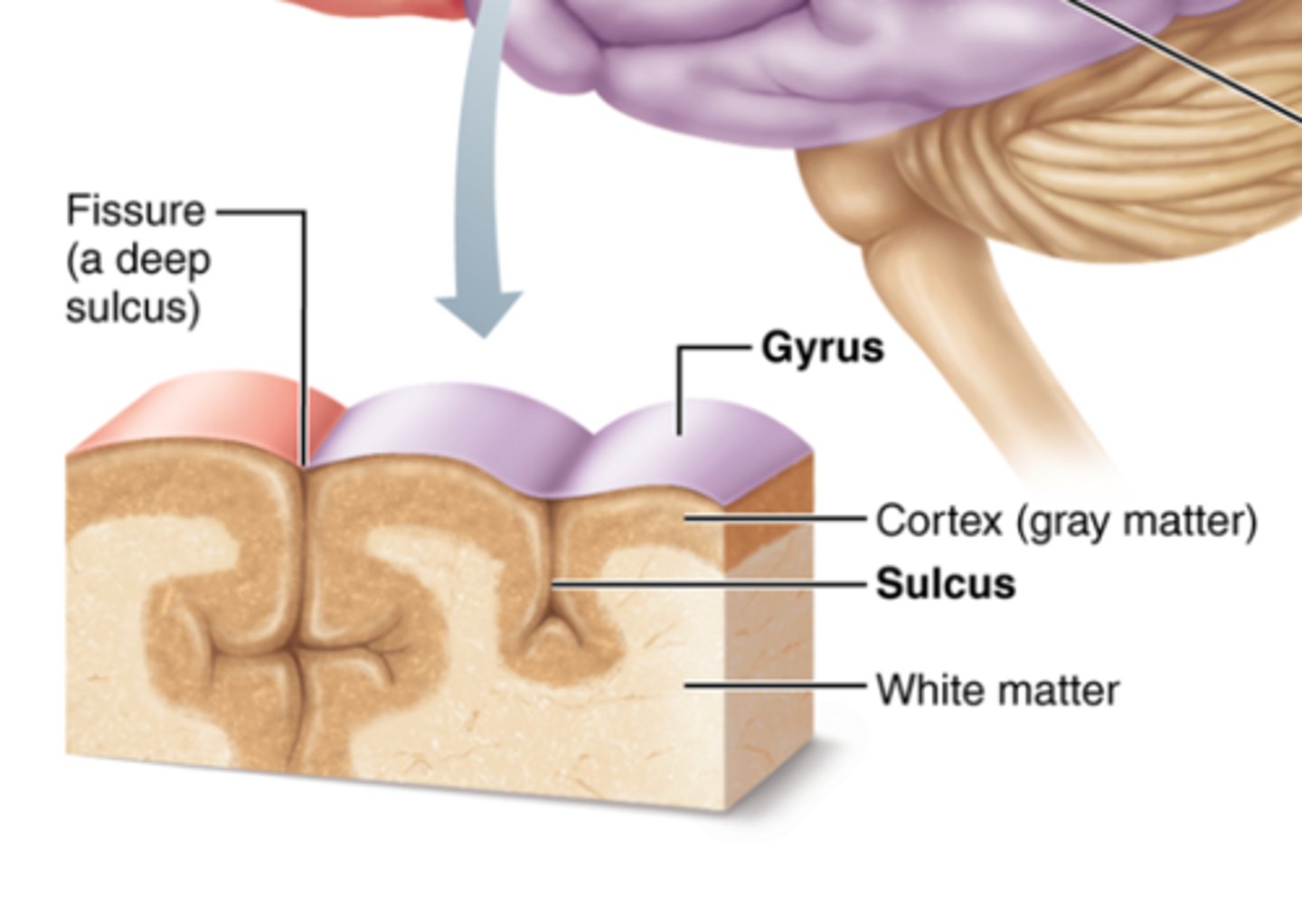
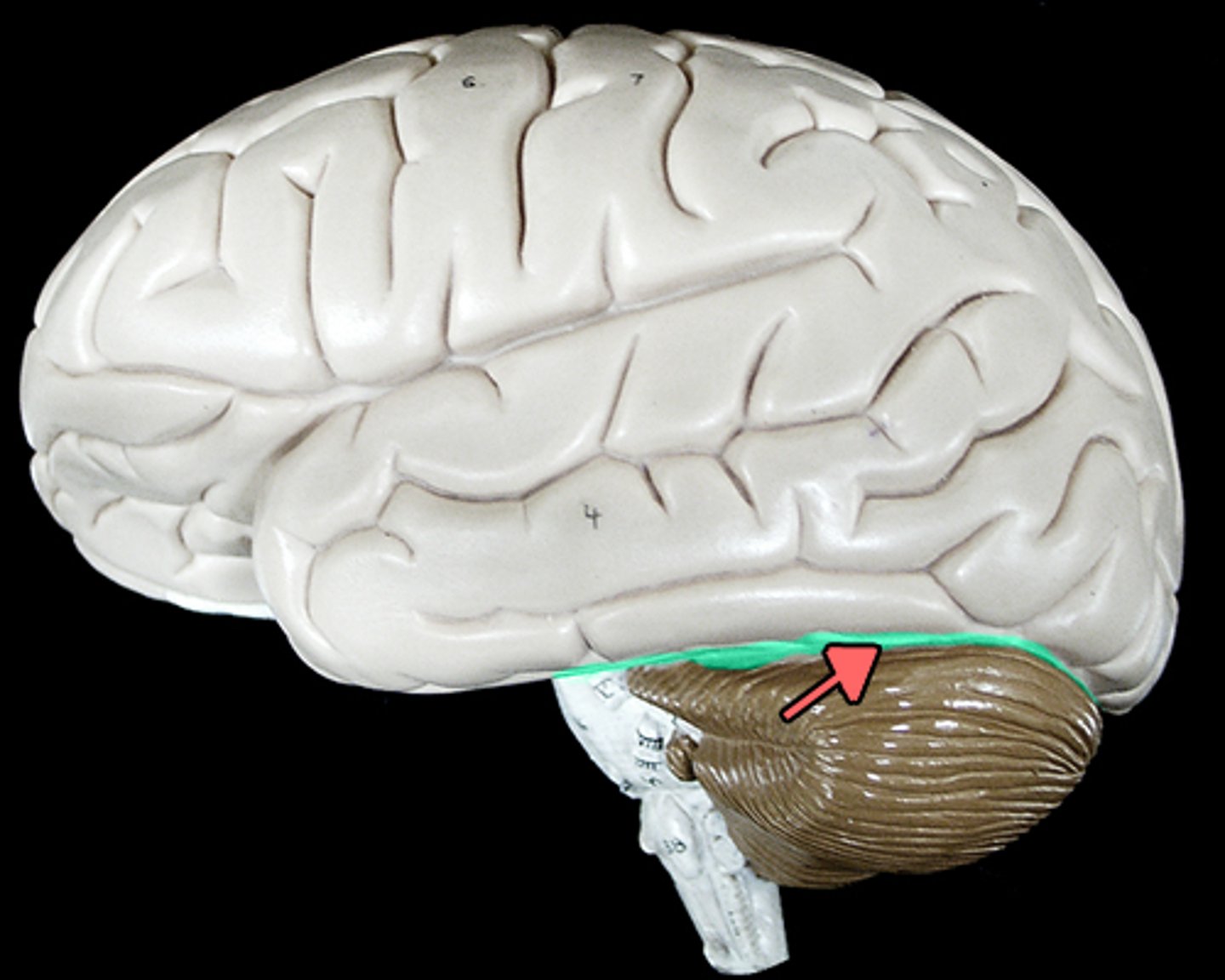
What is a fissure?
A deep groove in the brain that separates large regions or lobes
Longitudinal fissure → divides the brain into right and left hemispheres.
Transverse fissure → separates the cerebrum from the cerebellum
“Fissure = far deeper than a sulcus.”
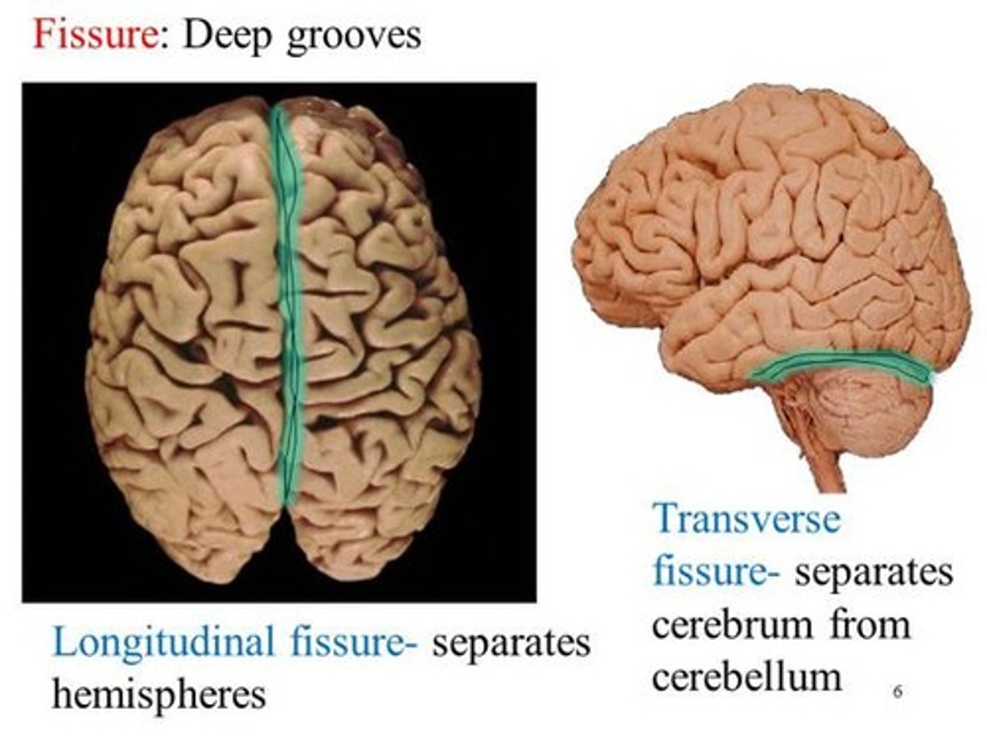
What major structure separates the left cerebral hemisphere from the right?
Longitude fissure
“Long line = Longitudinal fissure splits left and right.”
What major structure separates the cerebrum from the cerebellum?
Transverse Fissure
“Transverse = across” → it cuts across the brain, separating top (cerebrum) from bottom (cerebellum).
Describe the central sulcus.
- A deep groove (sulcus) that separates the frontal lobe from the parietal lobe.
- It runs roughly from the top of the brain down the sides, dividing the motor and sensory regions.
Function:
It serves as a landmark — the line between areas that control movement (in front) and sensation (behind).
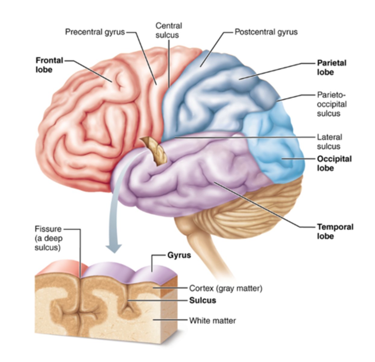
Describe the precentral gyrus.
Location:
Lies in front of the central sulcus (in the frontal lobe).
Function:
- Known as the primary motor cortex.
- Controls voluntary muscle movements — each region corresponds to a specific part of the body (motor homunculus).

Describe the path of motor control from the brain to skeletal muscle. Be sure to include the basal ganglia and cerebellum's role in addition to the primary motor and premotor cortex. Be able to locate upper and lower motor neuron (where does each start and end). Be able to follow the path a motor response takes through the brain and out to the muscle
1. Movement planned → Premotor Cortex & SMA → Primary Motor Cortex
2. Basal Ganglia modulates → Thalamus → Motor Cortex
3. Cerebellum adjusts → Thalamus → Motor Cortex
4. Primary Motor Cortex (UMN) fires → corticospinal/corticobulbar tract → LMN
5. LMN → skeletal muscle contraction
What is the function of the cerebral cortices: Primary somatosensory cortex/somatosensory association area. Which cerebral lobes are they found in?
Primary somatosensory cortex
Function: It receives and interprets sensory information from the body — such as touch, pressure, pain, and temperature.
Location: postcentral gyrus of the parietal lobe
Somatosensory association area
Function: It interprets and integrates sensory input, allowing you to recognize objects by touch (like knowing it’s your phone without looking).
Location: Found posterior to the primary somatosensory cortex in the parietal lobe.
What is the function of the cerebral cortices: Primary visual cortex/visual association area. Which cerebral lobes are they found in?
Primary visual cortex
Function: It receives visual input from the eyes — detects light, shapes, colors, and movement.
Location: Occipital lobe
Visual association area
Function: It interprets and analyzes visual information — recognizing faces, objects, and patterns.
Location: Surrounds the primary visual cortex in the occipital lobe.
What is the function of the cerebral cortices: Primary auditory cortex/ association area. Which cerebral lobes are they found in?
Primary Auditory cortex
Function: It receives sound information from the ears — detects pitch, volume, and basic auditory signals
Location: Found in the superior temporal gyrus of the temporal lobe
“Primary hears the sound.”
Auditory Association area
Function: It interprets and makes sense of sounds — understanding speech, music, or recognizing familiar sounds
Location: Surrounds the primary auditory cortex in the temporal lobe
“Association understands what you hear.”
What is the function of the cerebral cortices: Olfactory cortex. Which cerebral lobes are they found in?
Olfactory Cortex
Function: It receives and processes smell information from the nose. It also contributes to odor recognition and links smells to memory and emotion
Location: Found in the temporal lobe (specifically in the piriform cortex) and partly in the frontal lobe (orbitofrontal cortex for odor identification).
“Olfactory = odors → smell and remember.”
What is the function of the cerebral cortices: Gustatory cortex. Which cerebral lobes are they found in?
Function: It processes taste information from the tongue — detects sweet, sour, salty, bitter, and umami flavors.
Location: Found in the insula (primary gustatory cortex) and partially in the frontal lobe (for taste perception and decision-making about food).
“Gustatory = gulp → taste and enjoy.”
What is the function of the cerebral cortices: Frontal Eye Field. Which cerebral lobes are they found in?
Function: It controls voluntary eye movements, especially scanning, tracking, and shifting gaze.
Location: Found in the frontal lobe, near the premotor cortex (just anterior to the precentral gyrus).
What is the function of the cerebral cortices: Wernicke's Area. Which cerebral lobes are they found in?
Function: It comprehends language — helps understand spoken and written words. Damage can cause receptive aphasia (speech sounds fluent but makes little sense).
Location: Found in the posterior part of the superior temporal gyrus of the temporal lobe, usually in the left hemisphere.
“Wernicke = Word understanding.”

What is the function of the cerebral cortices: Broca's Area. Which cerebral lobes are they found in?
Function: It controls speech production and language expression. Damage can cause expressive aphasia (difficulty forming words, but comprehension remains intact).
Location: Found in the inferior frontal gyrus of the frontal lobe, usually in the left hemisphere.
“Broca = Speak Boldly.”
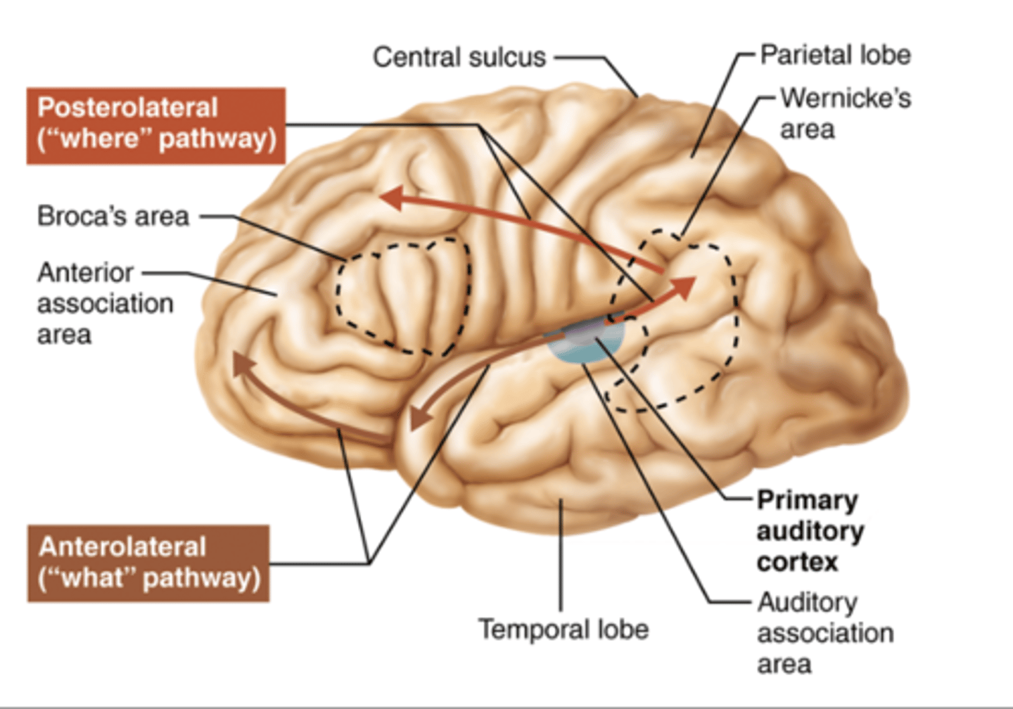
How do the primary cortices and association areas work together?
Primary cortices: Receive raw sensory input (or generate basic motor commands).
Example: Primary visual cortex detects light and shapes; primary auditory cortex detects sound.
Association areas: Interpret and integrate the input, giving it meaning.
Example: Visual association area recognizes a face; auditory association area understands words or music.
What is the function of the limbic system?
The limbic system is a group of interconnected brain structures that regulate emotions, memory, motivation, and some aspects of behavior.
"emotional brain*
What is the function of the reticular system?
The reticular system is a network of neurons in the brainstem that controls alertness, attention, and some autonomic functions.
Describe the pathway of incoming sensory information. Include first, second and third order neurons including location of cell bodies and axon terminals.
First-Order Neuron
Function: Carries sensory information from receptors (skin, muscles, etc.) to the spinal cord or brainstem.
Cell body location: Dorsal root ganglion (DRG) of a spinal nerve (or cranial sensory ganglion for cranial nerves).
Axon terminals: Synapse in the spinal cord (posterior horn) or medulla oblongata, depending on the pathway.
Second-Order Neuron
Function: Carries information up the spinal cord or brainstem to the thalamus.
Cell body location: Posterior horn of the spinal cord or nuclei in the medulla oblongata.
Axon terminals: Synapse in the thalamus (specifically in the ventral posterior nucleus).
Key feature: Axons often cross (decussate) to the opposite side of the CNS.
Third-Order Neuron
Function: Carries information from the thalamus to the cerebral cortex for conscious perception.
Cell body location: Thalamus.
Axon terminals: Primary somatosensory cortex in the parietal lobe (postcentral gyrus).
Describe the pathway of outgoing motor commands. Include upper and lower neurons including location of cell bodies and axon terminals
1. Upper Motor Neuron
Function: Carries motor commands from the brain to the spinal cord.
Cell body location: Primary motor cortex (precentral gyrus) in the frontal lobe.
Axon pathway:
Travels down through the brainstem (corticospinal or corticobulbar tracts).
Most decussate (cross over) in the medulla oblongata (forming the lateral corticospinal tract).
Axon terminals: Synapse on lower motor neurons in the anterior (ventral) horn of the spinal cord (or cranial nerve nuclei in the brainstem).
2. Lower Motor Neuron
Function: Carries motor commands from the spinal cord to the effector (muscle).
Cell body location: Anterior (ventral) horn of the spinal cord or cranial nerve motor nucleus.
Axon terminals: Synapse on skeletal muscle fibers at the neuromuscular junction.
What is the function of the projection fibers?
Projection fibers connect the cerebral cortex with lower brain regions and the spinal cord. They carry information vertically — either sensory input going up to the cortex or motor commands going down from the cortex.
Ascending fibers: carry sensory info to the cortex.
Descending fibers: carry motor commands to the brainstem and spinal cord.
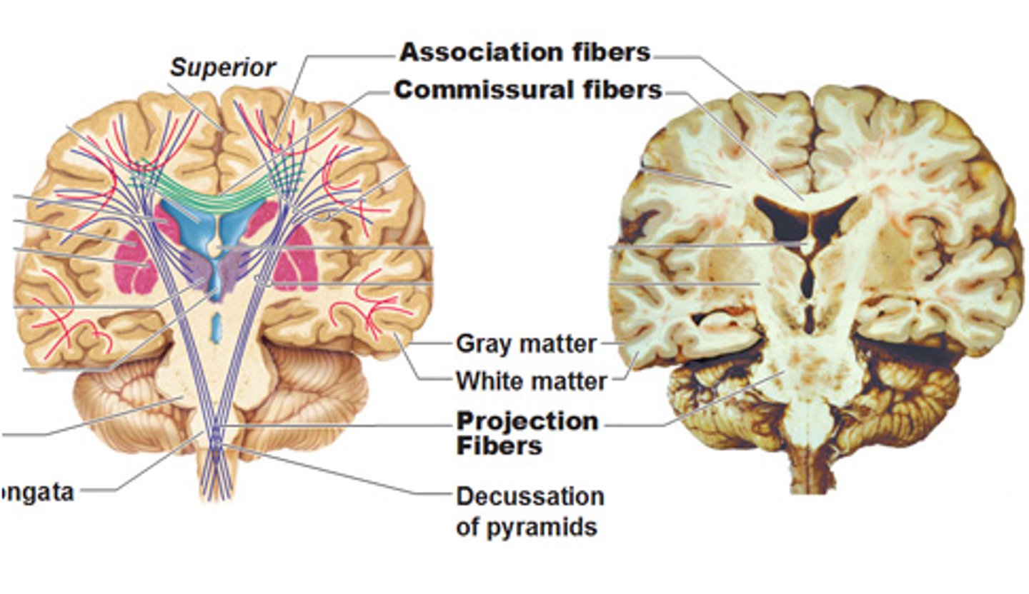
What is the function of the commissural fibers?
Commissural fibers connect the left and right cerebral hemispheres, allowing them to communicate and coordinate activities.
What is the function of the association fibers?
Association fibers connect different areas within the same cerebral hemisphere, allowing regions of one side of the brain to share information and work together.
Short association fibers (arcuate fibers): connect neighboring gyri.
Long association fibers: connect different lobes within the same hemisphere.
Function: Integrate information — for example, linking sensory input with memory or language areas.
A fly just landed on your arm. Describe how the signal travels from the arm, to the brain. What areas of the brain process the information, and what parts of the brain send a response to move your arm.
1. Sensory Detection:
Touch receptors (mechanoreceptors) in your skin detect the fly’s movement.
The signal (action potential) travels along a sensory neuron.
2. Sensory Pathway to Brain
First-order neuron:
- Cell body in dorsal root ganglion (PNS).
- Carries the signal from the arm → spinal cord.
Second-order neuron:
- Cell body in spinal cord or medulla oblongata.
- Axon crosses to the opposite side and travels to the thalamus.
Third-order neuron:
- Cell body in thalamus.
- Axon projects to the primary somatosensory cortex (parietal lobe).
3. Processing the Sensation
- The primary somatosensory cortex detects where you were touched.
- The somatosensory association area interprets the feeling (“A fly landed on my arm”).
4. Motor Response
Decision-making: The prefrontal cortex decides to brush it off.
Planning: The premotor cortex plans the movement sequence.
Command: The primary motor cortex (frontal lobe) sends the command down the descending motor pathway.
5. Motor Pathway to the Arm
Upper motor neuron: Cell body in primary motor cortex.
Axon travels down through brainstem → spinal cord.
Lower motor neuron: Cell body in anterior horn of spinal cord. Axon exits via spinal nerve to skeletal muscles of the arm → muscles contract → you brush away the fly.
Basel Nuclei/Basel Ganglia
Subconscious control of skeletal muscle tone, coordination of learned movement patterns. May initiate motor movement. Provides a general pattern of rhythm for undergoing movements.
Cerebrum
Logic, learning & memory, "conscious" brain.
Cerebellum
Body posture, fine tune movements

Corpora quadrigemina (superior and inferior colliculus)
Process visual and auditory sensations
The largest nuclei. Divided into the superior and inferior colliculi

Hypothalamus
Thirst, hunger, emotion, hormone production. Control of autonomic function, secretion of hormones.
Limbic System (a functional group - not an anatomical structure)
Establishes emotional states, links conscious, intellectual functions of cerebral cortex, facilitates memory storage and retrieval.
Mammillary bodies
Control of feeding reflex (chewing, licking, swallowing)
Medulla oblongata
Breathing, heart rate, visceral activities, sensory and motor nuclei of cranial nerves
Pons
Higher levels of respiratory control, sensory and motor nuclei of cranial nerves
Thalamus
Acts as a filter for ascending sensory information that is projected to the primary cortex and basal nuclei.
Ventricles
Filled with CSF
Describe the anatomy spinal cord, include the following items:
a. Cervical enlargement
b. Lumbar enlargement
c. Conus medullaris
d. Cauda equina
e. Filum terminal
f. White matter
g. Grey matter
a. Cervical enlargement - Contains extra neurons to supply the upper limbs.
b. Lumbar enlargement - Contains extra neurons to supply the lower limbs.
c. Conus medullaris - Marks the end of the actual spinal cord.
d. Cauda equina - Nerves continue to travel downward to exit at their respective vertebral levels to innervate lower limbs and pelvic organs.
e. Filum terminal - Anchors the spinal cord to the coccyx, providing longitudinal stability.
f. White matter - Conducts sensory information to the brain and motor information from the brain.
g. Grey matter - Neuronal cell bodies, dendrites, and unmyelinated axons. Processes incoming sensory information and outgoing motor commands.
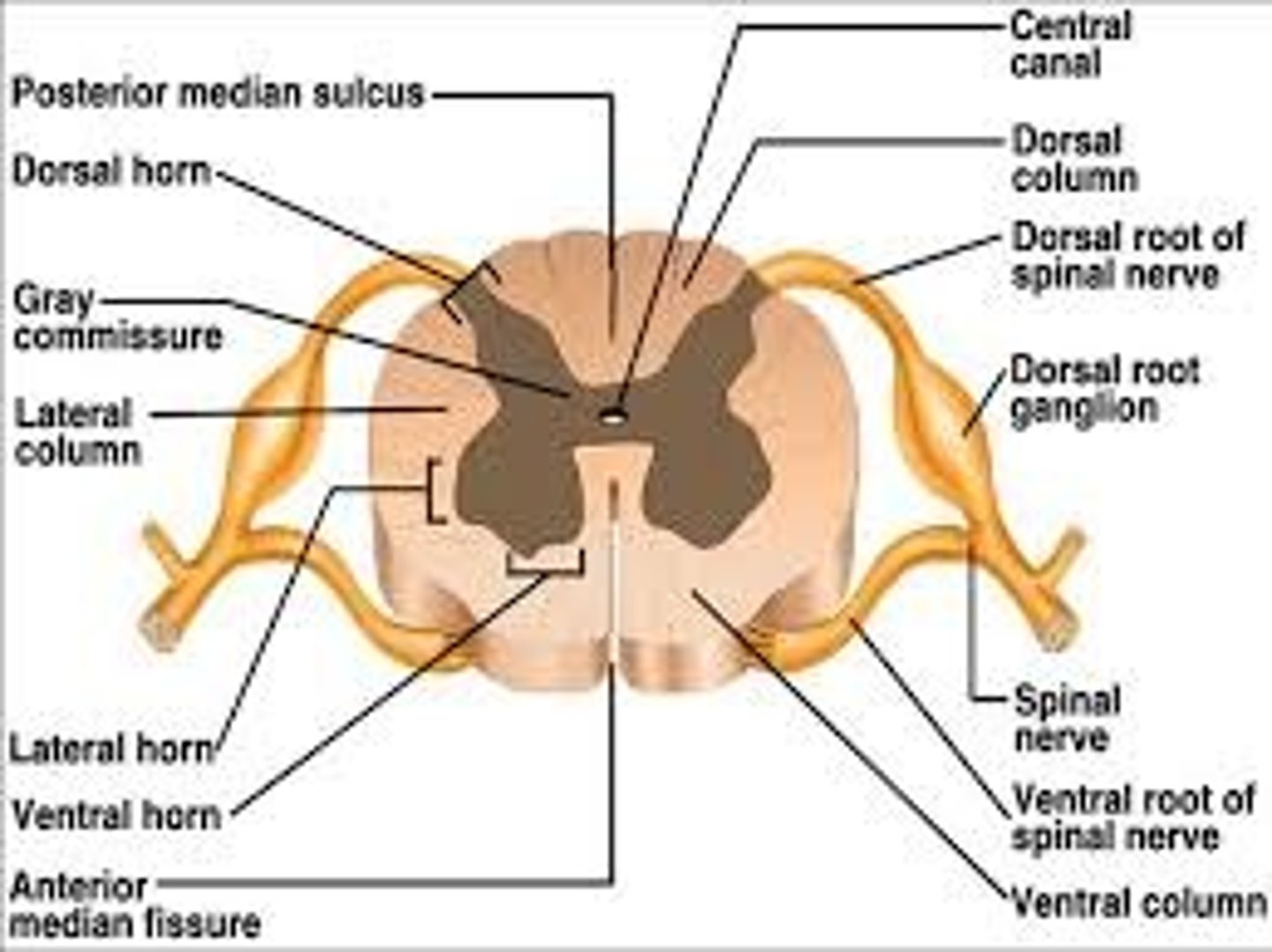
What can be found in each of these anatomical structures?
a. Anterior, posterior and lateral grey horn
b. Dorsal and ventral roots
c. Dorsal root ganglia.
d. Central canal
a. Anterior, posterior, and lateral grey horn
Anterior Horn: Somatic motor neurons (cell bodies)
Posterior horn: Interneurons and sensory neuron axon terminals
Lateral Horn: Cell bodies of autonomic (sympathetic) neurons
b. Dorsal and ventral roots
Dorsal Root: Sensory (afferent) axons entering the spinal cord
Ventral Root: Motor (efferent) axons leaving the spinal cord
c. Dorsal root ganglia
Contains: Cell bodies of sensory neurons
d. Central canal
Contains: Cerebrospinal fluid (CSF)
Describe the following structures:
a. Epidural space
b. Denticulate ligament
a. Epidural space
Location: Between the dura mater (outermost meningeal layer) and the vertebral canal (bone).
Contents: Fat tissue, Connective tissue, Blood vessels
Function: Cushions and protects the spinal cord
Acts as a space for epidural anesthesia during procedures like childbirth
b. Denticulate ligament
Location: Extensions of the pia mater that run laterally along the spinal cord.
Appearance: Tooth-like projections (hence the name “denticulate”)
Function: Anchors the spinal cord to the dura mater
Provides stability to prevent excessive movement of the spinal cord within the vertebral column
Describe the three meninges found in the spinal cord.
1. Dura Mater
Location: Outermost layer
Composition: Tough, dense connective tissue
Function: Protects the spinal cord from mechanical injury
Forms the dural sac, which ends at S2 vertebral level
2. Arachnoid Mater
Location: Middle layer, just beneath the dura mater
Composition: Thin, web-like membrane
Function: Covers the spinal cord without dipping into sulci. Forms the subarachnoid space (between arachnoid and pia mater) that contains cerebrospinal fluid (CSF)
3. Pia Mater
Location: Innermost layer, in direct contact with the spinal cord
Composition: Delicate, transparent connective tissue
Function: Follows the contours of the spinal cord
Forms denticulate ligaments that anchor the cord laterally to the dura. Extends as the filum terminale to anchor the cord to the coccyx
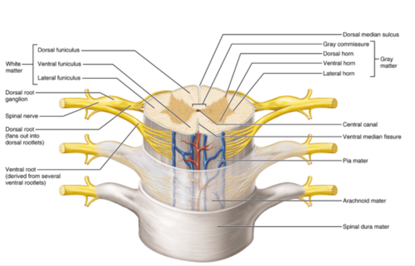
Describe the anatomy of a typical peripheral nerve. Include the following:
a. Axon
b. myelin
c. nerve fascicle
d. endoneurium, perineurium, and epineurium.
Peripheral Nerve Anatomy: A peripheral nerve is a bundle of axons that transmit sensory and motor information between the CNS and the body. It is organized in layers of connective tissue for protection and support.
a. Axon - Transmits action potentials from the neuron’s cell body to other neurons, muscles, or glands.
b. Myelin - Increases conduction speed of nerve impulses. Protects and electrically insulates axons
c. Nerve Fascicle - Organizes axons into manageable bundles and allows targeted distribution of nerve fibers.
d. endoneurium, perineurium, and epineurium.
d. Connective Tissue Layers
Peripheral nerves are protected by three layers of connective tissue:
Endoneurium: Surrounds individual axons. Provides support and protection for each axon.
Perineurium: Surrounds a nerve fascicle. Creates a barrier to protect axons from harmful substances and maintains the internal environment
Epineurium: Outermost layer that surrounds the entire nerve, including all fascicles and blood vessels. Provides structural support and protects the nerve from mechanical stress
What is a ganglia? A nucleus? A fiber tract?
Ganglia: A cluster of neuron cell bodies in the peripheral nervous system (PNS).
Function: Acts as a relay or processing center for sensory or autonomic signals.
Ex: Dorsal root ganglion & Autonomic ganglia
Nucleus: A cluster of neuron cell bodies in the central nervous system (CNS).
Function: Processes specific types of information.
Ex: Thalamic nuclei & Red nucleus
Fiber Tract: A bundle of axon fibers in the CNS that travel together carrying information.
Function: Transmits signals from one region of the CNS to another.
Ex: Corticospinal tract & Spinothalamic tract
Ganglion = PNS neuron cell bodies
Nucleus = CNS neuron cell bodies
Fiber tract = CNS axons
What is meant by a mixed nerve?
A mixed nerve is a peripheral nerve that contains both sensory (afferent) and motor (efferent) fibers.
Describe the pathway of incoming sensory information to the spinal cord, and outgoing motor information from the spinal cord to the periphery. Include the location of cell bodies and axons of neurons within the following structures of the spinal cord:
a. Dorsal root, ventral root, dorsal root ganglia
b. Posterior grey horn, anterior grey horn
c. Spinal nerve
1. Incoming Sensory Information (Afferent Pathway)
- Sensory receptors in the skin, muscles, or organs detect a stimulus (e.g., touch, pain).
- The sensory neuron carries the signal toward the CNS:
Cell body location: Dorsal root ganglion
Axon pathway:
Peripheral axon: from the receptor to the dorsal root ganglion
Central axon: enters the spinal cord via the dorsal (posterior) root
Once inside the spinal cord:
The axon enters the posterior grey horn and may:
Synapse with interneurons in the grey matter
Ascend to the brain in white matter tracts (if the signal is going to the brain)
2. Outgoing Motor Information (Efferent Pathway)
- Motor commands originate from motor neurons in the anterior (ventral) grey horn of the spinal cord.
Cell body location: Anterior grey horn
Axon pathway: leaves the spinal cord through the ventral (anterior) root
- The ventral root axons combine with sensory axons in the spinal nerve to form a mixed nerve.
- The axons travel to skeletal muscles or glands, producing movement or action.
The somatosensory cortex is found here:
A. Pons
B. Diencephalon
C. Medulla
D. Occipital lobe
E. None of the above
E. None of the above
The grey matter of the spinal cord is dominated by:
A. Unmyelinated axons
B. Cell bodies of neurons, neuroglia, and unmyelinated axons
C. Schwan cells and satellite cells
D. Myelinated axons
B. Cell bodies of neurons, neuroglia, and unmyelinated axons
A synapse is:
A. A junction between a neuron and a cell
B. Site of neuronal communication
C. Site of neurotransmitter release
D. Separated by a synaptic cleft
E. All of the above
E. All of the above
True/False: Grey matter mainly contains bundles of axons.
False
True/False: The ventral root of the spinal cord contains bundles of sensory axons.
False
True/False: Projection fiber tracts connect the left and right cerebral hemispheres.
False