Anat and phys exam 1
 Call Kai
 Learn
Learn Practice Test
 Spaced Repetition
 Match
Match Flashcards
 Knowt Play
1/189

Earn XP
Description and Tags
Dr. F delval
Name | Mastery | Learn | Test | Matching | Spaced | Call with Kai | Chat |
|---|
No analytics yet
Send a link to your students to track their progress
190 Terms
2 ways anatomy is divided
Macroscopic and microscopic
Macroscopic
“gross” anatomy, can see with naked eye; dissections, organs, organ systems
Microscopic
Study of cells and tissue through a microscope
Cytology
Study of cells
Histology
Study of tissue
Anatomy vs physiology
Study of structure, study of function
Anatomy (smallest to largest)
Cell, tissue, organ, organ system, organism
Cell
Smallest unit of life
Tissue
Group of cells that perform the same function (muscle, nerve)
Organs
2 or more types of tissue in same structure (skin, kidney, intestine, liver)
Organ Systems
2 or more organs working together (respiratory, digestive, repro, cardiovascular)
Parts of a cell
cell membrane (plasma membrane), cytoplasm, nucleus, organelles
Plasma membrane (cell membrane)
Includes the lipid bilayer (outside hydrophilic, inside hydrophobic), and contains integral proteins like carriers, channels, and receptors
Cytoplasm
gel-like fluid in the cell, very dynamic and constantly moving things
Nucleus
Center of the cell that has RNA and DNA
Organelles of the cell
Nucleus, Endoplasmic Reticulum (rough and smooth), Golgi Apparatus, Mitochondria, Lysosomes, Centrosome
Rough ER
Has ribosomes, MAKES proteins
Smooth ER
Makes lipids
Golgi Apparatus
Packages proteins/fats and exports them out of the cell
Mitochondria
Makes ATP through citric acid cycle, generates aerobically (more efficient) or anaerobically, catabolism of carbs, lipids, and proteins
Lysosome
Intracellular digestion
Centrosome
Spindle pole, organizes during cell division
Cell growth
Hyperplasia or hypertrophy, increase cell number or size
Hyperplasia
Increase number, reproduction of cells
Hypoplasia
Tissue doesn’t develop completely, less cells
Aplasia
Tissue doesn’t develop at all
Hypertrophy
Increase size of a cell with increased functional capacity
Atrophy
Decrease size of cell, not being used so tissue decreases in size
Mitosis
Asexual cell reproduction
Meiosis
s3xual cell reproduction (knowt won’t let me type s3x)
Metabolism
Utilizing of nutrients, everything that builds-up or breaks down nutrients, anabolism or catabolism
Anabolism
Uses ATP, builds nutrients
Catabolism
Releases ATP, breakdown nutrients
Properties of Cell Physiology
Irritability (excitability, reacts to stimuli), transport (gets material in/out of cells)
Organic
CHOPSN
Proteins
Macromolecules lined with Amino Acids, can be structural (part of cells, tissues, organs) or reactive (enzymes, receptors, hormones, contractile, antibodies)
Protein structure
Primary, secondary, tertiary, quaternary
Primary protein
Sequence of Amino Acids
Secondary protein
Conformation of the polypeptide chain
Tertiary protein
Folding of chain
Quaternary protein (most important)
Joining of 2 or more polypeptide chain; makes protein have a specific shape that goes into specific receptor, antibodies make 3d model of protein to combat disease
Lipids
Macromolecule, fats, cell membranes
Carbohydrates
Used for energy, glucose and glycogen
Glucose vs Glycogen
Main source of energy, how the energy is stored (polymer, liver/muscle storage)
Nucleic Acid
DNA, RNA, genetics and protein synthesis
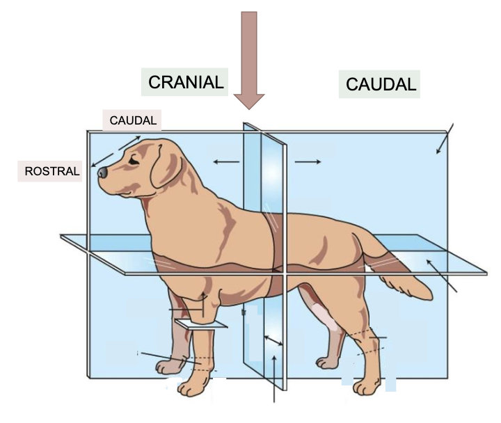
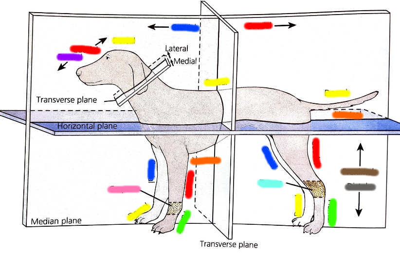
Transverse plane
Separates Cranial (front) and Caudal (back); can also separate rostral (close to nose) from caudal (close to head)
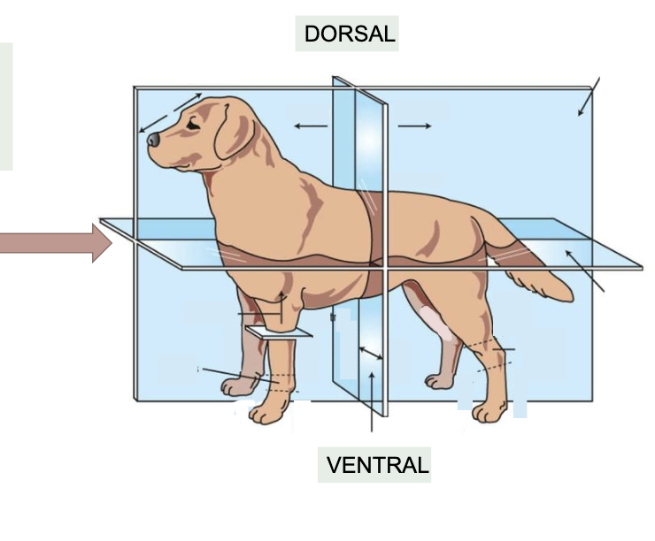
Horizontal plane
Separates dorsal (top) from ventral (bottom)
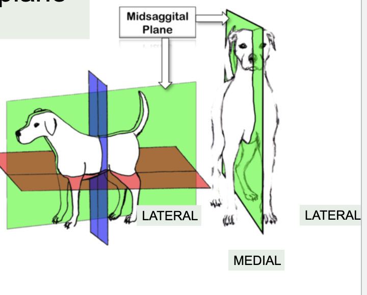
Median (midsagittal) plane
Separates body left and right
Sagital Plane
Planes parallel to the median plane

Cranial
Towards head, front half (blue)

Caudal
Towards tail, back half (red), can also be used towards the top of the head

Dorsal
Towards back, top half (yellow)

Ventral
Towards stomach, bottom half (orange)

Rostal
Used on head only (along with caudal in some scenarios), towards nose (purple)

Lateral
Away from median plane (what splits into left and right)

Medial
Towards Median plane (what splits into left and right)
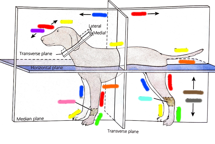
Distal
Used on limbs only, away from body trunk, gray

Proximal
Used on limbs only, closer to body trunk, brown
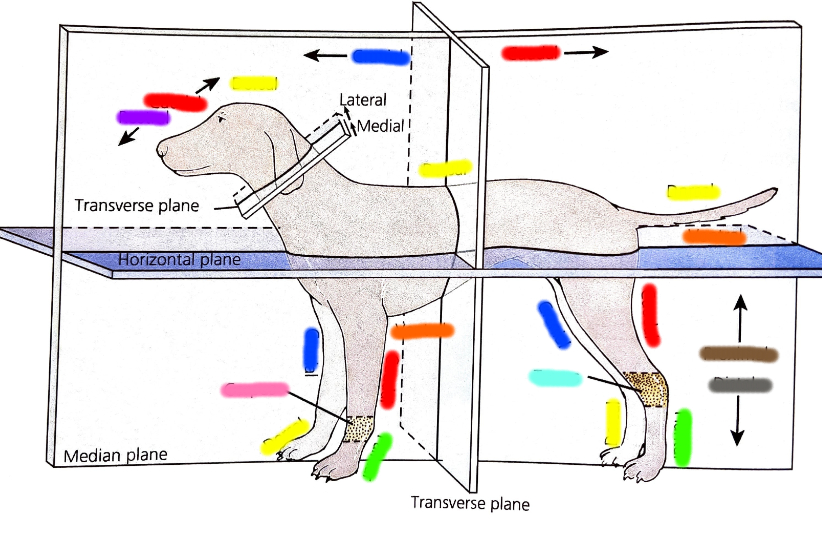
How to name directional terms
[body part A] is [directional term showing what A is closer to] to [body part B]; Example: Forelimb is cranial to hindlimb
Superficial
Away from animal’s core
Deep
Towards animal’s core

Palmer
Caudal aspect of front limb, palm, front green

Plantar
Caudal aspect of back limb, palm, back green

Prone
Animal lying on belly
Supine
Animal lying on back
Gamete
Sex cell, haploid, oocyte (F) + spermatozoan (M) = zygote
Zygote
Fertilization of egg and sperm
Zona pellucida
Glycoprotein layer that surrounds egg. Hardens when single sperm enters cell (if 2 sperm enters, twins)
Mitosis
Cell division, forms the morula (ball of
cells) (2 → 4 → 8 →16) 16 total once
mitosis is complete and comes a blastocyst, the collection of cells on the inside becomes animal
Blastocyst (blastula)
Hollow ball of cells with blastocele (cavity)
Inner cell mass
Inside of blastocyst that forms embryo, multiplies and
divides into 3 germ layers
Trophoblast
Outside of blastocyst and becomes placenta
Order of fertilization
1st, mitosis in uterine tube/oviduct (2,4,8 cell stage), travels to uterus for morula 16-cell stage and blastocoel, hatches once implantation occurs and sticks to uterus wall
Blastocoel
Cavity in blastocyst
Gastrulation
After blastulation, forms 3 germ layers of inner cell mass (ectoderm, endoderm, mesoderm)
Ectoderm(gastrulation)
Outer layer that forms from epiblasts; Becomes epidermis, hair, hooves, epithelium of sense organs, and nerve tissue (basically surface stuff and nerve tissues)
Endoderm (gastrulation)
Inner layer that forms from hypoblast cells, grows down and forms primitive gut (archenteron), becomes epithelium of gut, respiratory tract, and lower urinary tract (bottom stuff)
Mesoderm (gastrulation)
Middle layer, forms between other two, divided into somatic and splanchnic layers. has coelom; becomes all connective tissue, muscles, and epithelium of gonads and joints
Two layers of mesoderm
Somatic and Splanchnic
Somatic layer
Next to ectoderm; creates bones, ligaments, blood vessels, and connective tissues of the limbs
Splanchnic layer
Next to endoderm; makes heart, gut muscles and blood vessels
Coelom
Cavity forms between somatic and splanchnic in mesoderm, precursor to body cavities
Neurolation
Formation of the spinal chord. Ectoderm dorsal to Notochord (neural plate) thickens and folds into a groove, with both the meeting to form a tube. Thickens at cranial end to become brain, hollow tube filled with CSF(cerebrospinal fluid), spine forms, peripheral nerves grow laterally

CSF
Cerebrospinal fluid
Notochord
Develops from mesoderm to make first rod-like support structure in vertebrates, replace by vertebrae of the spin in later embryo, small remnant as nucleus pulposus inside intervertebral disks
Embryo development
Germ layers form tissues, tissues form organs, organs form organ systems, fetus forms when organs are in place (1st trimester)
Four basic types of primary tissue
Epithelial, connective, muscle, neural/nerve
Naming of epithelial cells
By number of layers and shape of cell
Simple
One layer thick epithelial, allows for material exchange
Stratified
Two layer thick epithelial, allows for protection
Main types of epithelial cells
Simple squamous, simple cuboidal, simple columnar, transitional, pseudo-stratified, ciliated columnar, stratified squamous (nonkeratinizing), stratified squamous (keratinizing)
Simple squamous (epithelial)
Single lay of thin, flat cells with IRREGULAR outlines, forms continuous thin membranes, allows for passage of different substances; includes endothelium, mesothelium, and mesenchymal
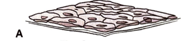
Endothelium simple squamous
Simple layer of squamous cells forming the inner lining of the heart, blood vessels, lymph vessels
Mesothelium simple squamous
Simple squamous epithelium lining the body cavities (pleura and peritoneum)
Mesenchymal simple squamous
Simple squamous lining of the subarachnoid spaces (in the brain) and chambers of the eye
Epithelial
Sheet-like lining, covers organs, makes glandular tissue, secretory cells of gland, lines viscera organs and blood vessels
Simple cuboidal (epithelial)
Equal height/square (appear square), widely distributed, found in outer covering of the ovary, glandular tissues, like thyroid and kidney
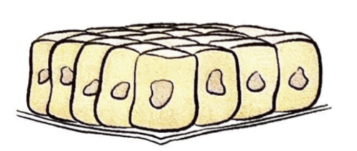
Simple columnar (epithelial)
Taller than wide, lining for digestive tract, can be either absorptive or secretory or both, some secrete mucus fir protection, can be ciliated

Ciliated simple columnar
In oviduct/uterine tube, moves tubular contents in single direction (like if something gets stuck in your throat)
Transitional (epithelium)
Stratified with varied appearance, lining of muscular urinary bladder (when empty, bladder contracts and has many layers, when full, bladder stretches and only has 2-3 layers)
