Anatomy Lecture Exam 1 Material
 Call Kai
 Learn
Learn Practice Test
 Spaced Repetition
 Match
Match Flashcards
 Knowt Play
1/238

There's no tags or description
Looks like no tags are added yet.
Name | Mastery | Learn | Test | Matching | Spaced | Call with Kai | Chat |
|---|
No analytics yet
Send a link to your students to track their progress
239 Terms
The first level of structural organization
Chemical level
The second level of structural organization
Cellular level
The third level of structural organization
Tissue level
The fourth level of structural organization
Organ level
The fifth level of structural organization
Organ system
The sixth level of structural organization
Organism
What does the chemical level of an organism entail?
Atoms that form molecules
What does the cellular level of an organism entail?
Cells and their organelles
What does the tissue level of an organism entail?
Cells grouped to achieve a common goal
What are the four primary tissue types?
Epithelial, connective, nervous, muscle
What does the organ level of an organism entail?
A discrete structure with more than one type of tissue
What does the organ system level of an organism entail?
Organs working together for a common goal
What does the organism level of an organism entail?
Result of all levels working together
Process by which cells become specialized to perform specific functions
Differentiation
These are undifferentiated cells
Stem cells
Embryonic stem cells that can become any adult cell
Pluripotent stem cells
These stem cells can become any cell including placenta
Totipotent stem cells
These stem cells are partially specialized, found in many adult tissues
Tissue specific stem cells
Toward the head end or upper part of a structure or the body; above
Superior (cranial)
Away from the head end or toward the lower part of a structure or the body; below
Inferior (caudal)
Toward or at the front of the body; in front of
Anterior (ventral)
Toward or at the back of the body; behind
Posterior (dorsal)
Toward or at the midline of the body; on the inner side of
Medial
Away from the midline of the body; on the outer side of
Lateral
Between a more medial and a more lateral structure
Intermediate
Closer to the origin of the body part or the point of attachment of a limb to the body trunk
Proximal
Farther from the origin of a body part or the point of attachment of a limb to the body trunk
Distal
Toward or at the body surface
Superficial (external)
Away from the body surface, more internal
Deep (internal)

What is this direction?
Superior
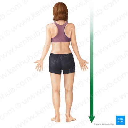
What is this direction?
Inferior

What is this direction?
Anterior

What is this direction the patient is facing (right, ignoring midline)?
Posterior
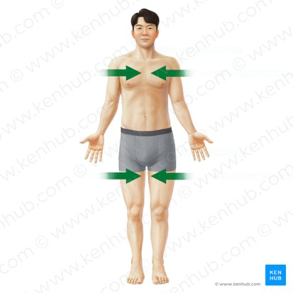
What is this direction?
Medial
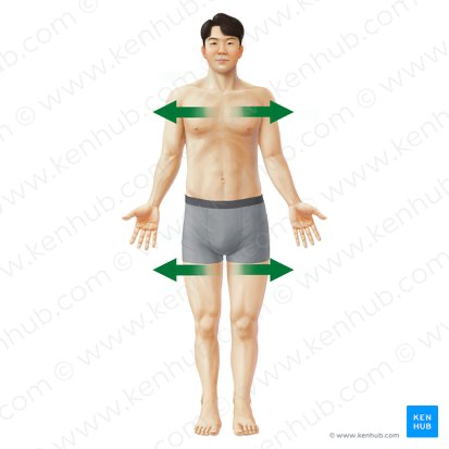
What is this direction?
Lateral

What is this direction?
Proximal
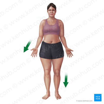
What is this direction?
Distal
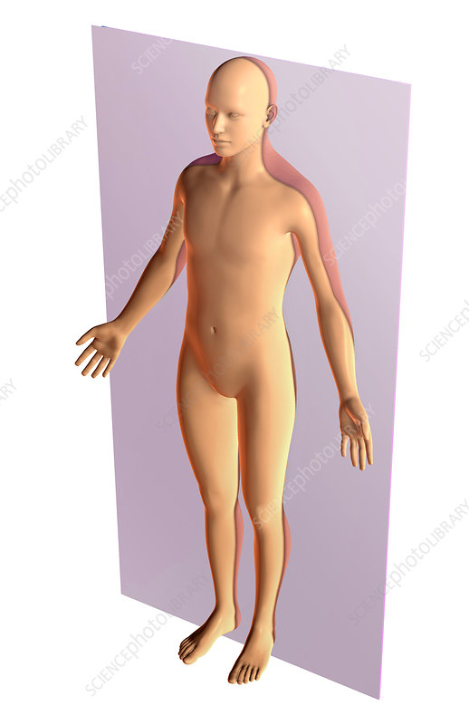
What is this plane?
Coronal plane
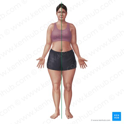
What is this plane?
Madian plane
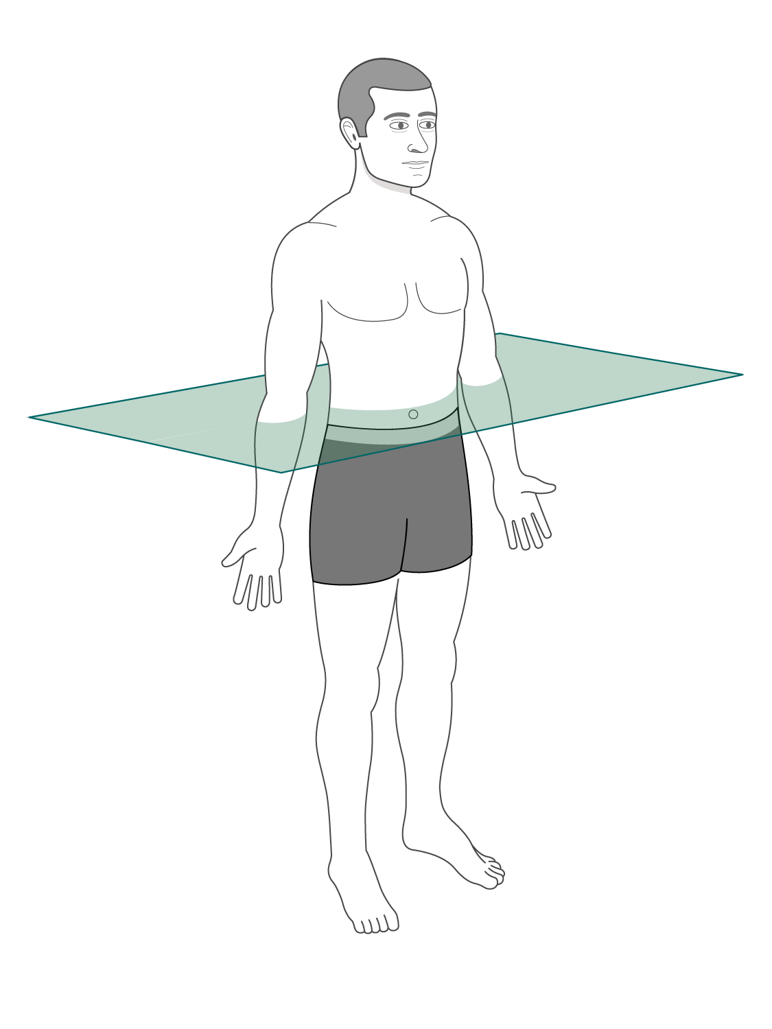
What is this plane?
Transverse plane
These two cavities are in the dorsal body cavity.
Cranial cavity and the vertebral cavity
This is a ventral body cavity.
Thoracic cavity
The thoracic cavity is composed of:
Two lateral parts and the mediastinum
The two lateral parts of the thoracic cavity contain:
A lung surrounded by a pleural cavity
The heart surrounded by the pericardial sac
Mediastinum
This cavity is divided into two parts.
Abdominopelvic cavity
This cavity contains the liver, stomach, intestines, kidneys, and other organs.
Abdominal cavity
This cavity contains the bladder, some reproductive organs, and rectum.
Pelvic cavity
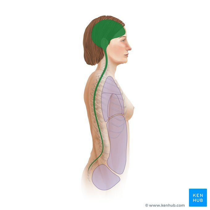
What is this cavity?
Dorsal cavity
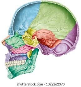
What is this cavity?
Cranial cavity

What is this cavity?
Vertebral cavity

What is this cavity?
Thoracic cavity

What is this cavity?
Abdominopelvic cavity
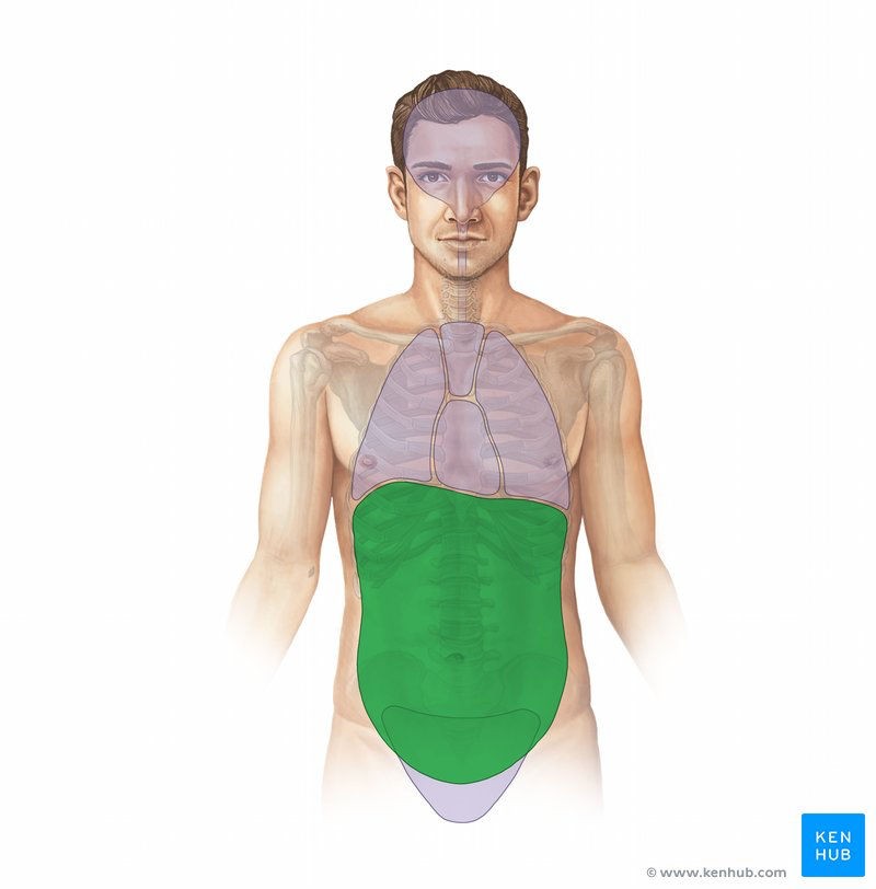
What is this cavity?
Abdominal cavity

What is this cavity?
Pelvic cavity
These cavities are serous cavities.
Ventral cavities
Serous cavities are lined by these:
Serous membranes
The serous membrane does this for the body:
Secretes fluid and reduces friction
This is a single layer of flattened cells
Mesothelium
Membrane covering the inside of each ventral cavity
Parietal serosa
The membrane covering visceral organs is called:
Visceral serosa

What is this membrane?
Visceral pericardium

What is this cavity?
Pericardial cavity with serous fluid

What is this membrane?
Parietal pericardium

What is this membrane (blue)?
Parietal pleura

What is this membrane (green)?
Visceral pleura
Used to examine small structures through a microscope.
Microscopy
Illuminates tissue with a beam of light
Light microscopy
Uses beams of electrons
Electron microscopy
Looks at surface topography
Scanning EM
Looks at structures within cells
Transmission EM
Deflects electrons in the beam to different extents
Heavy metal salt stain
Minor distortions of preserved tissues
Artifacts
Electromagnetic waves of very short length that is best for visualizing bones and abnormal dense structures.
X-rays
Takes successive X rays around a person's full circumference
Computed (axial) tomography
Forms images by detecting radioactive isotopes injected into the body
Positron Emission Tomography
Produces high-quality images of soft tissues
Magnetic resonance imaging (MRI)
Body is probed with pulses of high-frequency sound waves that echo off the body's tissues
Sonography (ultrasound imaging)
Three major components of a cell
Plasma membrane, Cytoplasm, Nucleus
Forms the boundary of each cell
Plasma membrane
This component is made of cytosol + organelles.
Cytoplasm
Composed of phospholipid bilayer interspersed with proteins, carbohydrates, and cholesterol.
Plasma membrane
Types of membrane proteins
Integral proteins and peripheral proteins
Movement of larger molecules
Endocytosis, exocytosis
Mechanism by which molecules or particles not able to pass (either directly or via transporters) through membranes enter cells.
Endocytosis
“Cell eating”- large particles - non-specific
Phagocytosis
“Cell drinking”- smaller particles - non-specific
Pinocytosis
Substance binds to receptor molecules on membrane; Invaginates and forms a coated pit; Pinches off to become a coated vesicle; specific molecules
Receptor-mediated endocytosis
Mechanism that moves substances out of the cell
Exocytosis
Lies internal to plasma membrane
Cytoplasm
Jelly-like fluid in which other cellular elements are suspended; Consists of water, ions, enzymes (control chemical reactions), and non-enzyme proteins
Cytosol
2 categories of organelles
Membrane bound and non-membrane bound
“Network within the cytoplasm”- flattened envelopes; membrane bound
Endoplasmic reticulum (ER)
Ribosomes stud the external surfaces; synthesis of proteins to be secreted out of cell; continuous with nuclear envelope
Rough ER
Consists of tubules in a branching network; No ribosomes are attached; therefore, no protein synthesis; Lipid (fat) synthesis, detoxifying enzymes, calcium storage; membrane bound
Smooth ER
A stack of three to ten disk-shaped envelopes; receives transport vesicles from ER; Post-translational processing of proteins; Sorts products of rough ER and sends them to proper destination; membrane-bound organelle
Golgi apparatus
Two membranes (inner/outer) and matrix (inner membrane) folded into cristae; generates metabolic energy = ATP; Enzymes of Citric acid cycle (Kreb’s cycle) in matrix; Electron transport chain on cristae of inner membrane (enzymes involved with ATP production); membrane bound
Mitochondria
Membrane-walled sacs containing acid hydrolases (enzymes); Digest unwanted substances (both intracellular substances plus substances brought into cells)
Lysosomes
Membrane-walled sacs of oxidase enzymes (peroxidases)
Peroxisomes
Constructed of proteins and ribosomal RNA; Site of protein synthesis; non-membrane organelle
Ribosomes