GBS 709 Week 3
 Call Kai
 Learn
Learn Practice Test
 Spaced Repetition
 Match
Match Flashcards
 Knowt Play
1/56

There's no tags or description
Looks like no tags are added yet.
Name | Mastery | Learn | Test | Matching | Spaced | Call with Kai | Chat |
|---|
No analytics yet
Send a link to your students to track their progress
57 Terms
secretory pathway + 3 major functions w/ examples of molecules involved for each
starts at ER, delivers transmembrane and soluble proteins to compartments and for secretion from cell
transports proteins for release from cell
biologically active peptides (hormones, neurotransmitters)
developmentally active factors (morphogenic, growth factors)
degradative proteins (proteases, lipases)
delivers proteins to cell surface
environment sensing receptors, signal transducers
molecules in cell-cell and cell-matrix communication
delivers proteins to intracellular compartments
processing enzymes (glycosylases, isomerases, proteases)
internal regulators (channels, ATPases)
organelle identifiers, trafficking proteins
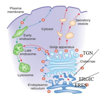
8 components of secretory pathway
plasma membrane
secretory vesicles
secretory granules (optional)
trans-Golgi network (TGN)
golgi (multi-compartment organelle)
ER
ER-Golgi Intermediate Compartment (ERGIC)
ER Exit Site (ERES)
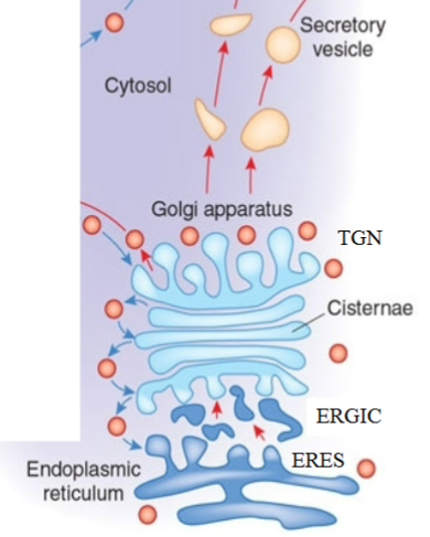
How are “correct” newly synthesized proteins moved to ER membrane and ER lumen? (from ~100,000 made in cell)?
ER is entry point for secretory pathway
all proteins (except few mitochondrial proteins) are synthesized on cytoplasmic ribosomes

How to target proteins into the Secretory Pathway?
ER is only entry compartment for all proteins that are secreted or reside in all subsequent secretory and endo-lysosomal compartments
proteins that enter secretory pathway have signal sequences that interact with Signal Recognition Particle (SRP) that targets them to ER
occurs before protein is fully synthesized (co-translational targeting)
once ~30 AAs of nascent chain emerge from ribosome, it is bound by SRP and translation is temporarily arrested (represents ~ 70 AAs total, since 30-40 AAs are buried within ribosome)
SRP-ribosome-nascent protein complex docks to SRP receptor protein complex and ribosome receptor on ER membrane. Nascent peptide inserts into translocon spanning ER membrane
after nascent peptide inserts into translocon, SRP is released and translational arrest is relieved, allowing translation of ER bound protein to resume and continue to translocate through translocon pore. SRP and SRP receptor recycled for another cycle of protein insertion (co-translocational, ribosome has to sit on membrane while being translated)
after insertion into translocon, nascent peptide continues to elongate through pore in a hairpin loop orientation with signal sequence held within translocon
signal sequence cleaved by signal peptidase, and peptide continues to elongate in a linear fashion and is released into ER lumen or retained in ER

Signal Recognition Particle (SRP) ****
ER Targeting Signals (not exact conservation of sequence)
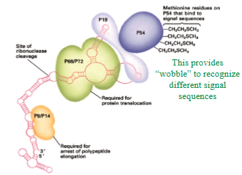
always starts with + and neutrally charged amino acids (which one) and ends with acidic amino acids, hydrophobic (15-20) AA’s not important which ones
methioninne brush: has many methionines, accommodate hydrophobic core, provides wobble in binding surface
How are different topological orientations attained in single-pass transmembrane proteins?
Targeting signals: regions of protein that facilitate insertion into membranes
at extreme N-terminus, but can be internal (signal sequence is a Start Transfer sequence (in addition to SRP binding))
Start and Stop Transfer sequences: 20-30 hydrophobic AAs (membrane anchor sequences)

ER
is a single compartment!
has thousands of entry points for proteins (where ribosome attaches and nascent chair begins translocation through translocon)
once translocated (or inserted into ER membrane), proteins have access to entire ER lumen/network
core glycosylation
occurs in the ER, co-translational
vast majority of transmembrane and secretory proteins are glycosylated, required for folding, stability and/or function
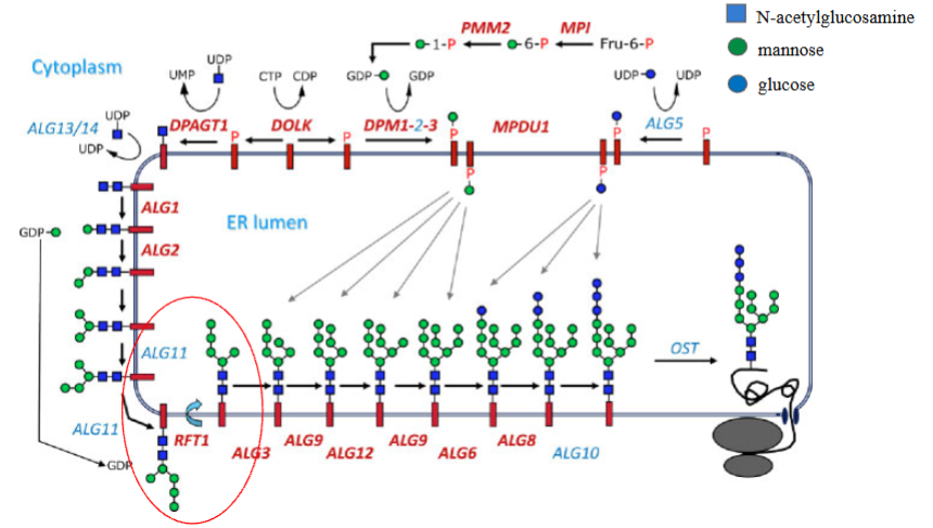
large oligosaccharide linked to lipid (dolichol) → entire glyco-chain transferred to asparagine residues in nascent peptide, occurs in ER lumen
defects cause Congenital Disorders of Glycosylation (CDG) as it is essential for protein quality control
removal/addition of terminal glucoses regulates folding cycles of proteins

3 steps for formation of dolichol precursor in core glycosylation
initial sugars (N-acetulglucosamine and mannose) added to dolichol on cytosolic side of ER
partial oligo-chain ”flips” across membrane of ER
additional sugars (mannose and glucose) added within lumen of ER
Congenital Disorders of Glycosylation (CDG)
low muscle tone or floppiness (hypotonia)
poor growth, failure to thrive
developmental delays
liver disease with elevated liver enzymes
abnormal bleeding or blood clotting
misaligned or crossed eyes (strabismus)
seizures
stroke-like episodes
calnexin-calreticulin cycle
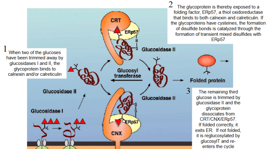
when 2 of glucoses have been trimmed away by glucosidases I and II, glycoprotein binds to calnexin and/or calreticulin
glycoprotein exposed to folding factor, ERp57 (thiol oxidoreductase that binds to calnexin and calreticulin). If glycoproteins have cysteines, formation of disulfide bonds catalyzed through formation of transient mixed disulfides with ERp57
remaining third glucose trimmed by glucosidase II and glycoprotein dissociates from CRT/CNX/ERp57
if folded correctly, it exits ER
if not folded, it is reglucosylated by glucosylT and re- enters cycle
folding sensor
UDP-glucose:glycoprotein glucosyl-transferase (UGGT) in ER lumen mediates quality control
only interacts with unfolded proteins, glycosylates them, and ‘tags’ protein for renewed interaction with CRT and CNX to allow protein folding
if folding does not occur within a predetermined time period, protein will be targeted for degradation. Identity of “timer” is unknown
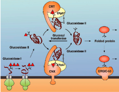
CRT and CNX quality control mechanisms works together with glucosidase II, UGGT and ERp57 to: (3 things)
increase folding efficiency of glycoproteins by extending exposure of proteins to folding machinery
prevent premature oligomeric assembly
prevent deployment of potentially malfunctioning proteins that could be detrimental to the cell and/or organism
ERAD (ER-associated degradation)
targets misfolded/unassembled proteins for degradation
glycoprotein is retro-translocated through translocon and degraded by proteasome
proteins are degraded in cytosol after being retro-translocated from ER lumen! ERAD is a misnomer!

How to get correctly folded proteins out of ER and transport them to next compartment of secretory pathway?
protein exit from ER occurs only at ERES where cargo proteins are sorted into vesicles, 50-100 ER exit sites (ERES)
ER exit is first time vesicular transport used in pathway; all subsequent transport is vesicular

how are proteins selected for vesicular traffic?
“fuzzy” buds – coating machinery that mediates cargo selection
cargo selection requires a “hold” of cargo proteins in a patch of ER membrane that will bud out as a vesicle
SORTING AND BUDDING
mechanism provided by cytosolic coat proteins that “hold” proteins while they form a lattice encloses newly forming “coated” vesicle

General coating mechanism (formation of vesicles from ERES)
coating initiated by guanine nucleotide exchange factor that activates small GTPase by facilitating GDP to GTP exchange (Sec12 activates Sar1)
GTPase recruits coat (Sar1 recruits Sec23/Sec24 – first layer of coat)
Sec24 binds cargo proteins to be sorted/sequestered into bud (transmembrane proteins bind Sec24 directly, and soluble proteins bind transmembrane cargo receptors that bind Sec24)
Sec23/Sec24 recruit Sec13/31 (second layer of the coat)
have to activate GTPAse, GTPAse recruits first layer, first layer recruits second layer
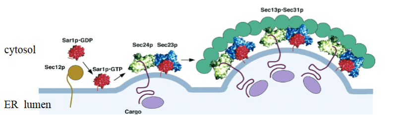
Cargo proteins have been sorted into ER-derived coated vesicles that fuse to make what larger structures?
ERGIC, moves cargo to Golgi
What would happen to newly synthesized proteins destined for secretion if Sec12 didn’t activate Sar1?
get stuck in lumen because of no vesicles
What would happen to newly synthesized proteins destined for secretion if Sec23 was mutated and couldn’t recruit Sec13/31?
the coat would not form properly in vesicle formation and protein will be stuck in ER
What would happen to Sec24 (and the other coat components) if Sec12 didn’t activate Sar1?
it would be free-floating in the cytosol
Golgi complex
main station in secretory pathway
series of flattened compartments (cisterna) adjacent to nucleus
composed of 3 regions: cis (entry), medial (modifications), trans (exit)
each cisterna is a separate compartment and has different proteins
major function in terminal glycosylation and proteolytic processing, terminal oligosaccharide processing of cargo proteins
How do proteins move through Golgi - cisternal maturation “escalator” model
Anterograde - Cargo inside cisternae
Retrograde - Golgi enzymes in COPI vesicles
cargo enters Golgi by being delivered to cis-most cisterna and stays within that cisterna as it’s being remodeled by continuous entry of distal Golgi proteins and exit of proximal enzymes
each cisterna has different protein composition

How do you selectively move Golgi enzymes between cisternae?
Vesicle formation at the Golgi:
activate small GTP-ase protein (Arf1 being activated by Gea)
GTPase recruits the coat (heptameric coatomer)
cytosolic tails of transmembrane cargo proteins (or cargo receptors for soluble proteins) bind coat
Which way are the ER-derived vesicles going and what do they carry?
going through the Golgi network and has cargo proteins (proteins destined for secretion or cell surface)
Which way are the Golgi-derived vesicles going and what do they carry?
vesicles carry enzymes from higher cisterna going to lower cisterna
How many types of Golgi-derived vesicles will there be in a Golgi stack? And what would each type carry?
as many vesicles as cisterna as each cisterna buds vesicles
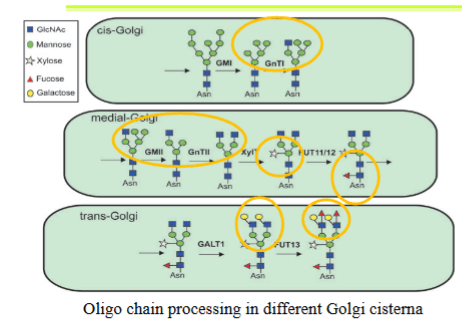
oligosaccharide chain processing
linear in cis to trans direction since glycosyl transferases are concentrated in different cisternae
composition of oligo chain dependent on protein (rules of what sugar will be added to which protein or which chain if a protein is glycosylated on multiple asparagines are unclear)
oligosaccharides promote folding and stability of glycoproteins
sugars are removed one at a time and other sugars are added one at a time (different from en masse transfer from dolichol intermediate)
endosomal pathway
starts at PM and recycles transmembrane and soluble proteins back to surface OR delivers transmembrane and soluble proteins to lysosomes
all cargo proteins move from the ER through Golgi together
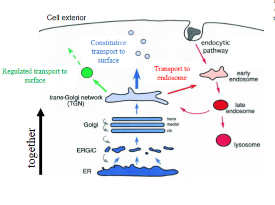
trans-Golgi network
secretory and endosomal pathways are connected at the TGN (trans-Golgi network)
proteins are sorted into different destinations at TGN
single tubulated compartment composed of distinct subdomains that mediate cargo protein sorting to distinct destinations
3 destinations
Regulated secretion
protein has to have an aggregation signal that diverts it into regulated secretion pathway (hydrophobic motifs promote aggregation)
occurs only in some cells (via secretory granules), only in response to a stimulus

Constitutive secretion
default if a protein doesn’t have specific signals to route it into the end-lysosomal pathway or into the regulated secretion pathway
occurs in all cells, all the time as no stimulus needed

Endo-lysosomal pathway
protein has to be tagged with mannose-6 phosphate

Where would a protein that normally was targeted to granules via aggregation go if its signal sequence was not recognized by SRP?
if SRP did not bind to ER and is not recognized, translation will not be halted so it will not be moved to ER membrane and will stay in cytosol and be degraded
Where would a protein that normally was secreted constitutively go if an aggregation sequence was engineered into it?
will start aggregating and then be diverted into regulated secretion pathway and end up stored in granules
Where would a protein that normally was targeted to granules via aggregation go if its aggregation sequence was removed?
constitutive pathway
Lysosomal proteins
inserted into ER like all the other soluble and transmembrane proteins
exit through ERES with other proteins, move via ERGIC to the Golgi, traverse the entire Golgi complex with other proteins
sorted to endosomes at the TGN.
Lysosomes
degradative organelles
contain acid hydrolases that degrade lipids, proteins, carbohydrates, nucleic acids, at low pH
degrade items from outside of the cell that are delivered through endocytosis
degrade components from inside the cell that are delivered through autophagy
have low internal pH due to proton ATPase that pumps H+ from cytosol into lysosomal lumen
many lysosomes in one cell – separate compartments
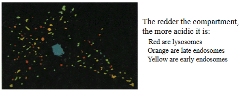
Why is acidic lumen of lysosomes and acidic pH maxima of hydrolases advantageous?
enzymes will be ineffective if lysosomes damaged and hydrolases released into cytosol which has neutral pH
gangliosides
complex glycolipids containing a ceramide or sphingosine
lipid-anchoring domain attached to a complex oligosaccharide chain; synthesized in ER and glycosylated in Golgi before being delivered to the PM
defects in enzymes 1-6 (required to degrade gangliosides) cause neurological diseases

4 steps for tagging and sorting of lysosomal proteins
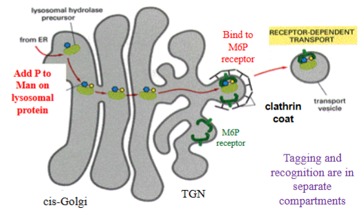
newly synthesized lysosomal glycoprotein tagged on terminal mannose of its oligo chain with phosphate in cis-Golgi
M6P-tagged lysosomal protein moves together with all other proteins to TGN.
at TGN, tag recognized by M6P receptor (transmembrane protein) that binds lysosomal protein-M6P and “holds” it in a patch coated on cytosolic side with clathrin coat
forming bud will pinch off as a clathrin-coated vesicle that will fuse with endosomes
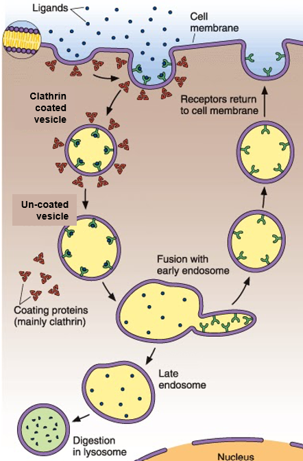
4 steps for delivery of lysosomal proteins to endosomes and release
lysosomal proteins tagged with M6P sorted into clathrin-coated vesicles at TGN
vesicles move in cell and attach and fuse with late endosomes, delivers M6P-tagged protein bound to M6P-R to late endosome
late endosomes are acidic and interaction between M6P-tagged protein and M6P-R is destabilized; lysosomal protein releaed into lumen of endosome
transmembrane M6P-R sorted into clathrin-coated vesicles that recycle it to TGN for repeated rounds of transport

endocytosis + 4 types
internalize components from outside cell and deliver them to lysosomes for degradation
protection against pathogens (phagocytosis)
nutrient uptake (LDL, transferrin)
signaling and receptor downregulation
drug delivery
4 types: phagocytosis, pinocytosis, receptor-mediated endocytosis, caveolae
4 types of endocytosis
phagocytosis: large particles; microplastics, bacteria, drug beads; involves pseudopodium and phagosomes
pinocytosis: small particles; chemicals, salts, nutrients, sugar, AAs; involves vesicles
receptor-mediated endocytosis: selected proteins; LDL-R, Tfn-R, EGF-R; PDGF-R; involves clathirin-coated vesicles, proteins, and coated pits
caveolae: selected proteins; ibumin, folic acid, viruses (SV40); involves caveolin-coated vesicles, proteins, and flask-shaped pits

3 steps of phagocytosis
Detection: immune cell (macrophage or neutrophil) detects bacteria by recognizing opsins (antibodies that tag bacteria) that bind to Fc receptors on immune cell
Engulfment: cell extends pseudopodia around bacterium. These projections form a cup that seals pathogen inside a phagosome
Maturation and Digestion: phagosome fuses with lysosome to form phagolysosome where pathogen will be digested
Pathogens internalized at cell surface, but lysosomes deeper inside cell. Phagosome has to move into cell to fuse with lysosome, facilitated by motors that attach phagosome and move it along microtubules

Chédiak-Higashi Syndrome
phagosome-lysosome fusion impaired due to microtubule polymerization defect
neutrophils and monocytes have large acidic granules with acid hydrolyses (abnormal lysosomes) but poor bacterial destruction, have recurrent infections
other defects such as progressive neuropathy
clathrin-mediated endocytosis
Cargo Selection: specific ligands bind to trans-membrane receptors on surface of plasma membrane. only ligands that have receptors will be endocytosed
Coat Assembly: receptors with bound ligand bind adaptor proteins via their cytoplasmic tails. adaptor proteins recruit clathrin, which assembles into a basket-like lattice
Membrane Invagination: growing clathrin lattice bends cell membrane inward to form a deep coated pit
Uncoating: Chaperone proteins (HSC70 and auxilin) release clathrin coat so vesicle can fuse with an early endosome (EE, a sorting station) and deliver its cargo.
Maturation: EE undergo remodeling to become late endosomes LE (get more acidic and get acid hydrolases)
Sorting from EE: select components are recycled from EE back to the PM. This process controls key physiological functions like development, movement, growth, signaling, etc.
Degradation: LE mature further or fuse with pre-existing lysosomes to deliver cargo for degradation
Clathrin lattice
made of clathrin triskelions
trickelion has 6 proteins (3 clathrin heavy chain and 3 clathrin light chains)
triskelions pack together to make a clathrin coat

clathirin heavy vs light chains
3 Clathrin Heavy Chains: Large subunits (~ 190 kDa each) joint at their C-termini to form a central hub and extend outward to form ”arms" of triskelion.
3 Clathrin Light Chains: Small subunits (~ 25 kDa each) bind non-covalently to each heavy chain near central hub

Vesicle Scission
GTPase dynamin pinches neck of invaginated pit, releasing fully formed clathrin-coated vesicle into cell
scission mediated by GTPase dynamin which uses energy of GTP hydrolysis to “strangle” vesicle off PM
GTPase dynamin localizes to necks of clathrin-coated vesicles –collar-like structure

dynamin mutations
dynamin with mutations that inactivate its ability to hydrolyze GTPase (functionally dead dynamin) localizes to necks of clathrin-coated vesicles but can’t pinch them off
causes Synaptic Vesicle Recycling: After exocytosis empties neurotransmitters into synapse, CCV must retrieve membrane from PM or neurons would run out of synaptic vesicles
Charcot-Marie-Tooth (CMT): changes in the DNM2 gene encoding dynamin-2. Forms such as axonal CMT2M and intermediate CMTDIB cause damage to peripheral nerves and result in distal muscle atrophy and sensory loss.
LDL particles (low-density lipoprotein particles)
microscopic (~20 nm) complexes of protein/cholesterol shell surrounding core of strongly hydrophobic cholesteryl ester molecules, carry cholesterol to all cells through bloodstream.

Familial Hypercholesterolemia
body unable to remove low density lipoprotein (LDL) particles from blood
causes high blood levels of LDL that facilitate narrowing of arteries from atherosclerosis at an early age
LDL receptors regulate blood cholesterol levels and control the cell's internal production of cholesterol
caused by lack of functional LDL receptors LDL particles were taken into the cells via a cell surface receptor
receptor-mediated endocytosis
Lumens
in compartments and vesicles, analogous to the “outside” of cell
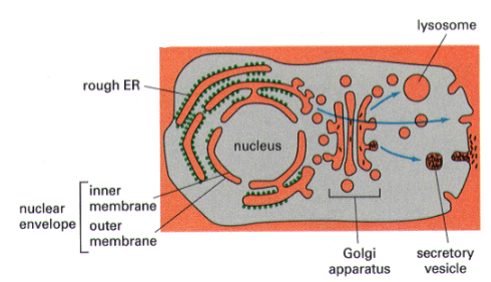
topology
maintained during vesicle transport
once inside lumen of compartment, soluble proteins do not translocate into cytosol and remain in a lumen of all subsequent compartments and vesicles
once inserted in certain orientation into ER membrane, transmembrane proteins do not flip across membrane

which organelles have distinct compartments and which ones have single compartments?
single: ER, TGN, plasma membrane
distinct: ERGIC, golgi (but each a single compartment), secretory vesicles, secretory granules, early/late endosomes, lysosomes
3 cytosolic coats
COPII: export from the ER for all cargo proteins
COPI: intra-Golgi transport for recycling of compartment components (ex: glycosyl-T)
Clathrin: export from TGN for lysosomal proteins, internalization of specific receptors from PM, export of recycling receptors from early endosomes back to PM, export of m6P receptor from early endosomes to TGN